202 Korean J Radiol 10(2), April 2009
Atypical Pulmonary Metastases from a True Malignant Mixed Tumor of the Parotid Gland
A 58-year-old male patient presented with a recurrent true malignant mixed tumor of the parotid gland. Patchy pulmonary opacities were identified with a chest radiograph. Subsequently, a CT scan of the chest showed pulmonary parenchymal consolidation with amorphous calcifications. This abnormality was confirmed to be the result of a metastatic true malignant mixed tumor by using CT-guided biopsy. The current case demonstrated an extremely rare example of atypical pulmonary metastases from a true malignant mixed tumor of the parotid gland showing an air-space pattern and calcification.
alignant mixed tumors of the salivary glands are classified into three distinct histologic types: 1) carcinoma ex pleomorphic adenoma, 2) benign metastasizing pleomorphic adenoma, or 3) true malignant mixed tumor (carcinosarcoma) (1). Of these tumors, carcinoma ex pleomorphic adenoma represents approximately 99% of cases (1). A true malignant mixed tumor is a very rare tumor composed of both malignant epithelial and malignant mesenchymal elements. The most common malignant epithelial component is squamous cell carcinoma or adenocarcinoma, whereas the most common malignant mesenchymal component is chondrosarcoma, followed by fibrosarcoma, leiomyosarcoma, osteosar- coma, and liposarcoma (2). A true malignant mixed tumor represents only 0.04% to 0.16% of salivary gland tumors and 0.4% of malignant salivary gland neoplasms (2).
About 65% of cases occur in the parotid gland but it has also been described in the submandibular and minor glands of the palate (3). Although they do develop de-novo, an association with pleomorphic adenoma has been reported in approximately one third of patients (4).
A true malignant mixed tumor is an aggressive high-grade malignancy and has a high propensity for both local and regional recurrence and metastasis (5). However, the incidence and imaging appearance of pulmonary metastasis from a true malignant mixed tumor of salivary glands have not previously been described in English litera- ture sources. Herein we report a case of a true malignant mixed tumor of the parotid gland that developed pulmonary metastases with atypical radiologic appearance. The imaging appearance and histopathologic features of the atypical pulmonary metastases are discussed.
CASE REPORT
A 58-year-old male patient presented with a one-week history of bloody otorrhea from the right side. In the past 34 years, the patient had undergone four surgeries to remove the masses from his right parotid gland. Histopathologic examinations Wen-Chiung Lin, MD1
Chao-Shiang Li, MD3 Chih-Kung Lin, MD2 Hsian-He Hsu, MD1 Tsun-Hou Chang, MD1 Tom, Yun-Cheng Chen, MD3 Guo-Shu Huang, MD1
Index terms :
Atypical pulmonary metastases Computed tomography (CT) Parotid gland, true malignant
mixed tumor
DOI:10.3348/kjr.2009.10.2.202
Korean J Radiol 2009;10:202-205 Received July 13, 2008; accepted after revision September 24, 2008.
Departments of 1Radiology, 2Pathology, Tri-Service General Hospital and National Defense Medical Center, Taipei, Taiwan, ROC; Department of 3Radiology, Renai Branch, Taipei City Hospital, Taipei, Taiwan
Address reprint requests to : Chih-Kung Lin, MD, Department of Pathology, Tri-Service General Hospital and National Defense Medical Center, No.325, Sec. 2, Cheng-Kung Rd, 114, Taipei City, Taiwan, R.O.C.
Tel. 886-2-87927244 ext. 16480 Fax. 886-2-87927245 e-mail: [email protected]
M
Atypical Pulmonary Metastases from Malignant Mixed Parotid Gland Tumor
Korean J Radiol 10(2), April 2009 203
A B
C D
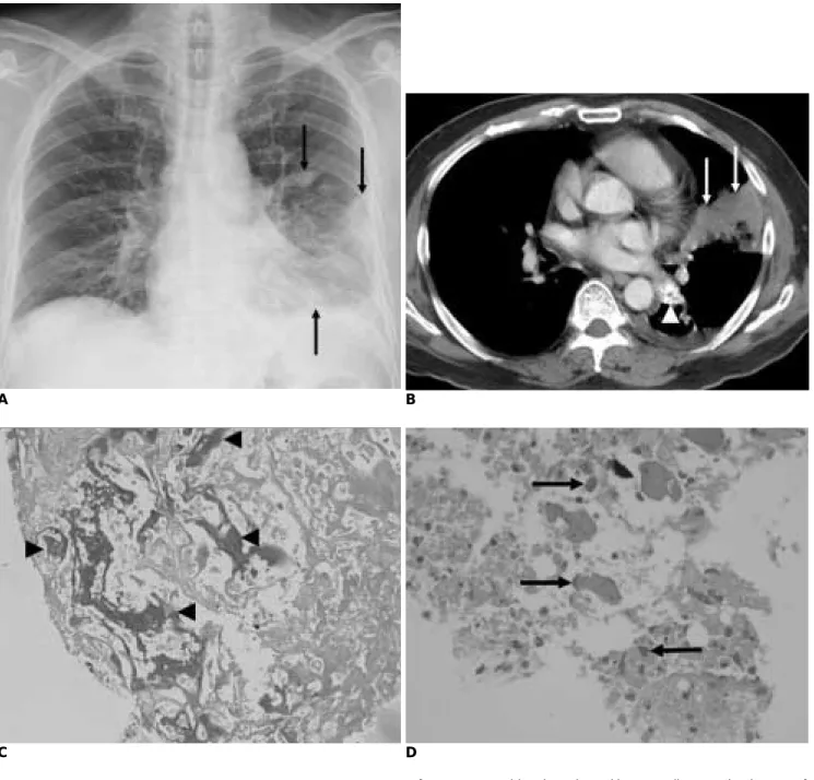
Fig. 1. 58-year-old male patient with true malignant mixed tumor of parotid gland metastasizing to lung.
A. Frontal chest radiograph showed patchy opacities in left middle and lower lung zones (arrows).
B. Contrast-enhanced CT scan of chest revealed parenchymal consolidation in left lingual segment and left lower lobe (arrows), with some amorphous calcifications (arrowhead).
C. Photomicrograph (Hematoxylin-Eosin staining; original magnifi- cation, ×200) of specimen obtained with CT-guided biopsy revealed metastatic malignant mixed tumor. Note presence of osteoid materials (arrowheads) within necrotic background.
D. Photomicrograph (Hematoxylin-Eosin staining; original magnifi- cation, ×400) of specimen showed malignant osteoblastic cells (arrows) surrounding osseous material.
E. Contrast-enhanced CT scan of neck nine months previously showed tumor of right parotid gland (arrows). Note calcification within this tumor (arrowheads).
E
performed after the first three surgeries indicated the presence of benign pleomorphic adenomas. The latest surgery was performed nine months ago to remove a 3.3- cm mass from the parotid gland. Following this surgery however, a histopathologic examination revealed a true malignant mixed tumor (carcinosarcoma) composed of poorly differentiated carcinoma and osteosarcoma.
Immunohistochemical studies were positive for P53, Ki67, and vimentin, weakly positive for cytokeratin, and negative for Actin and the S100 protein. The patient was given adjuvant radiotherapy after the latest surgery and was doing well except for symptoms associated with irradi- ation.
The patient underwent a follow-up high-resolution CT scan of the temporal bone for the presented symptoms and showed extensive opacification of the right mastoid air cells, posterior petrous bone, middle ear, and the external auditory canal. The imaging appearance raised the suspicion of a recurrent tumor. Excisional biopsy of the external auditory canal mass was performed and confirmed the diagnosis of a recurrent true malignant mixed tumor.
During this hospitalization, a chest radiograph indicated the presence of patchy opacities in his left lower lung field (Fig. 1A) not seen in previous chest radiographs; this warranted a CT scan for further characterization. A CT scan of the chest showed parenchymal consolidation with some amorphous calcifications (Fig. 1B); hence, the possibilities of fungal infection or granulomatous disease were considered initially. This patient had no fever and did not report associated symptoms of infectious or inflamma- tory respiratory tract diseases; therefore, a CT-guided biopsy of the pulmonary lesion was performed because of the concern of atypical pulmonary metastases. The biopsy generated two pieces of specimen that were used for histopathologic examination.
Microscopically, the specimen showed nearly total necrosis and the presence of atypical cells; within the mesenchymal element, some osteoid materials and foci of osteonecrosis were noted (Fig. 1C). Malignant osteoblastic cells surrounding osseous materials were also noted (Fig.
1D). Immunohistochemical studies were positive for vimentin, negative for TTF-1 (thyroid transcription factor- 1) and cytokeratin. A review of the surgical specimen from the patient’s right parotid gland nine months ago revealed similar findings. The specimen obtained during the lung biopsy showed no carcinomatous component, and did not demonstrate tumor emboli in pulmonary vessels. A radionuclide bone scan did not reveal increased activity at locations other than right neck and lung. The histopatho- logic analysis of the lung biopsy suggested a metastatic true
malignant mixed tumor. Retrospective reading of his neck CT nine months ago clearly depicted calcification within the right parotid gland tumor (Fig. 1E).
DISCUSSION
The lung is a common site for metastases. Large autopsy series of patients with extrathoracic malignancies reveal pulmonary metastases in 20-54% of cases (6, 7). Typical radiologic findings of pulmonary metastases may include multiple round variable-sized nodules and a diffuse thickening of interstitium, representing hematogenous metastases and lymphangitic carcinomatosis, respectively (7, 8). In daily practice, however, atypical radiologic features of metastases are often encountered that make distinguishing between metastases and other nonmalignant pulmonary diseases difficult. Herein we describe a case of atypical pulmonary metastases from a true malignant mixed tumor of the parotid gland demonstrating calcifica- tion and an air-space pattern of the pulmonary metastases.
Calcification of a pulmonary nodule is usually suggestive of its benign nature, most commonly a granuloma and less commonly a hamartoma. However, calcification or ossifi- cation can also occur in metastases from a variety of tumors including osteosarcoma, chondrosarcoma, synovial sarcoma, giant cell tumor of the bone, carcinomas of the colon, ovary, breast, and thyroid (7). In our case, the mesenchymal component of the true malignant mixed tumor of the parotid gland was osteosarcoma. As a result, the lung metastases show calcification or ossification; this observation was confirmed with a CT-guided biopsy. The calcification is often depicted only with CT, as in our case.
Metastases from an extrapulmonary adenocarcinoma may spread into the lung along the intact alveolar walls (lepidic growth) in a fashion similar to a bronchioloal- veolar carcinoma and exudative pneumonia (7, 8). This type of metastases may demonstrate an air-space pattern in radiologic studies. In one series, six of 65 patients with pulmonary metastases from an adenocarcinoma of the gastrointestinal tract had this pattern of metastasis (8). In addition, adenocarcinomas from the breast and ovary can also show this pattern of metastasis (9). The malignant epithelial component in our case was poorly differentiated carcinoma, and unusual scenario for the development pulmonary metastasis of air-space pattern. Rastogi et al.
(10) described a variety of atypical locations and presenta- tions of thoracic metastases from osteosarcoma in 16 patients; pulmonary metastasis of air-space pattern did not occur in their series. In our case, the extensive necrosis revealed by the lung biopsy may be one of speculative mechanisms of air-space pattern. Pulmonary infarction due Lin et al.
204 Korean J Radiol 10(2), April 2009
Atypical Pulmonary Metastases from Malignant Mixed Parotid Gland Tumor
Korean J Radiol 10(2), April 2009 205
to the presence of a tumor embolism may also demonstrate an air-space pattern (7). However, in our case, the limited pieces of specimen obtained at lung biopsy did not demonstrate foci of carcinomatous components and were negative for the presence of a tumor embolism. Although we could not explain the definitive mechanism of air-space pattern on the basis of limited histopathologic analysis, the current case demonstrated an extremely rare example of atypical pulmonary metastases from a true malignant mixed tumor of the parotid gland.
Although most cases of pulmonary metastases can be diagnosed radiologically on the basis of typical findings, atypical radiologic manifestations of pulmonary metastases make confident diagnosis difficult, as in our case. An awareness of the possibility of atypical pulmonary metastases in patients with known extrapulmonary malignancy was crucial to allow an accurate diagnosis. In cases displaying atypical radiologic features of pulmonary metastases, tissue diagnosis at bronchoscopy or percuta- neous transthoracic needle aspiration biopsy is
recommended.
References
1. Batsakis JG. Malignant mixed tumor. Ann Otol Rhinol Laryngol 1982;91:342-343
2. Gnepp DR. Malignant mixed tumors of the salivary glands: a review. Pathol Annu 1993;28:279-328
3. Fan KF, Connor SE, Macbean AD, Langdon JD. MRI findings of carcinosarcoma in the parotid gland. Oral Oncol 2006;42:323- 326
4. Kwon MY, Gu M. True malignant mixed tumor (carcinosar- coma) of parotid gland with unusual mesenchymal component:
a case report and review of the literature. Arch Pathol Lab Med 2001;125:812-815
5. Liess BD, Hirschi S, Zitsch RP 3rd, Frazier S, Konrad A.
Carcinosarcoma of the parotid gland: report of a case with immunohistochemical findings. Ann Otol Rhinol Laryngol 2007;116:702-704
6. Crow J, Slavin G, Kreel L. Pulmonary metastasis: a pathologic and radiologic study. Cancer 1981;47:2595-2602
7. Seo JB, Im JG, Goo JM, Chung MJ, Kim MY. Atypical pulmonary metastases: spectrum of radiologic findings.
Radiographics 2001;21:403-417
8. Gaeta M, Volta S, Scribano E, Loria G, Vallone A, Pandolfo I.
Air-space pattern in lung metastasis from adenocarcinoma of the GI tract. J Comput Assist Tomogr 1996;20:300-304
9. Foster CS. Mucus-secreting ‘alveolar-cell’ tumor of the lung: a histochemical comparison of tumors arising within and outside the lung. Histopathology 1980;4:567-577
10. Rastogi R, Garg R, Thulkar S, Bakhshi S, Gupta A. Unusual thoracic CT manifestations of osteosarcoma: review of 16 cases.
Pediatr Radiol 2008;38:551-558