submit.radiology.or.kr J Korean Soc Radiol 2013;68(3):213-216
213 INTRODUCTION
Malignant melanoma accounts for 1.5% of all reported can- cers with prevalence increasing in the past decade. Skin is the most common site for primary malignant melanoma, but it can involve virtually every organ system where melanin cells exist.
They can frequently metastasize to the liver, lung, brain and bone before the primary lesion in the skin is clinically evident (1). Here, we report a case of malignant melanoma representing superior mediastinal mass without extrathoracic primary lesion.
CASE REPORT
A 32-year-old man was presented with 3 weeks’ history of hoarseness and had been suffering from right ptosis for 6 months.
His laryngoscopic examination revealed right vocal cord palsy without vocal cord abnormality. He had no definite past medical history, except for vitiligo whenever he gets sunlight. He had a
history of resolved vitiligo on both cheeks and left medial malle- olar area, which was retrospectively noted.
Initial chest radiograph (Fig. 1A) showed a right apical mass- like opacity showing lobulated contour and obtuse angle with adjacent pleura. The patient subsequently had a chest CT scan, which demonstrated a lobulated mass (7.4 × 4.2 × 5.8 cm) with heterogenous enhancement in the right superior mediastinum (Fig. 1B). Tracheal deviation, and obliteration of ipsilateral tra- cheoesophageal groove by the mass was noted, and this could explain hoarseness with the involvement of ipsilateral recurrent laryngeal nerve. Two days later, magnetic resonance (MR) im- aging of the cervical spines was performed on a 1.5-T MR unit.
The superior mediastinal mass was mildly hyperintense to mus- cle on T1 and T2-weighted images (Fig. 1C). The mass con- tained a small area that was hyperintense on T1-weighted imag- es and hypointense on T2-weighted images. The mass showed invasion of right neural foramen of the 2nd and 3rd thoracic vertebrae and erosion of the adjacent right 2nd rib, which might
Case Report
pISSN 1738-2637
J Korean Soc Radiol 2013;68(3):213-216
Received July 25, 2012; Accepted December 6, 2012 Corresponding author: Dong Wook Sung, MD Department of Diagnostic Radiology, Kyung Hee University Hospital, 23 Kyungheedae-ro, Dongdaemun-gu, Seoul 130-702, Korea.
Tel. 82-2-958-8616 Fax. 82-2-968-0787 E-mail: [email protected]
Copyrights © 2013 The Korean Society of Radiology
Malignant melanoma most commonly occurs in the skin, and other organs are sec- ondarily involved. Malignant melanoma presenting in the mediastinum without an extrathoracic primary is very rare. Authors report a case of malignant melanoma of the superior mediastinum without clinical history of extrathoracic malignant mela- noma primarily and discuss its radiologic findings. CT shows lobulated heteroge- nous enhanced mass. Magnetic resonance shows mild hyperintense mass on T1 and T2-weighted images contained focal hemorrhage and necrosis, and invasion to neural foramen. In addition, positron emission tomography/computed tomography shows high standard uptake values uptake of the mass.
Index terms
Mediastinal Neoplasm Melanoma/Radiography Magnetic Resonance Imaging
Malignant Melanoma Presenting as Superior Mediastinal Mass without Extrathoracic Primary Melanoma
흉곽외 원발성 흑색종 없이 상종격동 종양으로 발견된 악성 흑색종
Myung-Won You, MD, Dong Wook Sung, MD, Young Kyung Lee, MD
Department of Diagnostic Radiology, Kyung Hee University Hospital, Seoul, Korea
Malignant Melanoma Presenting as Superior Mediastinal Mass without Extrathoracic Primary Melanoma
submit.radiology.or.kr
J Korean Soc Radiol 2013;68(3):213-216
214
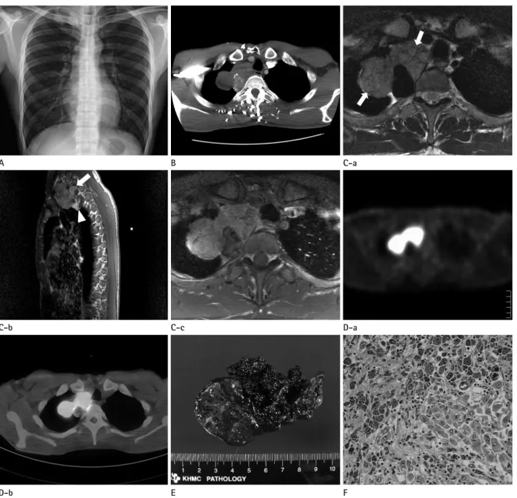
Fig. 1. Imaging findings of superior mediastinal mass in a 32-year-old man.
A. Chest radiography shows lobulated contour of mass on the right lung apex with left side deviation of trachea. The lesion shows homogenous opacity, obtuse angle with adjacent pleura and smooth interface with right upper lung. Cervicothoracic sign is positive (mass opacity above the clavicle), so the lesion would be located in the posterior portion of superior mediastinum.
B. Chest CT shows well margined lobulated and homogenously enhanced mass on right superior mediastinum. Right tracheoesophageal groove is obliterated, left side tracheal deviation and compression or invasion of great vessels (right common carotid, subclavian arteries and internal jugular vein). Some calcifications are visible on the mass.
C. Axial T2-weighted MR image at the upper T2 level (a), shows heterogenous mass (arrows) with intermediate to high signal intensity (com- pared with adjacent muscles). (b) The mass lesion has also slightly hyperintense area on sagittal T1-weighted image. Invasion of T1-2 and T2-3 neural foramina is noted (arrow). Higher signal intensity area at the lower portion of the mass (arrowhead) correlated with the calcifications on the CT scan. (c) Contrast enhanced T1-weighted MR image with fat suppression shows heterogeneous enhancement of mass.
D. (a, b) Positron emission tomography/computed tomography shows high intense signal (standard uptake values 8.25) at right apex, and not uptake at the portion of necrotic area.
E. The gross pathologic examination reveals a dark brownish lobulating mass. Multiple tan-brown areas of soft to firm tissues with areas of hem- orrhage and necrosis are noted at the cut-section of the mass.
F. The histologic specimen shows large pleomorphic cells with prominent nucleoli with some of which contain melanin pigments (H&E, x 200).
C-b
D-b A
C-c
E B
D-a
F C-a
Myung-Won You, et al
submit.radiology.or.kr J Korean Soc Radiol 2013;68(3):213-216
215
(7), melanoma with unknown primary (MUP) can be explained by two hypotheses. One theory supports de novo melanoma through malignant transformation of ectopic melanocytes and the other, more reliable theory states an undetected primary melanoma that may undergo regression by the host immune re- sponse, after it has metastasized. Also, Webb (8) explained that the direct spread of the tumor into the anterior medistinum can occur through communicating lymphatics in the neck and axil- la. Considering authors’ case had vitiligo, occult primary skin le- sion and secondary mediastinal metastasis would be more of a possibility.
Author’s case shows similar MR findings with those of the re- ported case by Takao et al. (4). Lower central portion of the mass was hyperintense on T1-weighted and hypointense on T2- weighted images in both of the cases. This portion correlated with melanin-rich area on the pathologic exam. With the larger amount of melanin, the more T1 shortening can occur. There- fore, the prediction of the degree of pigmentation within the tu- mor would be possible by using quantitative evaluation of the signal intensities (4).
The metastatic melanoma patients with vitiligo and MUP pa- tients shows better prognosis than metastatic melanoma with known primary. Author’s case is also currently doing well with 4 years disease-free state after surgical excision of the mass.
REFERENCES
1. Karuppiah SV, Buchan KG. Primary malignant melanoma:
a rare cause of mediastinal mass. Jpn J Thorac Cardiovasc Surg 2006;54:396-398
2. Loewenthal B, Shiau MC, Garcia R. Metastatic melanoma:
an unusual diagnosis for a large anterior mediastinal mass.
Radiographics 2004;24:1714-1718
3. Lau CL, Bentley RC, Gockerman JP, Que LG, D’Amico TA.
Malignant melanoma presenting as a mediastinal mass.
Ann Thorac Surg 1999;67:851-852
4. Takao H, Shimizu S, Doi I, Watanabe T. Primary malignant melanoma of the anterior mediastinum: CT and MR find- ings. Clin Imaging 2008;32:58-60
5. Fishman EK, Kuhlman JE, Schuchter LM, Miller JA 3rd, Magid D. CT of malignant melanoma in the chest, abdo- men, and musculoskeletal system. Radiographics 1990;10:
have caused right side ptosis. Positron emission tomography/
computed tomography scan (Fig. 1D) showed high standard uptake values (SUV) uptake (max SUV: 8.25) of the mass and no evidence of metastatic diseases, other than the thoracic le- sion. There was no evidence of primary melanoma outside the mediastinum.
The patient underwent total excision of the right superior me- diastinal mass (Fig. 1E, F). Gross pathologic examination showed an irregular dark brownish mass measuring 8.3 × 4 × 4 cm and weighing 54 g. Serial sections revealed multiple lobulated tan- brown areas of soft to firm tissue with areas of hemorrhage and necrotic foci. Histologic analysis of the mass revealed large pleo- morphic cells with prominent nucleoli, some of which contain melanin pigments. A Fontana-Masson stain confirmed the pres- ence of melanin pigment. Immunohistochemical staining was positive for S100 protein and HMB45. The diagnosis was made to malignant melanoma.
DISCUSSION
Melanoma most commonly represents as skin lesions, al- though there have been various incidences where they have been found in other parts of the body. Primary malignant melanoma has been reported from the brain, bronchus, rectum, gastroin- testinal tract and esophagus, but melanomas outside the skin are usually secondary deposits (1). Metastatic involvement of medi- astinal and hilar lymph nodes is less common than pulmonary parenchymal involvement (2). Further, primary malignant mel- anoma of the mediastinum is extremely rare, with only a few re- ports (1, 3-6).
Author’s case is similar to the case reported by Lau et al. (3) in that both patients had a history of vitiligo in common. However, the patient of Lau et al. had two skin lesions removed previously basal cell carcinoma and squamous cell carcinoma. They con- cluded that the malignant melanoma in the mediastinum could be secondary deposit from regressed primary lesion rather than de novo melanoma. Our patient had no history of skin malig- nancy at all, including malignant melanoma. However, melano- ma seen in the mediastinum is almost always metastatic (3). In spite of no detection of primary melanoma, author’s case is more likely to be metastasis from regressed or occult primary skin le- sion rather than de novo melanoma. According to Clerico et al.
Malignant Melanoma Presenting as Superior Mediastinal Mass without Extrathoracic Primary Melanoma
submit.radiology.or.kr
J Korean Soc Radiol 2013;68(3):213-216
216
and analysis of 24 patients. Med Oncol 2012;29:2978- 2984
8. Webb WR. Hilar and mediastinal lymph node metastases in malignant melanoma. AJR Am J Roentgenol 1979;133:
805-810 603-620
6. Kalra A, Kalra A, Palaniswamy C, Gajera M, Rajput V. Pri- mary malignant melanoma presenting as superior medias- tinal mass. Int J Surg Case Rep 2011;2:239-240
7. Clerico R, Bottoni U, Paolino G, Ambrifi M, Corsetti P, De- virgiliis V, et al. Melanoma with unknown primary: report
흉곽외 원발성 흑색종 없이 상종격동 종양으로 발견된 악성 흑색종
유명원 · 성동욱 · 이영경
악성 흑색종은 주로 피부에서 가장 흔히 발생하며 그 이외의 부위는 대개 이차성으로 발생한다. 흉곽외 원발성 흑색종 없 이 종격동에 발견된 악성 흑색종은 매우 드물다. 저자들은 흉곽외 악성 흑색종의 과거력이나 현 병력이 없었던 환자에서 상종격동 종양으로 발견된 악성 흑색종의 증례를 CT, magnetic resonance, positron emission tomography/computed to- mography의 영상의학적 소견과 함께 보고하고자 한다.
경희대학교병원 영상의학과