Malignant Melanoma of Unknown Primary Origin Presenting as Cardiac Metastasis
3
0
0
전체 글
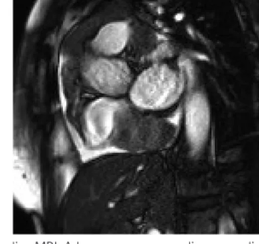
(2) Eun Young Lee, et al. 279. Fig. 1. Chest computed tomography. An inhomogeneous enhancing mass in the right atrium (arrow) and a massive amount of pericardial effusion were identified.. Fig. 2. Two-dimensional cardiac echocardiogram. In a subcostal view of the heart, a large mass measuring 42×31 mm was visualized in the right atrium.. Discussion Malignant melanoma has aggressive biological behavior and the greatest tendency for metastasis to the heart.1) Although autopsy studies reported an incidence of 50% to 71%, cardiac metastasis is diagnosed in less than 1% of patients with malignant melanoma because less than 10% of these patients present with cardiac symptoms.2) Such metastases most frequently occur after multifocal hematological dissemination and may develop anywhere in the heart.3) Melanotic metastases can invade the wall of any of the 4 cardiac chambers, and the RA is involved most frequently.4) The clinical signs and symptoms of cardiac metastasis are unclear and non-specific, although when present, the clinical signs and symptoms include fatigue, weakness, pericardial effusion, congestive www.e-kcj.org. Fig. 3. Cardiac MRI. A large mass surrounding ascending aorta spread into transverse sinus and around pulmonary trunk and right pulmonary artery.. heart failure, cardiac arrhythmia, superior vena cava syndrome, right ventricular outflow and inflow obstruction, and transient ischemic attack.5) However, patients with malignant melanoma who have cardiac metastases may present symptoms only caused by tumors in other organ systems. Although cardiac involvement occurs during the course of the disease, it is rare that the initial manifestation is cardiac metastasis. A tumor’s anatomic location and extent of invasion determine the feasibility of surgical intervention, which should optimally be performed during the early stages of the disease.6) A complete resection of an intracardiac melanoma prevents potential morbidities that are associated with progressive intracardiac growth, such as superior vena cava syndrome, right ventricular outflow and inflow obstruction, dysrhythmia, cardiac tamponade, and heart failure.6) Even when total resection is not possible, conservative surgery can relieve symptoms and prevent imminent cardiac failure. Conservative surgery improves the quality of a patient’s life, as in our patient’s case. Although more than 90% of melanomas have a cutaneous origin,7) melanomas may sometimes present metastatically in the absence of a primary lesion, termed melanomas of unknown primary origin. Most authors estimate that 2-6% of patients are diagnosed with metastatic melanoma of unknown primary site. In particular, such a metastatic melanoma in the heart such as in this patient without a known primary cutaneous origin is a rare presentation and chiefly an anecdotal finding of metastatic melanoma. Several reported cases can be found in the literature about cardiac involvement with cutaneous primary malignant melanoma and multiple metastasis in Korea, as well as worldwide.8)9) As far as we know, malignant cardiac melanoma without a primary origin has not yet been reported in Korea. The survival of patients with unknown primary melanoma was demonstrated to be similar to that of patients with known primary tuhttp://dx.doi.org/10.4070/kcj.2012.42.4.278.
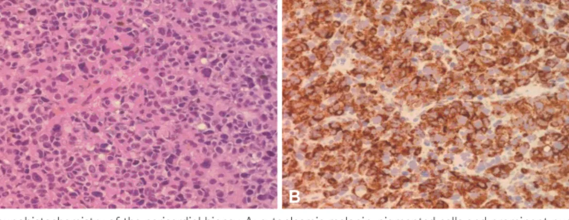
(3) 280 Malignant Melanoma of Unknown Primary Origin Presenting as Cardiac Metastasis. A . B . Fig. 4. Pathology and immunohistochemistry of the pericardial biopsy. A: cytoplasmic melanin-pigmented cells and prominent nucleoli were found (×200). B: in immunohistochemistry, antibodies to HMB45 were positive (×200).. mors when corresponding stages were compared.10-12) Those patients with metastases to any other visceral sites are described to have a 1 year survival rate of 41% and a median survival of approximately 6 months.11)13)14) Accordingly, we expected that the outcome of this patient might be very unfavorable. Although total resection was not possible because of extensive cardiac metastasis, a pericardial window operation could resolve her symptoms. After surgery, chemotherapy was planned, including cisplatin, dacarbazine, casmustine, and tamoxifen. However, the patient refused further chemotherapy after completion of two sessions. The patient has survived for 14 months after diagnosis. She is attending our hospital’s outpatient oncology department. As only few reports about cardiac metastasis of malignant melanoma without cutaneous origin have been published, we are uncertain if conservative surgery is associated with prolonged survival and if the role of surgery for survival is worth further investigation.. References 1. Ozyuncu N, Sahin M, Altin T, Karaoguz R, Guldal M, Akyurek O. Cardiac metastasis of malignant melanoma: a rare cause of complete atrioventricular block. Europace 2006;8:545-8. 2. MacGee W. Metastatic and invasive tumours involving the heart in a geriatric population: a necropsy study. Virchows Arch A Pathol Anat Histopathol 1991;419:183-9. 3. Savoia P, Fierro MT, Zaccagna A, Bernengo MG. Metastatic melanoma of the heart. J Surg Oncol 2000;75:203-7. 4. Malouf JF, Thompson RC, Maples WJ, Wolfe JT. Diagnosis of right atrial metastatic melanoma by transesophageal echocardiographic-guided transvenous biopsy. Mayo Clin Proc 1996;71:1167-70.. http://dx.doi.org/10.4070/kcj.2012.42.4.278. 5. Qu G, Kaur JS, Seward JB. Metastatic melanoma presenting as cardiac mass and hemobilia. Am J Med Sci 2003;325:157-9.. 6. Basarici I, Demir I, Yilmaz H, Altekin RE. Obstructive metastatic malignant melanoma of the heart: imminent pulmonary arterial occlusion caused by right ventricular metastasis with unknown origin of the primary tumor. Heart Lung 2006;35:351-4. 7. Chang AE, Karnell LH, Menck HR. The national cancer data base report on cutaneous and noncutaneous melanoma: a summary of 84,836 cases from the past decade: the American College of Surgeons Commission on Cancer and the American Cancer Society. Cancer 1998;83: 1664-78. 8. Lee KS. Resection of cardiac metastasis of malignant melanoma. Korean Circ J 2000;30:1170-4. 9. Kim OG, Hong JM, Lee SJ, Hong JS. Cardiac metastasis of malignant melanoma: a case report. Korean J Thorac Cardiovasc Surg 1999;32: 840-3. 10. Baab GH, McBride CM. Malignant melanoma: the patient with an unknown site of primary origin. Arch Surg 1975;110:896-900. 11. Schlagenhauff B, Stroebel W, Ellwanger U, et al. Metastatic melanoma of unknown primary origin shows prognostic similarities to regional metastatic melanoma: recommendations for initial staging examinations. Cancer 1997;80:60-5. 12. Katz KA, Jonasch E, Hodi FS, et al. Melanoma of unknown primary: experience at Massachusetts General Hospital and Dana-Farber Cancer Institute. Melanoma Res 2005;15:77-82. 13. Manola J, Atkins M, Ibrahim J, Kirkwood J. Prognostic factors in metastatic melanoma: a pooled analysis of Eastern Cooperative Oncology Group Trials. J Clin Oncol 2000;18:3782-93. 14. Balch CM, Soong SJ, Gershenwald JE, et al. Prognostic factors analysis of 17,600 melanoma patients: validation of the American Joint Committee on Cancer melanoma staging system. J Clin Oncol 2001;19: 3622-34.. www.e-kcj.org.
(4)
수치
관련 문서
We experienced a patient with upper thoracic primary IDEM spinal cord melanoma who was diagnosed to be with hydrocephalus and without intracranial lesions.. Initial symptoms of
Usually, in case of metastatic cancer the pa- tient reported a history of malignant melanoma and in the ovary, there is not a benign cystic teratoma [4].. At the pres- ent, no
In this report, treatment with pembrolizumab after radical surgery was not effective for this patient who had a primary cervical melanoma that metastasized to bone and lung
He was found to have multiple primary heart tumors obstructing the right su- perior pulmonary vein in the left atrium, which were diagnosed as malignant peripheral nerve
