Korean J. Environ. Biol. 35(3) : 335~340(2017) https://doi.org/10.11626/KJEB.2017.35.3.335
INTRODUCTION
A brown algal genus Dictyota J.V. Lamouroux (Dictyotales, Dictyotaceae) is common in tropical to warm temperate regions worldwide (De Clerck and Coppejans 1999; De Clerck et al. 2006; Guiry and Guiry 2017). This genus was established based on D. dichotoma by Lamouroux (1809).
At that time, twenty-three species were involved in the genus, but later, most of them were moved to other genera (Papenfuss 1977).
Many species of Dictyota show substantial morphologi- cal variations in the generic characteristics, distinguishing it from similar genera (Dilophus J. Agardh, Glossophora J.
Agardh, Pachydictyon J. Agardh and Glossophorella Niza- muddin et Campbell) within the tribe Dictyoteae (Hörnig et al. 1992; De Clerck and Coppejans 2003). This suggested a necessary taxonomic revision of the generic delineation of Dictyota (Hörnig et al. 1992; Phillips 1992; De Clerck and
Coppejans 2003). Thus, Dictyota merged the four morpho- logically similar genera on the basis of molecular phylo- genetic data (De Clerck et al. 2006; Silberfeld et al. 2014).
Instead, two new genera Canistrocarpus De Paula et De Clerck and Rugulopteryx De Clerck et Coppejans which are supported by molecular analysis and combined reproductive and vegetative features were established (De Clerck et al.
2006). Dictyota and the two genera have been recognized in the Dictyoteae (Guiry and Guiry 2017). Dictyota is charac- terized by parenchymatous flattened thalli with a single api- cal lenticular growing cell oriented transversely (De Clerck et al. 2006).
A total of 92 species are currently accepted based on the emended generic delineation of Dictyota (Guiry and Guiry 2017), and of these, five species have been recorded in Ko- rea (Lee and Kang 1986; Lee and Hwang 2010; Kim et al.
2013). During the present study, a brown alga belonging to the tribe Dictyoteae was collected from Geoje, located on the southern coast of Korea. Morphological observation and molecular analysis were made to identify this alga. The present study reports on this D. bartayresiana newly re-
* Corresponding author: Ki Wan Nam, Tel. 051-629-5922, Fax. 051-629-5922, E-mail. [email protected]
ⓒ2017. Korean Society of Environmental Biology.
Dictyota bartayresiana, a Brown Algal Species Newly Recorded in Korea
Pil Joon Kang and Ki Wan Nam*
Department of Marine Biology, Pukyong National University, Busan 48513, Republic of Korea Abstract - A marine brown alga was collected from Geoje, located on the southern coast of Korea. This species shares the generic features of Dictyota and is morphologically characterized by the combined features of erect or prostrate thallus with smooth margin, dichotomous to subdichotomous branches not twisted at the terminal part, absence of surface proliferations, 1-3 cell layered cortex and unilayered medulla, and lack of involucrum in sporangia subtended by a single stalk cell. In a phylogenetic tree based on rbcL sequences, this species nests in the same clade with Dictyota bartayresiana. The genetic distance between both sequences within the clade was calculated as 1.2%, considered to be within the intra-species range for the genus. Based on these morphological and molecular analyses, this Korean alga is identified as D. bartayresiana described originally from the West Indies. This is the first record of D. bartayresiana in Korea.
Key words : marine alga, the first record, morphology, rbcL sequences
<Original article>
criteria. Sections of the thallus were mounted in 20% corn syrup for permanent preparation.
Total genomic DNA was extracted from silica-gel-pre- served samples using the DNeasy Plant Mini Kit (Qiagen, Hilden, Germany) according to the manufacturer’s pro- tocol. Before extraction, dried material was crushed with liquid nitrogen using a mortar and pestle. Concentrations of extracted DNA were assessed using gel electrophoresis on a 1% agarose gel. PCR amplifications were performed in a TaKaRa PCR Thermal Cycler Dice with an initial de- naturation step at 94℃ for 5 min followed by 35 cycles at 94℃ for 1 min, 56℃ for 1 min, and 72℃ for 2 min and a final extension at 72℃ for 7 min. The reaction volume was 20 μL, consisting of 20 ng of genomic DNA, 2 μL of 10×
PCR buffer, 2 μL of 200 μM dNTP, 1 μL of each forward and reverse primer, and 0.5 units of Taq polymerase (Takara Korea, Seoul, Korea). Amplifications were examined using gel electrophoresis in a 1% agarose gel, and amplified rbcL region products were purified using a QIAquick Gel Extrac- tion Kit (Qiagen, Hilden, Germany). The PCR primer was also used for sequencing. It is as follows: rbcL (forward:
5ʹ GGAGGATTAGGGTCCGATTCC 3ʹ, reverse: 5ʹ CTTC CGTCAATTCCTTTAAG 3ʹ) (Lin et al. 2001). The PCR products were moved to Macrogen Sequencing Service for sequencing (Macrogen, Seoul, Korea). Sequences for the rbcL region were aligned using BioEdit (Hall 1999). Phylo- genetic analyses were performed using the neighbor-joining (NJ), maximum-likelihood (ML) and maximum parsimony (MP) methods. Bootstrap values were calculated with 1,000 replications. RbcL sequences of other species were obtained from GenBank. Dictyopteris polypodioides was used as an outgroup.
tened, attached by mat-like basal filamentous rhizoids, with- out stoloniferous holdfasts, crisp in texture, dark brown (Fig. 1A); iridescence absent; branches dichotomous to subdichotomous, without dentate margin, not twisted at the terminal part, without surface proliferations (Fig. 1B); inter- dichotomies 1-2 cm length, 0.3-0.8 cm width, with a 2.5- 3.3 ratio; branch apex rounded, with a single lenticular grow- ing cell oriented transversely (Fig. 1C); cortex pigmented, 1-2 cell layered in the upper portion, 1-3 cell layered in the middle portion, 2-3 cell layered in the lower portion (Fig.
1F); cortical cells 10-20 μm×10-25 μm; medulla not pig
mented, unilayered throughout the thallus (Fig. 1F); medul- lary cells rectangular, with thick walls, 800-1,100 μm×
900-1,200 μm at the upper portion, 1,200-1,400 μm×900- 1,200 μm at the lower portion; sporangia formed solitary only on single sides of branches, subtended by a single stalk cell, without involucrum, 80-120 μm in diam (Fig. 1D, E).
Antheridia and oogonia were not observed during the pres- ent study.
The morphological features, such as the numbers of lay-
ers in the medulla and cortex and the relative abundance of
surface proliferations, had been adopted for the traditional
generic classification within the tribe Dictyoteae (De Clerck
and Coppejans 1997, 1999). However, these features are
not practical in delineating the genera, because of substan-
tial variations (Hörnig et al. 1992; Phillips 1992; De Clerck
and Coppejans 2003). Therefore, the four similar genera
Dilophus, Glossophora, Pachydictyon, and Glossophorella
were merged to Dictyota based on molecular phylogenetic
data (De Clerck et al. 2006; Silberfeld et al. 2014). Instead,
two new genera, Canistrocarpus and Rugulopteryx, were
proposed with an emendation to the broad generic concept
of Dictyota (De Clerck et al. 2006). Dictyota involves the
species characterized by parenchymatous, flattened thalli with apical lenticular growing cells oriented transversely, while the two genera are morphologically supported by the combination of cortex and medulla structure, presence or absence of surface proliferations, numbers of stalk cells and developmental state of the involucrum in sporangia, and
antheridial and oogonial features (De Clerck et al. 2006).
Our Korean specimens share the morphological features of Dictyota.
In certain species, cell sizes and ratios in the mature parts of thalli are useful (Weber-Peukert 1985; Hörnig et al. 1992).
Numbers of sporangial stalk cells and sizes of sporangia
Fig. 1. Dictyota bartayresiana. (A) Habit of vegetative thallus with subdichotomous branches; (B) Details of branches without surface prolif-erations and dentate margin; (C) Branch apex with a single lenticular growing cell(arrows) and smooth margin; (D) Sporangia formed solitary on only single sides of branches; (E) Sporangium subtended by a single stalk cell(arrow); (F) 1-3 cell layered cortex and uni- layered medulla in transverse section of branches.
A B
C D
E F
and spores were also evaluated for taxonomic characters (Phillips 1992). Of these, the number of sporangial stalk cells is especially significant for discriminating among no- toriously difficult species (Phillips 1992). Our Korean alga shows the cortical cell dimension of 10-25 μm and a ratio of 1.2-1.8. It has sporangia of 80-120 μm in size subtended by a single stalk cell and lacking a conspicuous involucrum of sterile cells. Surface proliferations are not found in the
thalli. De Clerck and Coppejans (1997) also reported these features in D. bartayresiana after verification of the type specimen.
According to Tsuda (2004), three growth forms, such as compact prostrate clumps, loose prostrate mats with narrow branches and extremely long inter-nodes and straggly twist- ed thalli, are found in D. bartayresiana from Micronesia.
The specimens in this study showed the second loose mat
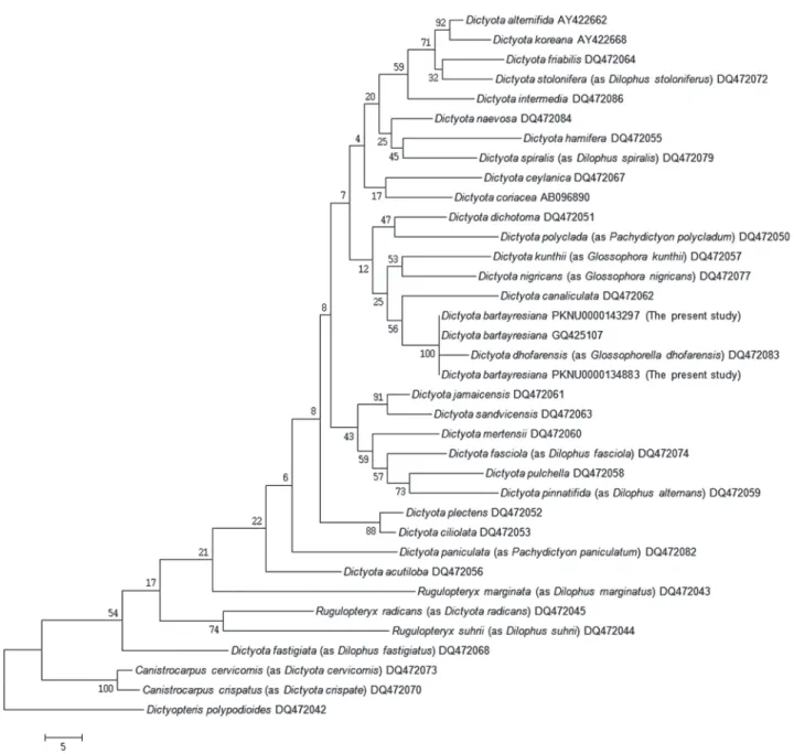
Fig. 2. Phylogenetic tree of Dictyota species obtained from ML analysis based on rbcL sequences. Bootstrap percentages(1000 replicatessamples) are shown above branches. Scale bar=0.02 substitutions/site.
form in habit (Fig. 1A, B), even though not typical. The branches are not so much narrow and long at the inter-nodes (Fig. 1A, B). Unlike in Micronesian species, terminal bran- ches, which are spirally twisted, were also not observed in the Korean specimens (Fig. 1A, B).
The alga collected from Geoje, Korea, during the present study is referable to D. bartayresiana based on the mor- phological features. This is also supported by molecular analysis (Figs. 2, 3). In the phylogenetic tree based on rbcL
sequences, this Korean alga nests in the same clade with D.
bartayresiana. Genetic distance between both sequences within the clade was calculated as 1.2%. Considering that the inter-species divergence ranged from 1.8% to 10.6% in Dictyota (the present study), this value is involved in the intra-species range. Based on these morphological and mo- lecular analyses, this Korean alga is identified as D. bartay- resiana described originally from Antilles, the West Indies.
This is the first record of D. bartayresiana in Korea.
Fig. 3. Phylogenetic tree of Dictyota species obtained from MP analysis based on rbcL sequences. Bootstrap percentages(1000 replicates samples) are shown above branches. Scale bar=0.02 substitutions/site.
De Clerck O and E Coppejans. 1997. The genus Dictyota J.V.
Lamour(Dictyotaceae, Phaeophyta) from Indonesia in the Herbarium Weber-van Bosse, including the description of Dictyota canaliculata spec. nov. Blumea 42:407-420.
De Clerck O and E Coppejans. 1999. Two new species of Dic- tyota(Dictyotales, Phaeophyta) from the Indo-Malayan region. Phycologia 38:184-194.
De Clerck O and E Coppejans. 2003. Morphology and sys- tematic of two aberrant species of Dictyota(Dictyotaceae, Phaeophyta), including a discussion on the generic bound- aries in the tribe Dictyoteae. pp. 275-284. In Proceedings of the 17th International Seaweed Symposium(Chapman ARO, RJ Anderson, VJ Vreeland and IR Davison eds.).
Oxford University Press, New York.
De Clerck O, F Leliaert, H Verbrugeen, CE Lane, JC De Paula, DI Payo and E Coppejans. 2006. A revised classification of the Dictyoteae(Dictyotales, Phaeophyceae) based on rbcL and 26S ribosomal DNA sequence data analyses. J. Phycol.
42:1271-1288.
Guiry MD and GM Guiry. 2017. AlgaeBase. World-wide elec- tronic publication, National University of Ireland, Galway.
http://www.algaebase.org; searched on 13 August 2017.
Hall TA. 1999. BioEdit: a user-friendly biological sequence alignment editor and analysis program for Windows 95/98/
NT. Nucleic Acids Symp. Ser. 41:95-98.
Hörnig I, R Schnetter and WF Prud’homme van Reine. 1992.
The genus Dictyota(Phaeophyceae) in the North Atlantic.
II. Key to the species. Nova Hedwig. 54:397-402.
Kim HS, SM Boo, IK Lee and CH Sohn. 2013. National List of Species of Korea 「Marine Algae」. Jeonghaengsa, Seoul.
sources. Incheon.
Lin S-M, S Fredericq and MH Hommersand. 2001. Systemat- ics of the Delesseriaceae(Ceramiales, Rhodophyta) based on large subunit rDNA and rbcL sequences, including the Phycodryoideae, subfam. nov. J. Phycol. 37:881-899.
Papenfuss GF. 1977. Review of the genera of Dictyotales (Phaeophycophyta). Bull. Jap. Soc. Phycol. 25(suppl.):
271-287.
Phillips JA. 1992. Taxonomy and reproduction in Australian species of Dilophus(Dictyotales, Phaeophyta). Aust. Syst.
Bot. 5:657-694.
Silberfeld T, F Rousseau and B de Reviers. 2014. An updated classification of brown algae(Ochrophyta, Phaeophyceae).
Crypt. Algol. 35:117-156.
Silva PC, PW Basson and RL Moe. 1996. Catalogue of the benthic marine algae of the Indian Ocean. Univ. California Publ. Bot. 79:1-1259.
Tsuda RT. 2004. Dictyota(Phaeophyceae) from Micronesia. pp.
41-55. In Taxonomy of Economic Seaweeds with Refer- ence to the Pacific and Other Locations, Volume IX(Abbott IA and KJ McDermid eds.). Hawaii Sea Grant College Pro- gram, University of Hawaii. Honolulu.
Weber-Peukert G. 1985. Ontogenetische, aütokologische und taxonomische Untersuchungen an ausgewählten Arten der Gattung Dictyota(Dictyotales, Phaeophyceae). Nova Hed- wig. 42:123-149.
Received: 20 August 2017 Revised: 11 September 2017 Revision accepted: 11 September 2017