Jae Woo An, Pil Joon Kang and Ki Wan Nam
150Korean J. Environ. Biol. 36(2) : 150~155(2018) https://doi.org/10.11626/KJEB.2018.36.2.150
INTRODUCTION
Derbesia Solier, which belongs to Bryopsidales J.H. Schaf
fner (Lam and Zechmann 2006; Verbruggen et al. 2009), is a siphonous green algal genus with a thallus composed of erect and prostrate filaments and multiflagellate zoospores.
This genus was established based on D. marina (Lyngbye) Solier (1846), which was originally described from Quivig, Faeröes, Denmark (Lyngbye 1819). At that time, only the general features of filaments and sporangia with gross mor
phology were given to the type species (Lyngbye 1819). How
ever, many studies on life history, with great interest, incre
ased our knowledge of Derbesia (Kornmann 1938; Feldmann 1950; Ziegler and Kingsbury 1964; Page 1970; MacRaild and Womersley 1974; Mayhoub 1976; Kobara and Chihara 1981, 1984). It was first clarified by Kornmann (1938) that
Derbesia marina and Halicystis ovalis (Lyngbye) Areschoug are sporophytic and gametophytic phases of the one species.
This suggested that Derbesia shows an alternation of hetero
morphic generations with a Halicystis phase, which had been regarded as an independent genus characterized as having a coenocytic thallus consisting of an erect vesicular part and a rhizoidal part, although some authors (Kornmann 1966, 1970; Sears and Wilce 1970) have shown that direct repro
duction of the sporophyte of D. marina can occur, as in Pedo
besia established based on D. clavaeformis from Australia (MacRaild and Womersley 1974; Silva et al. 1996).
Derbesia is distinguished from the most similar genus Pedobesia MacRaild & Womersley in having basal septum at the portion of branching and heteromorphic life history with a gametophytic Halicystis stage and in lacking calcium carbonate deposited inside the cell wall (MacRaild and Wom
ersley 1974; Woolcott et al. 2000, Table 2). This genus is globally distributed from the tropics to Arctic waters, and includes about 20 species worldwide (Guiry and Guiry 2018).
* Corresponding author: Ki Wan Nam, Tel. 0516295922, Fax. 0516295922, Email. [email protected]
ⓒ2018. Korean Society of Environmental Biology.
New Record of Two Derbesia Species (Chlorophyta) in Korea
Jae Woo An, Pil Joon Kang and Ki Wan Nam*
Department of Marine Biology, Pukyong National University, Busan 48513, Republic of Korea
Abstract - Two siphonous green algae were collected from the eastern coast of Korea. These species share the typical features of Derbesia sporophytes, such as erect and prostrate siphonous filaments and the presence of basal septum in lateral branches. One is characterized by the combined fea- tures of a relatively small tufted sporophytic thalli arising from a rhizoidal base, subdichotomous- ly branched filaments with a basal septum and one pyrenoid per subspherical to lenticular chloro- plast. The other shows a larger sporophytic thallus, sparsely subdichotomously branched filaments with a basal septum and one pyrenoid per spherical chloroplast. In a phylogenetic tree based on
rbcL sequence, the two above-mentioned Korean algae nest in the same clades as Derbesia minimaand D. indica, respectively. The genetic distance between the sequences within the clades was 0.5- 0.8%, which is considered to be included in the intra-specific range for the genus. These two sipho- nous Korean algae are identified as D. minima and D. indica, respectively, based on the morpho- logical and molecular analyses. These species are newly recorded in the Korean marine algal flora herein.
Keywords : Derbesia minima, D. indica, green algae, first record, Korea
<Original article>
10% formalin seawater, and pressed on herbarium sheets. A portion of the material was dried and preserved in silica gel for molecular analysis. Sections of the thallus were mount
ed in 20% corn syrup for permanent slides.
Total genomic DNA was extracted from a silicagelpre
served sample using the DNeasy Plant Mini Kit (Qiagen, Hil
den, Germany) according to the manufacturerʼs protocol.
Before extraction, dried material was crushed with liquid nitrogen using a mortar and pestle. Concentrations of extract
ed DNA were assessed using gel electrophoresis on a 1%
agarose gel. Extracted DNA was used for amplification of rbcL regions using following primers (forward: 5ʹTCCAA AAACTGAAACTAAAGCAGG3ʹ, reverse: 5ʹAATTTCT TTCCAAACTTCACAAGC3ʹ) (Lam and Zechman 2006).
PCR amplifications were performed in a TaKaRa PCR Thermal Cycler Dice with an initial denaturation step at 94°C for 5 min followed by 35 cycles at 94°C for 1 min, 56°C for 1 min, and 72°C for 2 min and a final extension at 72°C for 7 min. The reaction volume was 20 μL, consisting of 20 ng of genomic DNA, 2 μL of 10x PCR buffer, 2 μL of 200 μM dNTP, 1 μL each of forward and reverse primer, and 0.5 units of Taq polymerase (Takara Korea, Korea). Amplifications were examined using gel electrophoresis in a 1% agarose gel and amplified rbcL region products were purified using a QIAquick Gel Extraction Kit (Qiagen, Hilden, Germany).
The PCR products were moved to Macrogen Sequencing Service for sequencing (Macrogen, Seoul, Korea).
Sequences for the rbcL region were aligned using BioEdit (Hall 1999). Phylogenetic analyses were performed using the maximumlikelihood (ML), neighborjoining (NJ) and unweighted pair group method using arithmetic averages (UPGMA) methods. Bootstrap values were calculated with
Specimens examined: NIBRCL0000112655, MGARB01 2361, MGARB012362, MGARB012363, MGARB012364 (Gisungri: 5.vi.2016).
Habitat: Epiphytic near the lower intertidal.
Morphology: Sporophytic thalli up to 1 cm high, usually epiphytic on other algae, tufted, light green in color (Fig.
1A, B), with erect and prostrate systems; many erect fila
ments arising from a prostrate system with rhizoidal base, siphonous, cylindrical, 30-60 μm diam., subdichotomously branched several times (Fig. 1C); filamentous lateral branch
es with a basal septum (Fig. 1C); chloroplasts numerous, subspherical to lenticular, relatively small, measuring 3-10 μm long and 2-5 μm wide, each with a pyrenoid (Fig. 1D).
Sporangia and gametophytes were not observed during the present study.
Derbesia indica
(R. Patel & Isabella) P.C. Silva 1996: 810.
Type locality: Mithapur near Port Okha, Gujarat, India (Silva et al. 1996).
Korean name: Nagogeongkinsil nom. nov. (
신칭: 나곡 엉킨실).
Specimens examined: NIBRCL0000112654, MGARB01 2365, MGARB012366, MGARB012367, MGARB012368 (Nagokri: 24.vii.2015).
Habitat: Epilithic or epiphytic near the intertidal.
Morphology: Sporophytic thalli up to 2 cm high, epilithic or epiphytic on other algae, tufted, light green in color (Fig.
2A, B), composed of erect and prostrate systems; many erect filaments arising from rhizoidal base of a prostrate system, siphonous, cylindrical, 50-80 μm diam., sparsely subdicho
tomously branched (Fig. 2C); filamentous lateral branches
with a basal septum (Fig. 2C); chloroplasts numerous, sphe
rical, measuring 5-15 μm diam., each with a pyrenoid (Fig.
2D). Sporangia and gametophytes were not observed during the present study.
Derbesia minima, which occurs in various localities includ
ing Japan, Australia, New Zealand and Micronesia (Guiry and Guiry 2018), was originally described from Indonesia (Huisman 2015). This species is characterized by the com
bined features of relatively small tufted sporophytic thalli arising from an irregularly lobed rhizoidal base, cylindrical erect branches with transverse and basal septum, ovoid or pyriform sporangia with a short pedicel and double basal septa, and each subspherical chloroplast with one pyrenoid (Huisman 2015; the present study). Of these features, the basal septum at point of branching is one of the important
generic features of Derbesia (MacRaild and Womersley 1974;
Woolcott et al. 2000). According to Huisman (2015), apart from this basal septum, transverse septa in vegetative siphons are also occasionally observed in this species from Austra
lia. D. minima is distinguished from similar species with a small thallus (lower than 5 mm in height), such as D. atten
uate E.Y. Dawson, D. furcate R.W. Ricker and D. padinae Trono, by the occasional presence of the septum (Dawson 1954; Trono 1971; Ricker 1987; Huisman 2015). Derbesia minima appears to be distinct in having a transverse septum, which is not a typical feature of the genus. These transverse septa were not found in Korean specimens. The Korean spe
cimens also differ from the species from Australia and Japan in branching of erect filaments. It shows subdichotomous branching several times, while Australian and Japanese spe
cies branches once or not at all (Yoshida 1998; Huisman
Fig. 1. Derbesia minima Webervan Bosse. A: Sporophytic plant being epiphytic on other algal species; B. Herbarium specimen of sporophytes; C: Subdichotomously branched filaments with a basal septum(arrows); D: Subspherical to lenticular chloroplasts(arrows) with a pyrenoid in thalli.
A B
C D
2015). However, other vegetative features are shared with D. minima. Although sporangia and gametophytes were not observed, the Korean specimens appear to be referred to D.
minima based on vegetative morphology. This is also sup
ported by molecular data.
Derbesia indica was originally described from India based on gametophytes (Patel and Isabella 1978). So, at that time, it was established as Halicystis indica, but later transferred to Derbesia (Silva et al. 1996). This species is characterized by the combined features of tufted sporophytic thalli arising from rhizoidal base, sparsely subdichotomously branched filaments with a basal septum and each spherical chloroplast with one pyrenoid (the present study). D. indica is similar to D. minima, D. attenuata, D. tenuissima (Moris & De Notaris) P. Crouan & H. Crouan, and D. boergesenii (M.O.P. Iyengar
& Ramanathan) Mayhoub in having one pyrenoid per chlo
roplast. However, D. indica is readily distinguished from D.
minima and D. attenuata by thallus size. D. indica shows a relatively large thallus (about 2 cm), while the latter two spe
cies have thallus height of about 2 mm and 5 mm, respective
ly. D. tenuissima differs from D. indica in lacking a cross septum (Wormersley 1984; Kim and Klochkova 2010) or in occasionally having a basal septum in lateral branches (Ab
bott and Huisman 2004; Huisman 2015). D. boergesenii is also distinguished from D. indica in branching pattern and size of lateral filaments. The former species shows irregular branching and a relatively small size of 15-36 μm diameter (Mayhoub 1976) rather than sparsely subdichotomously bran
ching and large size of 50-80 μm diameter. More important
ly, it is distinguished from D. indica in being fusiform rather than spherical chloroplast (Mayhoub 1976).
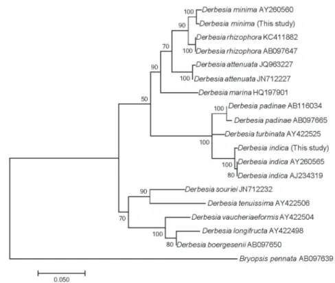
In a phylogenetic tree based on rbcL sequence, the Korean algae nest in the same clades as D. minima and D. indica, respectively (Fig. 3). According to Woolcott et al. (2000,
Fig. 2. Derbesia indica(R. Patel & Isabella) P.C. Silva. A: Habit of sporpophytic plant; B: Herbarium specimen of sporophytes; C: Sparsely subdichotomously branched filaments with a basal septum(arrow); D: Numerous spherical chloroplasts(arrows) possessing a pyrenoid.C D
Table 4), the genetic distance for rbcL sequence between Derbesia and Pedobesia is 13.9%. In the present study, the interspecific genetic distance in Derbesia was calculated as 1.4-11.9%. Based on these data, the genetic divergence bet
ween the sequences within the clades, which was 0.5-0.8%, is considered to be included in the intraspecific range for the Derbesia.
These morphological and molecular analyses indicate that the two siphonous Korean algae are identified as Derbesia minima and D. indica originally described from Indonesia and India, respectively. This study reports the first occurrence of the species in Korea.
ACKNOWLEDGEMENT
This work was supported by the Marine Biotechnology Program of the Korea Institute of Marine Science and Tech
nology Promotion (KIMST) funded by the Ministry of Oceans and Fisheries (MOF) (No. 20170431), and by a grant from the National Institute of Biological Resources (NIBR) fund
ed by the Ministry of Environment (MOE) of the Republic of Korea (NIBR201701204).
REFERENCES
Abbott IA and JM Huisman. 2004. Marine Green and Brown Algae of the Hawaiian Islands. Bishop Museum Press, Ho
nolulu, HI.
Dawson EY. 1954. Marine plants in the vicinity of the Institute Océanographique de Nha Trang, Viêt Nam. Pac. Sci. 8:372- Feldmann J. 1950. Sur lʼexistence dʼune alternance de généra469.
tions entre lʼHalicystis parvula Schmitz et le Derbesia ten
uissima(DeNot.) Crn. C.R. Acad. Sci. Paris Ser. D. 230:
322-323.
Guiry MD and GM Guiry. 2018. AlgaeBase. Worldwide elec
tronic publication, National University of Ireland, Galway.
http://www.algaebase.org. Accessed 15 April 2018.
Hall TA. 1999. BioEdit: a userfriendly biological sequence alignment editor and analysis program for Windows 95/98/
NT. Nucleic Acids Symp. Ser. 41:95-98.
Huisman JM. 2015. Algae of Australia: Marine Benthic Algae Fig. 3. Phylogenetic tree of selected taxa obtained from maximumlikelihood analysis based on rbcL sequences. Bootstrap percentages(1000
replicates samples) are shown above the branches. Scale bar=0.05 substitutions/site.
nomy of two species of Pedobesia(Bryopsidales, Chloro
phyceae) in Japan. Bot. Mag. Tokyo 97:151-161.
Kornmann P. 1938. Zur Entwicklungsgeschichte yon Derbesia und Halicystis. Planta 28:464-470.
Kornmann P. 1966. Eine erbliche Variante von Derbesia mari
na. Naturwissenschaften 53:161.
Kornmann P. 1970. Eine Mutation bei der siphonalen Grünalge Derbesia marina. Helgolander Wiss. Meeresunters. 21:1-8.
Lam DW and FW Zechman. 2006. Phylogenetic analyses of the Bryopsidales(Ulvophyceae, Chlorophyta) based on Rubisco large subunit gene sequences. J. Phycol. 42:669-678.
Lee IK and JW Kang. 1986. A check list of marine algae in Korea. Kor. J. Phycol. 1:311-325.
Lee YP and SY Kang. 2002. A Catalogue of the Seaweeds in Korea. Jeju National University Press, Jeju.
Lyngbye HC. 1819. Tentamen Hydrophytologiae Danicae. Sch
ultz, Copenhagen.
MacRaild GN and HBS Womersley. 1974. The morphology and reproduction of Derbesia clavaeformis(J. Agardh) De Toni (Chlorophyta). Phycologia 13:83-93.
Mayhoub MH. 1976. Sur lʼexistence dʼun Derbesia dans le cycle de lʼHalicystis boergesenii Iyengar et Raman(Chlorophyées, Derbesiales). C.R. Acad. Sc. Paris, Ser. D. 282:707-710.
Page JZ. 1970. Existence of a Derbesia phase in the life history of Halicystis osterhoutii Blinks and Blinks. J. Phycol. 6:
375-380.
Patel RJ and PK Isabella. 1978. A new species of Halicystis H.
Trono GC Jr. 1971. Some new species of marine benthic algae from the Caroline Islands, westerncentral Pacific. Micro
nesica 7:45-77.
Verbruggen H, M Ashworth, ST LoDuca, C Vlaeminck, E Coc
quyt, T Sauvage, FW Zechman, DS Littler, MM Littler, F Leliaert and O De Clerck. 2009. A multilocus timecalibrat
ed phylogeny of the siphonous green algae. Mol. Phyloge
net. Evol. 50:642-653.
Webervan Bosse A. 1913. Liste de algues du Siboga. I. Myxo
phyceae, Chlorophyceae, Phaeophyceae avec le concours de M. Th. Reinbold. Leiden.
Womersley HBS. 1984. The Marine Benthic Flora of Southern Australia. Part I. Government Printer, Adelaide.
Woolcott GW, K Knoller and RJ King. 2000. Phylogeny of the Bryopsidaceae(Bryopsidales, Chlorophyta): cladistic anal
yses of morphological and molecular data. Phycologia 39:
471-481.
Yoshida T. 1998. Marine Algae of Japan. Uchida Rokakuho, Tokyo.
Ziegler JR and JM Kingsbury. 1964. Cultural studies on the marine green alga Halicystis parvlllaDerbesia tenuissima.
I. Normal and abnormal sexual and asexual reproduction.
Phycologia 4:105-116.
Received: 24 April 2018 Revised: 18 May 2018 Revision accepted: 21 May 2018