1888. It is characterized by pulvinate thalli with spongy-ge- latinous structure, monostromatic base of apparently scat- tered filaments, branched and densely intricate erect fila- ments, 1-3 discoid phaeoplasts per cell, and plurilocular sporangia on terminal or lateral part of the erect filaments (Rienke 1888; Fletcher 1987; Peters 2003). Currently, six Microspongium species are recognized from worldwide: M.
alariae (P. M. Pedersen) A. F. Peters from Greenland (Ped- ersen 1981; Peters 2003), M. globosum J. Rienke from Ger- many (Rienke 1888), M. immersum (Levring) P. M. Ped- ersen from Norway (Levring 1937; Athanasiadis 1996), M. kuckuckianum V. Schiffner from Adriatic Sea (Schiffner 1916), M. radians (M. Howe) A. F. Peters from Peru (Daw- son et al. 1964) and M. stilophorae (P. L. Crouan & H. M.
Crouan) Cormaci et G. Furnari from Adriatic Sea (Crouan and Crouan 1867; Hauck 1884). None of these has been reported in Korea.
https://doi.org/10.11626/KJEB.2019.37.3.285
INTRODUCTION
The filamentous brown algal genus, Acinetospora, was described by Bornet in 1892. It is characterized by having sparsely branched uniseriate filaments forming entangled tufts, scattered meristematic zones, crampons, plurilocular sporangia ( acinetosporangia ), and monosporangia (Bornet 1892; Sauvageau 1899). Currently, four Acinetospora spe- cies are recognized from worldwide: A. crinita (Carmichael in Harvey) Sauvageau from Scotland (Womersley 1987), A. nicholsoniae Hollenberg from California, U.S.A. (Hol- lenberg 1971), A. filamentosa (Noda) Yaegashi from Japan (Noda 1970; Yaegashi et al. 2015), and A. asiatica Yaegashi, Yamagishi et Kogame from Japan (Yaegashi et al. 2015). Of them, A. crinita and A. asiatica have been reported in Korea (Kim 2010; Oteng’o et al. 2018).
The genus Microspongium was described by Reinke in
Original article
New records of two filamentous brown algae, Acinetospora filamentosa and Microspongium stilophorae from Korea
Antony Otinga Oteng’o, Jose Avila-Peltroche, So Young Jeong, Boo Yeon Won and Tae Oh Cho*
Department of Life Science, Chosun University, Gwangju 61452, Republic of Korea
Korean J. Environ. Biol.
37(3) : 285-292 (2019) ISSN 1226-9999 (print) ISSN 2287-7851 (online)
* Corresponding author Tae Oh Cho
Tel. 062-230-7161
E-mail. [email protected]
Received: 22 July 2019 Revised: 16 August 2019
Revision accepted: 19 August 2019
Abstract: Acinetospora filamentosa and Microspongium stilophorae are reported as
new records from South Korea based on morphological and molecular analyses. A.
filamentosa is mainly characterized by having the sparsely branched erect filaments, the scattered meristematic zones, forming crampons, plurilocular sporangia on both prostrate filaments and lower part of erect filaments, and spherical to oval unilocular sporangia formed either sessile or with a pedicel. M. stilophorae is an epiphytic thalli mostly on Stilophora sp. It is characterized by prostrate filaments with irregular cells, short erect filaments with short ramuli, phaeophycean hairs, uniseriate plurilocular sporangia on the terminal part of erect filament. Our molecular analyses of rbcL and cox1 genes reveals that A. filamentosa and M. stilophorae are nested within the clades of Acinetospora and Microspongium, respectively.
Keywords:
Acinetospora filamentosa, Microspongium stilophorae, Ectocarpales, Pha-
eophyceae
Samples of Acinetospora filamentosa were collected from west and south coasts of Korea. They were sorted into vou- cher herbarium specimens, silica gel samples, and formalin samples. Formalin samples were preserved in 4-5% for- malin/seawater. A sample of Microspongium stilophorae was collected from east coast of Korea. It was isolated from Dic- tyopteris pacifica and cultured in provasoli enriched seawa- ter (PES) medium in order to get enough material for mor- pho-anatomical and molecular analysis. Photomicrographs taken using an Olympus BX51TRF microscope (Olympus, Tokyo, Japan) and an Olympus DP71 camera. Permanent slides were mounted in 70% karo syrup. Representative specimens examined in this study were deposited in the herbarium of Chosun University (CUK) and National In- stitute of Biological Resources (NIBR), Korea.
2. Molecular study
Genomic DNA was manually extracted from silica-gel samples using extracted using a NucleoSpin Plant II Kit (Macherey-Nagel, D ü ren, Germany). The extracted DNA was stored at -20°C and used to amplify rbcL and cox1.
The rbcL gene was amplified using the primer combina- tions NDrbcL2-DRL1R and DRL2F-R3A (Kogame et al.
1999 ; Hwang et al. 2005) with HelixAmp Ready-2x-Go Series (NanoHelix Co., Ltd., Daejeon, Korea). The GazF2- GazR2 combination of primers used for cox1 (Saunders 2005; Lane et al. 2007). All PCR amplication were carried out with a Veriti 96-well Termal cycler (Applied Biosystem, ThermoFisher Scientific, USA). PCR products were pu- rified using a PCRquick-spin
TMPCR product purification kit (iNtRON Biotechnology, Inc, Seongnam, Korea). All rbcL and cox1 sequence data were compiled by the pres- ent study and obtained from GenBank and aligned with ClustalW (Thompson et al. 1994). New rbcL sequences obtained from Acinetospora filamentosa have been deposit- ed in EMBL/GenBank under the accession numbers MN
formed using MrBayes 3.2.6 (Huelsenbeck and Ronguist 2001; Ronguist and Huelsenbeck 2003). Markov chain Monte Carlo runs were conducted for 2 million genera- tions, each with one cold chain and three heated chains using the GTR + Γ + I evolutionary model and sampling and printing every 1,000 generations. Summary trees were generated using a burn-in value of 800.
RESULTS AND DISCUSSION
Acinetospora filamentosa (Noda) Yaegashi, Uwai et Kogame, 2015
실솜말(
신칭) (Figs. 1, 2)
Basionym: Ectocarpus filamentosus Noda 1970. Sci. Rep.
Niigata Univ. Ser. D. 7: 27.
Heterotypic Synonym: Ectocarpus ugoensis Konno in Konno et Noda 1974. Sci. Rep. Niigata Univ. Ser. D. 11: 80.
Material examined: NIBROR0000001612 & CUK12425 ( = MBRB0099TC12425) Chuja-hang, Chuja-myeon, Jeju-si, Jeju Special Self-govering Province, Korea (33°
57ʹ44.69ʺN, 126°17ʹ47.12ʺE), June 27, 2014, T. O. Cho, S.
Y. Jeong, D. B. Mostajo, J. G. Lee and S. Y. Park, at 1 m depth by hand; CUK12847 ( = MBRB0099TC12847), Dae- jin-hang, Daejin-dong, Donghae-si, Gangwon-do, Korea (37°34ʹ 47.64ʺN, 129°6ʹ51.13ʺE), August 01, 2014, T. O.
Cho, S. Y. Jeong, D. B. Mostajo, J. G. Lee and S. Y. Park, at 1 m depth by hand; CUK13048 ( = MBRB0099TC13048), Biyang-do, Hanrim-eup, Jeju-si, Jeju Special Self-govering Province, Korea (33°24ʹ 21.9ʺN, 126°13ʹ46.40ʺE), May 30, 2014, T. O. Cho, S. Y. Jeong, D. B. Mostajo and J. G. Lee, at 1 m depth by hand; CUK18942 ( = MBRB0099TC18942), Mo-hang, Byeonsan-myeon, Buan-gu, Jeollabuk-do, Korea (35°34ʹ 58.49ʺN, 126°30ʹ18.41ʺE), May 12, 2018, T. O.
Cho and B. Y. Won, at 1 depth by hand.
Habitat: Epiphytic and saxicolous at the tide pool in inter-
tidal zone.
Morphological observation: Plants are uniseriate, form- ing entangled tufts (Fig. 1A) to 10 cm or more in length attached to rocks and other seaweeds (e.g. Sargassum spp.).
Erect filaments are irregularly and sparsely branched at wide to right angles (Fig. 1B) and form straight to curved
“crampons” composed of 2-5 cells (Fig. 1C). Meristematic zones (Fig. 1D) are scattered, consisting of short cells. Cells of erect filaments are 20-80 μm in length and 18-28 μm in width, containing many discoid chloroplasts (Fig. 1E) with pyrenoids. Phaeophycean hairs (Fig. 1F) are found later-
Fig. 1. Acinetospora filamentosa
(CUK18942) from Mohang, Jeollabuk-do, Korea. A. Thalli forming entangled tufts; B. Erect filaments with crampons; C. Crampon on erect filament; D. Meristematic zone (arrows) on erect filaments; E. Cell with discoid chloroplasts; F. Phaeophy- cean hairs on erect filaments. Scale bars: A = 0.5 cm; B = 1 mm; F = 500 μm; C, D = 50 μm; E = 20 μm.
A B
C D
E F
ally or terminally on erect filaments. Unilocular sporangia are spherical to oval, 25-55 μm in length and 25-55 μm in width, sessile or on a pedicel, and are formed on erect fila- ments.
World distribution: Asia: Japan and Korea; Europe:
Greece (Guiry and Guiry 2019).
Identifier: Tae Oh Cho and Antony Otinga Oteng’o.
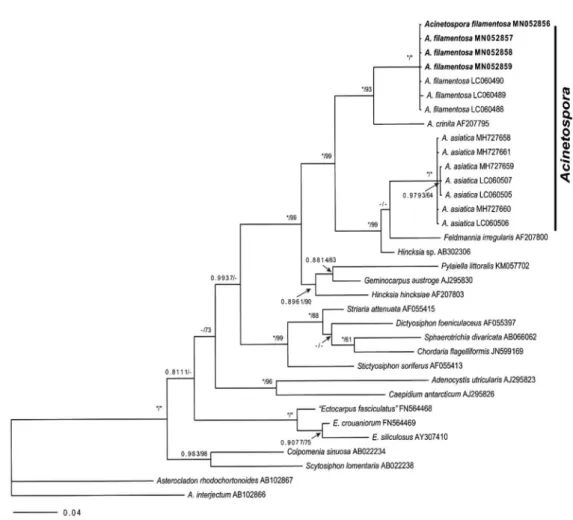
Phylogenetic analyses: The 1326-nucleotide portion of rbcL was aligned for Acinetosproa filamentosa. Phylogenetic analyses revealed that our Acinetospora samples from Korea were placed within a clade of Acinetospora filamentosa in rbcL (Fig. 2). There was only 0-0.008% gene sequence di- vergence between Genbank and our collection of Acineto- spora filamentosa. In addition, it revealed that Acinetospora filamentosa differs from A. asiatica by 4.4-4.7% and from A.
crinita by 3.0% gene sequence divergence respectively.
Remarks: Acinetospora filamentosa was a new combination
with Ectocarpus filamentosus as the basionym (Yaegashi et al. 2015). Our samples collected from Korea had vegeta- tive morphology similar to that of A. filamentosa. Our mo- lecular analyses based on rbcL gene show that our samples are nested in the clade of A. filamentosa. In this study, we report A. filamentosa as a new record from Korea and add this species in the list of Korean macroalgal flora.
Microspongium Reinke, 1888,
점말속(
신칭)
Microspongium stilophorae (P. L. et H. M. Crouan) Cormaci et G. Furnari, 2012
바늘점말(
신칭) (Figs. 3, 4)
Basionym: Ectocarpus stilophorae P. L. et H. M. Crouan, Florule du Finist é re…., 1867: 161, Paris and Brest. x + 262 pp., 31 [ + 1] pls, frontispiece.
Homotypic synonym (s): Streblonema stilophorae (P. L.
Crouan & H. M. Crouan) De Toni 1895; Streblonema stilo-
Fig. 2. Phylogenetic tree of Acinetospora species based on Bayesian and ML analysis with rbcL sequences. Value above branches
= Bayes-
ian posterior probabilities
>0.75/Maximum likelihood bootstrap values in %
>50. Values lower than BPP 0.75 or BS 50 are indicated by
hyphens (-). Values of BPP 1.00 or BS 100 are indicated by asterisks (*).
phorae (P. L. Crouan & H. M. Crouan) Kylin 1908 (comb.
illeg.).
Heterotypic synonym (s): Ectocarpus stilophorae f. caespi- tosus Rosenvinge 1893; Ectocarpus stilophorae v. caespitosus
(Rosenvinge) L. Newton 1931; Microspongium tenuissi- mum (Hauck) A. F. Peters 2003; Streblonema stilophorae v. caespitosum (Rosenvinge) De Toni 1895; Streblonema tenuissimum Hauck 1884.
Fig. 3. Microspongium stilophorae
(CUK19276) from Yangjeong-hang, Gyeongsangbuk-do, Korea. A. A ‘ball-like’ spongy spherical thallus; B.
Prostrate filaments of irregular cells (arrows) and short erect filaments (arrowheads); C. Laminar- and lobate-shaped phaeoplasts (arrows);
D. Uniseriate plurilocular sporangia (arrows) mostly on the terminal part of erect filaments. E. Uniseriate plurilocular sporangia. Scale bars:
A = 250 μm; B-D, E = 20 μm.
A B
C
D E
Material examined: NIBROR0000001610 & CUK19276 ( = MBRB0097TC19276) Yangjeong-hang, Uljin-eup, Uljin-gun, Gyeongsangbuk-do, Korea (37°00ʹ59.15ʺN, 129°24ʹ48.17ʺE), May 01, 2018, T. O. Cho and B. Y. Won, at 1 m depth by hand.
Habitat: Epiphytic on other seaweeds (e.g. on Stilophora sp., Nemalion sp. and Dictyopteris pacifica) at the tide pool in intertidal zone.
Morphological observation in culture: Cultured thallus isolated from Dictyopteris pacifica forms a spongy ball-like spherical tissue (Fig. 3A) formed by prostrate filaments of irregular cells in shape and size (Fig. 3B, arrows) and short erect filaments with short ramuli (Fig. 3B, arrowheads).
Erect filaments are formed by cells 1-5 times longer than wide and 3-8 μ m in diameter. The phaeoplasts (Fig. 3C, arrows) are one or two per cell. Phaeophycean hairs not frequent. Plurilocular sporangia lateral or mostly terminal, in uniseriate lodges, 3-8 μ m wide (Fig. 3D, arrows).
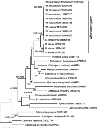
“M. tenuissimum”) and M. alariae is 2.9-3.6%. However, there was only 0.16-0.49% gene sequence divergence be- tween Genbank and our collection of M. stilophorae.
Remarks: Morphologically, our Microspongium sample is matched into the description of Microsporangium stilopho- rae. Microspongium tenuissimum and M. radians were con- specific based on cox1 gene (Mur ú a et al. 2018). Microspon- gium tenuissimum is currently a synonym of M. stilophorae (Cormaci et al. 2012; Guiry and Guiry 2019). Our mo- lecular data based on cox1 gene revealed that our Korean sample is nested in the same clade of M. stilophorae. In this study, we report Microsporangium stilophorae as a new re- cord from Korea and add this species to the list of Korean macroalgal flora.
ACKNOWLEDGEMENTS
This study was supported by a grant from the National Institute of Biological Resources (NIBR), funded by the Ministry of Environment (MOE) of the Republic of Ko- rea (NIBR 201801205) and by a grant from the research fund of Chosun University 2018. This research was also supported by Basic Science Research Program through the National Research Foundation of Korea (NRF) fund- ed by the Ministry of Education, Science and Technology (2019R1F1A1060346) and a grant from Marine Biotech- nology Program (20170431) funded by Ministry of Oceans and Fisheries of Korean Government to Tae Oh Cho.
REFERENCES
Athanasiadis A. 1996. Taxonomisk litteratur och biogeografi av Skandinaviska rödalger och brunalger. pp. 1-280, 1map.
Göteborg: Algologia.
Fig. 4. Phylogenetic tree of Microspongium species based on