대한관젏경학회지 체 13권 제 1 호 2009 Journal of Korean Arthroscopy Soc.
V이나me 13, Number 1, February, 2009
관절 연골 손상 정도에 따른 반월상 연골 부분절제술의 결과
가톨릭대학교 의과대학 정형외과학교 실 최남용•양영준•나기호•양혁재•송현석
The Results of Partial Meniscectomy according to the Grade of the Articular Cartilage Injury
Nam Yong Choi, M.D., Young Jun Yang, M.D., Ki Ho Nah, M.D., Hyuk Jae Yang, M.D., and Hyun Seok Song, M.D.
Department of Orthopedic Surgery, St PauCs Hospital, The C或h이ic University of Korea, Seoul, Korea
Purpose: To identify the damage of the articular cartilage and analyze the clinical results of the partial medial meniscectomy.
Materials and Methods: From January 2001 to December 2004, forty-eight patients in the degenerative arthritis and medial meniscal tear of knee who undertook arthroscopic partial meniscectomy and were able to keep pace with the times follow-up for more than 3 years were enrolled in this study. Six cases were men and 42 were women, and the mean age was 55.7 years (40-78 years). The patients were classified into two groups acc댢rding to the Outerbridge grade. Grade I and II were class访ed into group 1 and grade III and IV into group 2. Group 1 included 30 cases and group 2 included 18 cases. The result were analyzed according to the Tapper & Hoover classification and Lysholm knee scoring scale. And we measured the distance of medial joint space in standing anteroposterior (AP) view of both sides at the pre-operative and the last follow-up.
Results: According to the Tapper & Hoover classification, 28 cases were excellent, 7 cases good, 8 cases fair, and 5 cases poor.
Among group 1, 25 cases were excellent, 2 cases good, 2 cases fair, 1 case poor. However, among group 2, 3 cases were excellent, 5 cases good, 6 cases fair, 4 cases poor. Average Lysholm knee score was improved from 62.4 preoperatively to 94 postoperatively in group 1, and from 58 preoperatively to 77.9 postoperatively in group 2. The joint space narrowing showed no statistically significant difference in both knees standing AP view.
Conclusion: The arthroscopic partial meniscectomy for a patient with the degenerative articular cartilage was an effective treat
ment. However, we could not satisfy the results, in cases of Outerbridge grade III and IV. In grade III and IV, the partial meniscecto
my was not aggravated the cartilage damage.
KEY WORDS: Knee, Articular cartilage injury, Medial meniscal tear, Partial meniscectomy, Outerbridge
서 론
반월상 연골은 대퇴골 및 경골의 접촉 면을 증가시켜 관절 면에 가해지는 스트레스를 분산 감소시켜 관절 연골을 보호
* Address reprint request to Hyun Seok Song, M.D.
Department of Orth얘)edic Surgeiy, StPaul, s H域pital, The Catholic University of Korea, 620-56 JeoniK)ng-dong, D(Migdaemun-gu, Seoul 130-709, K顷心
Tel: 82-2-958-2159, Fax: 82-2-965-1456 E-mail: [email protected]
하는 기능이 있으며' 활액을 관절에 골고루 분산시켜 윤활 기 능을 촉진시키고, 관절 운동 시 활액막이 관절 사이에 끼이는 것을 방지하며, 체중전달 및 관절의 안정성에 기여하는 등 다 양한 기능을 수행한다®. 반월상 연골 손상에 대한 수술적 치 료의 목적은 동통의 감소와 수상 전의 활동 정도로 회복할 수 있게 하며 관절의 조기 퇴행 변화를 예방하는 것이다. 반월상 연골의 약 30%를 절제한 후에는 관절 연골에 미치는 부하가
3배까지 증가된다고 알려져 있다'®. 반월상 연골 절제술 시행 이후 관절염의 발생에 관련되는 요인으로는 제거된 반월상 연골의 양, 관절 연골의 동반된 퇴행성 변화의 정도, 슬관절의 안정성 및 정렬 등이 알려져 있다財s. 고령에서의 반월상 연
22 —
골 손상에 대한 수술적 치료에 대하여는 아직 이견이 많이 있 으나, 여러 연구에서 반월상 연골의 부분절제술로 만족스러 운 결과를 얻었다고 보고하고 있다用.
이에 저자는 내측 반월상 연골 부분절제술 시 확인된 관절 연골 손상의 정도를 확인하여, 이에 따른 수술 후 임상적 결과 를 분석하고자 하였다. 또한 반월상 연골 부분절제술에 의한 관절 연골 손상의 진행여부에 대한 조사를 하였다.
대상 및 방법
본원에서 20()1년 1월부터 2004년 12월까지 내측 반월상 연골 파열로 인해 관절경적 부분절제술을 시행하였으며, 관 절경 소견상 관절 연골의 손상이 동반되고, 술 후 3년 이상 추 시가 가능하였던 48예를 대상으로 후향적으로 분석하였다.
釦세 미만 및 동측 슬관절의 내측 및 외측 반월상 연골 모두 병변이 있거나 인대 동반 손상이 있는 경우, 아전절제술을 시 행한 예는 연구대상에서 제외하였다. 남자가 6예, 여자가 42
예였으며 수술시 평균 나이는 55.7세 (40〜78세) 였다. 전체 환 자군의 추시 기간은 평균 4년 2개월이었다.
전신마취하에 전내측, 전외측 삽입구를 이용하여 관절경하 에 내측 반월상 연골 부분 파열에 대하여 부분절제술을 시행 하였다(Fig. 1). 관절경 소견상 관절 연골의 손상은
Outerbridge 분류"에 의하여 grade I은 관절 연골의 연화
(softening)가 있는 경우, II는 1/2 인치 이하 직경의 균열
(fissure) 이나 파편 (fragmentation) 이 있는 경우, III는 1/2
인치 이상 직경의 균열이나 파편이 있는 경우, IV는 골이 노
출되어 있는 경우로 분류하였다. 연구대상은 Outerbridge
분류 grade I과 II는 1군으로, grade III와 IV는 2군으로 나
누었다. 제 1군이 30예였고 제 2군이 18예였다.
결과는 Tapper & Hoover 분류”〉(Table 1)및 Lysholm
의 슬관절 점수 척도로 측정하였다. Lysholm의 슬관절 점수
척도는 95~100점을 우수(excellent), 85~94점을 양호 (good), 60~84점을 보통(fair), 60점 이하를 불량(poor)으
로 분류하였다. 단순 방사선 검사로서, 술전과 최종 추시시 환 자가 기립하여 체중을 부하한 상태에서 양측 슬관절의 전후 면 촬영을 하였다. 대퇴내과와 경골 내과의 최단거리를 측정 하였다. 통계적 분석은 SPSS 10.1K를 사용하였으며 유의 수 준은 95% 신뢰수준으로 하였다. 각 군의 술전과 술후
Fig. 1. (A) Preoperative radiographs shows a standing anteroposterior view of Left knee in 63-year-old woman. (B) Magnetic reso
nance image shows the medial meniscus tear in coranal and saggital plane. (C) Arthroscopic photograph shows the tom medi
al meniscus. (D) Arthroscopic photograph shows the medial meniscus after partial meniscectomy.
—23 —
대한관절경학회지 제 13권 제 1 호 2009년
Table 1. Tapper & Hoover classification
Classification Description
Excellent Good
The patient had no symptoms and no disability related to his knee
The patient had minimum symptom, such as aching or weakness after heavy use or effusion after heavy exertion, but there was essentially no disability
Fair The patient had symptoms, such as trouble kneeling or climbing stairs; weakness, pain, or discomfort had became enough of a problem to interfere somewhat with everyday activities and the patient thought he had some disability; and he was active but could not participate in vigorous sports (such as skiing, tennis, foot
ball, and so forth)
Poor The symptoms were severe and included all of those listed under fair as well as the presence of pain at rest, limit motion, and locking. The patient was clearly disabled, and his activities, including walking, were definitely limited because of his knee.
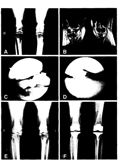
Fig. 2. (A) Preoperative radiographs shows a standing anteroposterior view of Left knee in 51-year-old woman. (B) Magnetic reso
nance ima흥e shows the medial meniscus tear in coranal and saggital plane. (C) Arthroscopic photograph shows the tom medi
al meniscus and Outerbridge grade IV of artic비ar cartilage. (D) Arthroscopic photograph shows the medial meniscus after partial meniscectomy. (E) Radiographs shows a standing anteroposterior view of left knee after 5 months. (F) Postoperative radiographs shows a standing anteroposterior view of left knee after TKA.
_ 24__
Lysholm 점수와 관절간격의 비교에는 paired t-test를 사
용하였으며, 두 군의 술후 점수 비교에는 모수 검정법으로
Student's t-test를, 비모수 검정법으로 Mann-Whitney U-test 를 사용하였다.
결 고卜
관절경하 내측 반월상 연골 부분절제술에 따른 합병증은 없 었다. 단순 방사선 검사상, 술전 평균 4.8 mm(6.8~2.9 mm)에
서 최종 추시시 평균 4.6 mm(6.8~l mm)로 내측 관절 간격의 감소는 유의하지 않았다(p=0.000). 수술 당시의 Outerbridge
분류상 IV에 해당하였던 1예에서 반월상 연골의 부분절제술 후 동통의 증가 및 관절염의 급속한 진행으로 인하여 술 후 5개
월에 인공관절 전치환술을 시행하였다(Fig. 2). 연구대상으로 한 48예의 Tapper & Hoover 분류에 따른 판정 결과는 우수
28예 (58.3%), 양호 7예 (14.6%), 보통 8예 (16.7%), 불량 5예 (10.4%) 였다. 제 1군은 우* 25예(83.3%), 양호 2예 (6.6%),
보통 2예(6.6%), 불량이 1예(3.3%)였으며 , 반면에 제 2군에
Group 1 (N=30) 6.6% 33%
6.6%^
齢 a Excellei
囊 Good 2 83 3匕』Fair 2
' » Poor 1
서는 우수 3예(16.6%), 양호 5예 (27.7%), 보통 6예(33.3%),
불량이 4예(22.2%)였다(Fig. 3).
기능적 평가상 Lysholm 점수는 1군에서는 술전 보통 21
예, 불량 9예로 술전 평균 62.4점 표준편차 7.0)에서 술후 우
수 21 예, 양호 6예, 보통 2예, 불량 1 예로 술후 평균 94.0점 표
준편차 9.0)으로 향상되 었다(p=0.000). 2군에서는 술전 보통
14예, 불량 4예로 술전 평균 58.0점 표준편차 7.6) 에서 , 술후
우수 3예, 양호 4예, 보통 10예, 불량 1예로 술후 평균 77.9점
(표준편차 16.2)으로 향상되 었다(p=0.000) (Fig. 4). 술후 점
수의 비교에서 1군이 2군에 비하여 우수하였다(t-test's p=0.001, U-test's p=0.000).
고 찰
반월상 연골을 절제하여 제거하는 경우, 추후 병발하게되 는 관절 연골의 손상에 대하여 여러 보고가 있다. Krause 등"
은 부분절제술 후 접촉면적 이 약 1/3로 감소되 어 관절 하중이
3배 정도 증가하였다고 보고하였으며, 이것이 관절 연골과 연
Gro 叩 2 (N=18)
Fig. 3. Chart shows a postoperative Tapper & Hoover classification.
Gr。叩 1 Group 2
Fig. 4. Chart shows a preoperative and postoperative Lysholm knee scoring.
—25 —
대한관절경학회지 제 13권 제1호 2009년
골하 골의 퇴행에 직접적으로 연관이 있다고 하였다. Baratz
등»은 15~34%의 절제술 후에 접촉면의 압력이 최고 350%
까지 증가된다고 하였다. 또한 관절경하 반월상 연골 부분절 제술 후 약 4~5년 추시 단순 방사선 검사에서 약 40%에서 관 절염이 관찰된다는 연구도 있다沁〉. 절제술 시행후 관절염 발 생에 따른 예후에 영향을 미칠 수 있는 요소로 제거된 반월상 연골의 양, 동반된 퇴행성 변화의 정도, 내측 또는 외측 구획 여부, 슬관절의 안정성 및 정렬 등이 알려져 있다心".
이미 관절 연골의 손상이 있는 경우에, 진단된 반월상 연골 파열의 치료에 대하여 여러 이견이 존재한다. McBride 등如>
은 초기 관절 퇴행성 변화가 있는 퇴행성 연골 파열에서의 부 분절제술은 이득이 없다고 하였으며, 이미 퇴행성 변화가 있 는 환자에서는 좋은 예후를 얻기 어렵다고 하였다. 그러나
Matsusue와 Thompson*은 나이가 젊을수록, 술 전 증상 기 간이 짧을수록, 퇴행성보다 비퇴행성 반월상 연골 파열시 환 자 만족도가 높다고 하였다. Bin 등3은 증상이 있는 반월상 연골 파열과 동반된 진행된 관절염 환자에서 관절경하 반월 상 연골 부분절제술이 효과적이라고 발표하였다. Pearse와 Craig">는 진행된 골관절염 환자에서 반월상 연골의 부분절 제술 후 증상의 호전이 있었고, 관절경 수술이 골관절염의 진 행을 촉진시키지는 않았다고 하였다. 또한 Shelbourne과 Dickens"〉는 안정된 슬관절의 내측 반월상 연골의 부분절제 술 후 12년 추시 결과에서 방사선 및 주관적 환자 만족도가 좋았다고 보고하였 다.
그러나 여러 보고에도 불구하고 관절 연골 손상이 있는 경 우에 있어서, 반월상 연골 부분절제술의 결과를 보다 과학적 으로 접근하려면, 수술 당시의 관절 연골 퇴행성 변화 정도에 따른 결과를 구분하여야 될 필요가 있다고 생각한다. Bin 등"
은 단순 방사선 검사상 내반 정렬(varus malalignment), 내
반 이동(varus thrust), 2 mm 이하의 내측 관절 간격
(medial joint space)과 같은 심한 퇴행성 변화가 동반된 환 자에서 반월상 연골 파열이 진단된 경우에는 관절경적 부분절 제술의 결과가 나쁠 수 있다고 하였다. 본 연구에서는 관절경 으로 확인된 Outerbridge 분류에 따른 관절 연골 손상 정도 가 반월상 연골 부분절제술의 결과에 영향을 미침을 알 수 있 었다. 저자들은 퇴행성 골관절염이 동반된 경우에 반월상 연골 파열이 진단되면 관절경을 이용한 반월상 연골 부분절제술이 도움이 될 것으로 판단되며, 관절 연골 손상 때문이 아닌 반월 상 연골 파열에 의한 증상인 경우에는 더욱 도움이 될 것으로 생각된다. 갑자기 관절내 삼출액이 동반된 급성기 증상인 경 우, 내측 관절연의 국소 압통, 잠김 현상 등의 기계적 인 증상이 있는 경우, 특별한 물리적 외상 이후에 나타난 증상인 경우, 영 상학적 검사상 이물체(loose body)가 있는 경우, 관절염이 심 하지 않은 경우, 환자가 수술적 치료를 원할 경우에 반월상 연 골의 부분절제술을 시 행하는 것이 좋을 것으로 사료된다.
본 연구의 제한점은 반월상 연골 파열 형태에 따른 분류, 부 분절제술의 정도, 연령 및 성별에 따른 세분화된 비교가 실시
되지 않았으며, 연구대상이 많지 않았다는 점이다.
결 론
관절 연골 손상이 동반된 환자에서 관절경적 반월상 연골 부분절제술은 증세 호전에 도움을 줄 수 있는 효과적인 치료 법이기는 하나, 관절 연골의 손상이 심한 경우에는 많은 호전 을 기대하기 어렵다고 생각한다.
REFERENCES
1) Baratz ME, Fu FH, Mengato R: Meniscal tears: the effect of meniscectomy and of repair on intraarticular con
tact areas and stress in the human knee. A preliminary report. Am J Sports Med, 14: 270-275, 1986.
2) Bhattacharyya T? Gale D, Dewire P, et al.: The clinical importance of meniscal tears demonstrated by magnetic resonance imaging in osteoarthritis of the knee. J Bone Joint Surg, 85-A: 4-9, 2(X)3.
3) Bin SI, Lee SH, Kim CW, Kim TH, Lee DH: Results of arthroscopic medial meniscectomy in patients with grade IV osteoarthritis of the medial compartment. Arthroscopy, 24: 26가-268, 2008.
4) Car돊en RW: Arthroscopic meniscectomy. Orthop Clin North Am, 10: 619-627, 1979.
5) Covall DJ, Wasilewski SA: Rt^ntgenographic changes after arthroscopic meniscectomy: Five-year follow-up in patients more than 45 years old. Arthroscopy, 8: 242-246,1992.
6) Hede A, Larsen E, Sandberg H: Partial versus total meniscectomy. A prospective, randomized study with long-term follow-up. J Bone Joint Surg, 74-B: 118-121, 1992.
7) Kim JM, Choi NY, Sun DH, Yim HJ: Frequency of meniscal tear according to ligament injury. J of Korean Orthop Assoc, 29: 610-617, 1994.
8) WR, Pope MH, Johnson RJ, Wilder DG: Mechanical changes in the knee after meniscectomy. J Bone Joint Surg, 58-A: 599-604,1976.
9) Matsusue Y, Thomson NL: Arthroscopic partial medial meniscectomy in patients over 40 years old: A 5- to 11~
year follow-up study. Arthroscopy, 12: 39*44, 1996.
10) McBride GG, Constine RM, Hofmann AA, Carson RW: Arthroscopic partial medial meniscectomy in the older patients. J Bone Joint Surg, 66-A: 547-551,1984.
11) Outerbridge RE: The etiology of chondromalacia patel
lae. J Bone Joint Surg^ 43-B:752, 1961.
12) Pearse EO, Craig DM: Partial meniscectomy in the pres
ence of severe osteoarthritis does not hasten the sympto
matic progression of osteoarthritis. Arthroscopy^ 19: 963- 968,2003.
—26 —
13) Rangger C, Klestil T, Gloetzer W, Kemmler G, Benedetto KP: Osteoarthritis after arthroscopic partial meniscectomy. Am J Sports Med, 23: 240-244, 1995.
14) Shelbourne KD, Dickens JF: Digital radiographic evalu
ation of medial joint space narrowing after partial menis
cectomy of bucket-handle medial meniscus tears in anteri
or cruciate ligament-intact knee. Am J Sports Med, 34:
1648-1655,2006.
15) Tapper EM, Hoover NW: Late results after meniscecto
my. J Bone Joint Surg, 51-A: 517-526, 1969.
16) Woj切s EM, Chan DB: Meniscus structure and function.
Instr Course Leet, 54: 323-330, 2005.
초 록
목적: 슬관절의 퇴행성 관절 연골 손상과 동반된 내측 반월상 연골 파열에서, 관절 연골 손상의 정도를 확인하고 이에 따른 내측 반월상 연골 부분절제술의 임상적 결과를 알아보고자 하였다.
대상 및 방법: 2001 년 1 월부터 2004년 12월까지 슬관절의 퇴행성 관절염과 동반된 내측 반월상 연골 파열에서 곤절경 적 부분절제술을 시행 후 3년 이상 추시가 가능하였던 48예를 대상으로 하였다. 남자가 6예, 여자가 42예였으며, 나이는
평균 55.7세(40~78세), 관절경 소견상 Outerbridge 분류 기준으로 두 군으로 나누었으며 제 1 군은 grade I과 II를, 제 2군
은 grade 川와 IV를 포함하였다. 제 1 군이 30예였고, 제 2군이 18예였다. 임상 결과는 Tapper & Hoover분류 및 Lysholm knee score를 측정하여 평가하였다. 술전과 최종 추시 양슬관절 기립 전후면 사진에서 내측 관절 간격을 측정하였다.
결과 Tapper & Hoover분류에 따른 평가에서 우수가 28예, 양호 7여I, 보통 8여I, 불량 5예였다. 제 1 군은 우수 25예, 양
호 2예, 보통 2예, 불량이 1예다. 반면에 제 2군에서는 우수 3예, 양호 5예, 보통 6예, 불량이 4예였다. 기능적 평가상
Lysholm 점수는 1 군에서는 술전 평균 62.4점에서 술후 평균 94.0점으로 향상되었으며, 2군에서는 술전 평균 58.0점에서 술후 평균 77.9점으로 향상되었다. 술후 점수의 비교에서 1 군이 2군에 비하여 우수하였다. 기립 전후면 단순 방사선 사진 상 관절 간격의 감소는 유의하지 않았다.
결론: 관절염과 동반된 반월상 연골 파열에서 관절경적 반월상연골 부분절제술은 증세 호전을 기대할 수 있는 효과적 인 치료법이었다. 그러나 관절 연골의 손상이 Outerbridge III나 IV인 경우에는 만족스런 결과를 얻기가 어려웠다.
Outerbridge III 및 IV의 관절연골 손상에서는 반월상 연골 부분절제술이 증세 호전이 만족스럽지는 않았으나 관절연골손 상을 악화시키지는 않았다.
색인 단어: 슬관절 , 관절 연골 손상, 내측 반월상 연골 파열, 부분절제술, Outerbridge
— 27 —