Bacterial Toxin-antitoxin Systems and Their Biotechnological Applications
Yoonji Kim and Jihwan Hwang*
Department of Integrated Biology, Pusan National University, Busan 46241, Korea
Received January 27, 2016 /Revised February 11, 2016 /Accepted February 22, 2016
Toxin-antitoxin (TA) systems are ubiquitous genetic modules that are evolutionally conserved in bac- teria and archaea. TA systems composed of an intracellular toxin and its antidote (antitoxin) are cur- rently classified into five types. Commonly, activation of toxins under stress conditions inhibits di- verse cellular processes and consequently induces cell death or reversible growth inhibition. These ef- fects of toxins play various physiological roles in such as regulation of gene expression, growth con- trol (stress response), programmed cell arrest, persister cells, programmed cell death, phage protection, stabilization of mobile genetic elements or postsegregational killing of plasmid-free cells. Accordingly, bacterial TA systems are commonly considered as stress-responsive genetic modules. However, mole- cule screening for activation of toxin in TA system is available as development of antimicrobial agents.
In addition, cytotoxic effect induced by toxin is used as effective cloning method with antitoxic effect of antitoxin; consequently cells containing cloning vector inserted a target gene can survive and false-positive transformants are removed. Also, TA system is applicable to efficient single protein pro- duction in biotechnology industry because toxins that are site-specific ribonuclease inhibit protein syn- thesis except for target protein. Furthermore, some TA systems that induce apoptosis in eukaryotic cells such as cancer cells or virus-infected cells would have a wide range of applications in eukar- yotes, and it will lead to new ways of treating human disease. In this review, we summarize the cur- rent knowledge on bacterial TA systems and their applications.
Key words :
Antitoxin, antimicrobial, anticancer, antiviral, toxin
*Corresponding author
*Tel : +82-51-510-2194, Fax : +82-51-514-1778
*E-mail : [email protected]
This is an Open-Access article distributed under the terms of the Creative Commons Attribution Non-Commercial License (http://creativecommons.org/licenses/by-nc/3.0) which permits unrestricted non-commercial use, distribution, and reproduction in any medium, provided the original work is properly cited.
Journal of Life Science 2016 Vol. 26. No. 2. 265~274 DOI : http://dx.doi.org/10.5352/JLS.2016.26.2.265
서 론
Toxin-antitoxin (TA) system은 플라스미드 상에서 처음 발 견되었고, 세포 분열 중에 플라스미드를 가지지 못한 세포가 생장하는 것을 막기 위한 역할을 하는 것으로 알려졌다[51].
기본적으로 TA system은 toxin과 antitoxin으로 이루어진 유 전적 모듈인데, 전형적인 병원성 균들이 분비하는 toxin들과 는 달리 TA system의 toxin은 분비되지 않고 자기세포 내에 존재하는 타겟을 공격하여 독성을 나타내고, 그들의 독성을 중화시키기 위한 antitoxin을 가지고 있다[26]. 대표적으로
Escherichia coli의 F 플라스미드에 존재하는 ccd operon(CcdB-CcdA TA system)은 플라스미드가 존재하는 세포에서 지속적으로 발현되어 Lon protease에 의해 분해되는 불안정 한 CcdA antitoxin을 보충해준다. 이런 경우 CcdB toxin과 CcdA antitoxin이 복합체를 이룬 채 toxin의 활성이 억제되어 있다. 하지만 세포 분열 과정 중 F 플라스미드를 상실한 세포
가 생기게 되면 ATP 의존적인 Lon protease에 의해 상대적으 로 불안정한 antitoxin이 빠르게 분해되고, 이때, 자유로워진 CcdB toxin이 DNA gyrase에 결합하여 이들의 활성을 억제함 에 따라 DNA 복제가 이루어지지 않게 되어 세포의 사멸을 유도한다[18]. 이처럼 TA system은 플라스미드 유지 기작에 관여하는 것으로 처음 알려졌지만[12, 18, 51], 염색체상에서도 엄청난 수의 TA system들이 발견됨으로써 이들의 역할을 규 명하기 위한 관심이 상당히 증가하였다. 대표적으로 Escher-
ichia coli에는 33개의 TA system이 존재하며[63] Mycobacte- rium tuberculosis에는 79개의 TA system이 존재하고[43], 평균적으로 균마다 염색체에 3.8개 정도의 TA system을 가지고 있다[14].
TA system은 상당히 광범위한 적용이 가능한 특성을 가져 이를 이용한 응용 연구 또한 다양하게 이루어지고 있다. 실질 적으로 TA 유전자를 클로닝용 벡터에 도입하여 벡터의 self-li- gation 결과 toxin의 발현이 유도되도록 하면 원하는 유전자가 toxin 유전자의 중간 부에 삽입되지 않은 클론들이 제거되어 기존 클로닝 방법에서의 단점을 보완할 수 있다[48]. 또한, E.
coli MazF toxin은 single-stranded RNA의 ACA 서열을 특이
적으로 자르는 리보핵산 가수분해 활성을 가짐에 따라 세포 내 대부분의 mRNA를 절단한다. 따라서 ACA 서열이 존재하 지 않도록 조작된 코돈을 갖는 유전자로부터 발현되는 단백질 의 생산을 유리하게 하여 효과적인 단일 단백질 대량 생산을
- Review -
A B
C D
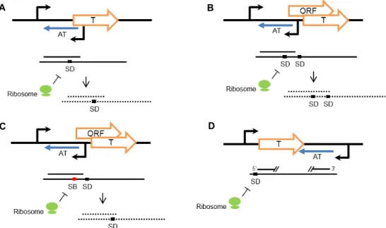
Fig. 1. Regulation mechanism of Type I TA modules. T, toxin. AT, antitoxin. SD, Shine-Dalgarno sequences. SB, Ribosome standby site. (A) Antitoxin that base-pairs with SD of toxin leads to mRNA degradation and blocks ribosome binding. (B) Antitoxin that base-pairs with SD of overlapping open reading frame (modulator) leads to mRNA degradation and inhibits modu- lator-dependent toxin translation. (C) Antitoxin that base-pairs with SB leads to mRNA degradation and of prevents ribosome loading onto SD of toxin. (D) Antitoxin that base-pairs with 5’ end and 3’ end of toxin mRNA blocks ribosome binding through conformational change of mRNA.
위해서 이용 가능하다[66]. 한편으로 TA system은 병원성 박 테리아에서 toxin의 발현과 활성만 유도하는 물질을 개발함으 로써 세균성 질병을 치료하기 위한 목적으로 이용될 수 있다 [25, 41, 58]. 또한, 정상적인 세포가 아닌 바이러스 감염 세포와 암세포에서만 toxin의 발현이 유도되도록 하여 진핵 세포의 사멸을 일으킴으로써 기존 치료제의 부작용을 줄임과 동시에 인간에게 나타나는 각종 바이러스성 질환 혹은 암의 치료에서 항바이러스제, 항암제 개발을 위한 핵심 요소로 이용될 수 있 을 것이다[36, 40]. 본 총설에서는 toxin의 다양성, 기능, 특징 등을 살펴보고 이를 응용한 생명공학 기술에 대해 논하고자 한다.
TA system의 분류와 조절원리
TA system은 antitoxin에 의해 toxin이 억제되는 방식과 antitoxin의 분자적 특성에 따라 현재 다섯 가지 유형으로 구분 된다. 공통적으로 TA system은 세포 내 toxin과 그들의 억제 자로 작용하는 antitoxin으로 구성되고, toxin은 단백질의 형태 로 antitoxin은 단백질 혹은 non-coding RNA의 형태로 작용 한다.
Type I TA system
Type I은 대부분이 toxin 유전자의 상부서열 혹은 하부서열 에 toxin에 비해 상대적으로 짧은 antitoxin 유전자가 반대 방 향으로 놓여있으며, toxin 유전자는 antitoxin 유전자 부위까지
포함하는 긴 전사체를 만드므로 toxin 전사체에 antitoxin 전사 체 서열과 상보적인 부분이 존재하게 된다. 일반적인 성장환 경에서는 antitoxin이 antisense RNA로서 이 부위에 결합하여 이중가닥 RNA를 만듦으로써 이것이 toxin의 번역을 막는다 [10]. 그러나 스트레스 상황에서는 세포 내 RNase들에 의해 상대적으로 불안정한 RNA antitoxin이 분해되면 결과적으로 toxin의 발현이 일어나게 된다. 대부분의 Type I toxin들은 작 은 소수성 단백질로서 세포 내막에 삽입되어 구멍을 형성하 고, 그 결과, 막 전위 상실 때문에 ATP 합성이 저해되어 독성 을 나타낸다. 이러한 활성을 지니는 Type I toxin 들의 예로 Hok, TisB toxin들이 있으나, 예외적으로 SymE toxin의 경우, RNA를 절단하는 활성을 보인다고 알려졌다[10, 22].
위에서 언급한 antisense RNA에 의한 toxin의 발현 조절방 식은 크게 네 가지로 나뉜다. 첫 번째 조절 기작은 toxin 유전 자의 상부서열에서부터 반대방향으로 전사되는 antisense RNA가 toxin 전사체의 리보솜 결합부위(Ribosome binding site, RBS)에 결합하는 경우이다. Antisense RNA의 결합은 mRNA의 절단을 유도함과 동시에 리보솜의 접근을 막아 tox- in의 번역을 억제하는데 이러한 기작은 SymE-SymR, Ibs- Sib system에서 나타난다(Fig. 1A) [10].
두 번째 조절 기작은 toxin과 antitoxin 외 다른 조절 유전자
가 toxin 유전자의 상부서열에 중첩되어 나타나고, antisense
RNA가 그 조절 유전자의 상부서열에서부터 반대 방향으로
전사되어 조절 유전자의 RBS에 결합하는 경우이다(Fig. 1B).
이 방식에서 대표적인 것이 Hok-Sok system으로, hok, sok 유 전자 코딩서열 외에도 mok이라는 조절유전자가 존재한다.
mok의 open reading frame (ORF)는 hok toxin의 ORF 와 중첩
되어 hok 유전자의 상부서열에 존재하며, sok antisense RNA 는 mok 유전자의 상부서열에서부터 반대 방향으로 전사된다.
따라서 hok의 발현은 mok의 발현에 의존적이게 되고, 첫 번째 조절 기작과는 다르게 sok antitsense RNA가 toxin 유전자의 RBS가 아닌 또 다른 ORF인 mok 유전자의 RBS에 결합함에 따라 결국 hok의 번역이 억제되는 것이다[12].
세 번째 조절 기작은 toxin 유전자와 중첩된 조절 유전자가 존재한다는 점에서 두 번째 조절 기작과 유사하지만 다른 원 리로 작용한다(Fig. 1C). 이런 예로써 TisB-IstR-1 system에서 는 tisA라는 조절 유전자가 tisB toxin의 상부서열에 중첩되어 있으며, tisAB mRNA 1차 전사체(+1)는 +42 mRNA로 가공되 었을 때, tisB의 ribosome standby stie가 드러나 tisB의 번역을 가능하게 한다. istR-1 antisense RNA는 이러한 tisB ribosome standby site 부위를 포함하면서 tisB 유전자와는 반대방향으 로 전사되기 때문에 tisB ribosome standby site에 istR-1 anti- sense RNA와 리보솜이 경쟁적으로 결합한다. 따라서 istR-1 antisense RNA가 결합하는 경우, 리보솜이 결합하여 tisB RBS 로 이동하는 것을 막아 tisB의 번역을 억제하는 것이다[53].
마지막 하나의 조절방식은 앞서 언급한 세 가지와 경우와 다르게 antitoxin의 위치가 toxin의 하부서열에서 나타난다 (Fig. 1D). Fst-RNA II system에서 RNA II antisense RNA는
fst 유전자의 하부서열에서부터 반대방향으로 전사되어 fsttoxin의 전사체와 결합함으로써 fst의 번역을 막는다. 이 때
fst mRNA와 RNA II antisense RNA는 상보적인 3’ 말단 서열을 가지며 각각의 5’ 말단에도 상보적인 두 부분의 직접 반복 서열이 존재하므로 이 서열끼리의 결합으로 생긴 구조적 변화 가 리보솜의 접근을 막아 fst toxin의 번역을 억제한다[56].
Type II TA system
Type II TA system은 TA system의 다섯 가지 유형 중에서 가장 광범위하게 연구되고 있다. 일반적으로 antitoxin의 ORF 가 toxin ORF의 앞쪽에 존재하는데, antitoxin과 toxin의 ORF 는 대다수가 4 bp 겹쳐진 형태로 존재하며 1 bp 겹쳐진 형태도 자주 나타난다[38]. 이러한 toxin과 antitoxin 유전자의 배열에 따라 toxin과 antitoxin 사이에 translational coupling이 일어 나고 따라서 bicistronic operon 형태로 존재하게 된다[61].
Type II의 조절 기작은 proteolysis와 관련되어 있다[4]. 정상 생육 조건에서, antitoxin 단백질은 toxin 단백질에 직접 결합 하여 toxin의 활성을 억제한다. 그러나 세포 내 단백질분해효 소들이 유도되는 스트레스 조건에서 상대적으로 불안정한 an- titoxin이 분해되면서 toxin이 노출되어 활성을 가지게 된다.
그뿐만 아니라 antitoxin은 앞서 말한 toxin의 불활성화 이외에 도 자가조절방식의 전사 억제자로 작용한다. 일반적으로
Type II TA 모듈의 프로모터 부위는 palindromic sequence를 가지며 이곳에 antitoxin뿐만 아니라 toxin-antitoxin 복합체가 결합하여 TA 모듈의 전사를 억제하는 것이다. 예외적으로 몇 몇 Type II TA system에서 antitoxin은 전사 억제자로서 기능 하지 않는다. 이러한 예로써 PaaR2-PaaA2-ParE2 system의 경 우, PaaA2 antitoxin 단독으로는 이들 오페론의 전사를 억제할 수 없으며, ParE2 toxin과 PaaA2 antitoxin의 복합체와 PaaR2 regulator가 오페론의 프로모터 부위에 결합하여 전사를 억제 한다[15].
대부분 antitoxin의 ORF가 toxin ORF의 앞쪽에 존재하는 것은 유리한 antitoxin의 생산을 위한 것으로 여겨진다[63]. 그 러나 예외적으로 antitoxin의 ORF가 뒤에서 나타나기도 한다.
HigB-HigA system이 그러한 경우로 higA antitoxin 유전자의 상부서열에 higB toxin 유전자가 위치한다[11].
Type II 유형의 toxin은 대다수가 mRNA를 절단함으로써 번역과정 혹은 전사 후 과정에 관여하며, 리보솜 소단위체와 결합하여 단백질 합성과정을 방해하거나 특이하게 DNA gy- rase의 활성을 저해하여 DNA 복제를 막기도 하고, 펩티도글 리칸 전구체를 인산화시켜 세포벽 합성을 저해하기도 한다.
대다수의 toxin들은 서열 특이적인 리보핵산 가수분해 활성 또는 서열 비특이적인 리보핵산 가수분해 활성을 가진다. 이 러한 toxin의 활성은 번역과정 중에 있는 mRNA를 절단하는 리보솜 의존적인 활성과 번역 과정 중에 있지 않은 free- mRNA를 절단하는 리보솜 비의존적 활성으로 나뉘게 된다 [21, 33, 60, 66].
또한, Doc toxin의 경우 30S 리보솜 소단위체와 직접적으로 상호작용함으로써 번역을 억제한다. Doc toxin은 아미노글라 이코사이드 계열의 항생제인 Hygromycin B (HygB)와 30S 리 보솜 소단위체에 경쟁적으로 결합하기 때문에, Doc toxin의 결합 부위는 HygB가 결합하는 16S rRNA에 보존된 helix 44 부위를 포함할 것으로 예측된다[26]. 한편 RatA (Ribosome as- sociation toxin A) toxin은 50S 리보솜 소단위체에 결합하여 50S와 30S 리보솜 소단위체의 결합을 저해하고, 결과적으로 70S 리보솜 소단위체의 형성을 막아 번역의 진행을 억제하게 된다[26, 65]. 반면, 특이하게도 E. coli의 HipA toxin은 kinase 활성으로 번역을 억제하는데, 이것은 glutamyl-tRNA synthe- tase (GltX)의 활성 부위에 보존된 Serine239 잔기를 인산화시 켜 aminoacylation을 억제하여 나타나는 현상이다[13].
반면에 ParE, CcdB toxin의 경우, 필수적인 Type II top- oisomerase의 subunit인 GyrA를 저해한다. 이렇듯 gyrase의 저해는 DNA 이중가닥의 절단을 초래하여 SOS 반응을 활성 화하고 세포 예정사를 유발한다[19, 32].
또한, ω-ε-ζ TA 모듈에서 ζ toxin의 경우 kinase 활성을 가지
는데, 펩티도글리칸 전구체인 UDP-N-acetylglucosamine
(UNAG)에서 N-acetylglucosamine의 3’-OH 부위를 인산화시
킨다. 인산화된 UNAG가 세포질 내에 축적되면 인산화되지
A B
C D
Fig. 2. Regulation mechanism of Type II TA modules. T, toxin. AT, antitoxin. SD, Shine-Dalgarno sequences. Type II antitoxin genes are located upstream of toxin genes (A, B) or downstream of toxin genes (C, D). Two genes, toxin and antitoxin, overlap by a few bases (A, C) or apart by a few bases (B, D).
않은 UNAG를 기질로 하여 펩티도글리칸 합성의 개시 단계를 촉매하는 효소인 MurA를 경쟁적으로 억제하고 결과적으로 박테리아 세포벽 형성을 방해하게 된다[34].
기존에는 Type II TA system을 단순히 toxin family를 아미 노산 서열과 구조의 유사성에 따라 구분하고 이에 특이적인 antitoxin family를 짝지어 하나의 TA family로 분류하였다 [38]. 그에 따라 Type II는 총 10개의 TA family로 구분되었다 [38]. 그러나 특정 family에 속하는 일부 toxin이 다른 family의 antitoxin과도 interaction 할 수 있다는 사실이 밝혀지면서 toxin, antitoxin family 각각에서 독립적인 연관성이 보고되었 고, 생물 정보학적인 접근 또한 이러한 연관 관계를 뒷받침하 고 있다[21]. 이에 따라, 현재 Type II TA system은 12개의 toxin family와 20개의 antitoxin family로 구분된다[24]. 이러 한 사실은 박테리아에서 기존 분류법으로 예측되는 Type II TA system보다 더 풍부하고 다양한 수의 TA system이 존재할 것이라는 가능성을 나타낸다.
Type III TA system
Toxin과 antitoxin이 각각의 프로모터로부터 전사되는 Type I TA system과는 달리, Type III의 toxin과 antitoxin은 단일 프로모터로부터 co-transcription 된다. 또한 Type I 유형 과는 다르게 RNA antitoxin이 toxin 단백질에 직접 결합하여 그것의 활성을 억제한다는 점이 특이하다. Type III 유형으로 처음 알려진 것은 Pectobacterium atrosepticum의 pECA1039 상 에 존재하는 ToxI-ToxN system이다. 이 ToxI-ToxN system에 서 ToxN toxin 유전자의 앞쪽으로는 직접반복서열(5.5 tan-
dem repeats of 36 nt)이 존재한다. ToxN toxin은 리보솜 비의 존적인 리보핵산 가수분해 활성을 가지기 때문에 toxI-toxN 전사체의 직접반복서열부위를 잘라 36 nt RNA antitoxin (toxI)을 만들어낸다. 따라서 toxI antitoxin 절편은 pseudoknot 구조를 형성하여 5’, 3’ 말단이 2개의 ToxN 분자와 각각 상호 작용하게 되고, 결과적으로 3개의 toxI antitoxin 절편과 3개의 ToxN toxin 단백질이 복합체를 이루어 toxI에 의해 ToxN의 활성이 억제되는 것이다.
Type III ToxI-ToxN system을 가지는 박테리아의 경우, 정 상적인 환경에서는 ToxN toxin의 활성이 toxI antitoxin에 의 해 저해되어 있다. 그러나 박테리오파지가 감염하게 되면 숙 주의 전사 혹은 번역 수준의 변화나 숙주 DNA의 분해로 인해 ToxN과 toxI 비율의 균형이 깨지거나 toxI가 불활성화될 수 있다. 이때 자유로워진 ToxN이 활성을 띠게 되어 박테리아와 박테리오파지의 RNA를 절단함에 따라 박테리아의 성장을 저 해하고 박테리오파지의 성숙을 막는 것으로 보인다[8].
Type IV TA system
Type IV TA system의 경우 toxin과 antitoxin 모두 단백질
로 작용한다는 점에서 Type II와 공통점이 있지만, toxin과 an-
titoxin 사이에 직접적인 상호작용이 없다는 점에서 차이가 있
다. Type IV 유형은 최근에 Escherichia coli의 YeeU-YeeV sys-
tem에서 처음으로 확인되었다[3]. YeeV toxin은 세포 분화와
형태유지에 관련된 FtsZ, MreB와 상호작용한다. FtsZ는 세포
격막에서 고리 구조를 형성하여 세포 분화에 필수적인 역할을
하는 GTPase로서[2], YeeV toxin에 의해 이들의 GTP 의존적
Table 1. Target and recognition site of Type II RNase toxins
Toxin Target Recognition site Organism Reference
MazF MazF MazF MazF MazF
mRNA mRNA mRNA mRNA mRNA
ACA UACAU UACAU GUUGC UUACUCA
Escherichia coli Staphylococcus aureus Bacillus subtilis Myxococcus xanthus Haloquadratum walsbyi
[66]
[68]
[39]
[35]
Unpublished data VapC
VapC VapC VapC
VapC (PAE0151) VapC (PAE2754)
tRNAfMet tRNAfMet tRNAfMet Total RNA Pentaprobes Pentaprobes
Anticodon stem-loop Anticodon stem-loop Unknown
Unknown
GGHG (H is U or G) GGHG (H is U or G)
Salmonella enterica Shigella flexneri Leptospira interrogans Sulfolobus solfataricus Pyrobaculum aerophilum Pyrobaculum aerophilum
[60]
[60]
[27]
[28]
[30]
[30]
PemK PemK PemK
mRNA mRNA Total RNA
UAH (H is C, A or U) UAUU
UACH (H is U or G)
Escherichia coli Staphylococcus aureus Xylella fastidiosa
[64]
[5]
[23]
MqsR mRNA GCU Escherichia coli [62]
Kid mRNA UAK (K is A or C) Escherichia coli [33]
HicA tmRNA AAAC Escherichia coli [21]
ChpBK mRNA ACY (Y is U, A, or G) Escherichia coli [67]
인 중합이 방해된다. MreB의 경우, 세포의 전형적인 막대 모양 을 유지하는데 필수적인 단백질로 나선형 구조로 중합되어 세포 길이를 따라 코일구조를 형성하는데[20], MreB 또한 YeeV toxin과의 상호작용으로 중합이 억제된다. Type II와는 달리 YeeU antitoxin은 YeeV toxin과 복합체를 형성하지 않으 며, YeeV toxin의 타겟인 FtsZ, MreB와 직접적으로 상호작용 함으로써 이들의 중합을 촉진한다. 마찬가지로 같은 Type IV 에 속하는 E. coli의 CptA-CptB system에서 CptA toxin은 MreB, FtsZ와 상호작용하여 세포골격을 형성하기 위한 이들 의 중합을 방해하며, CptB antitoxin은 MreB, FtsZ와 상호작용 하여 이들의 중합을 촉진하는 활성을 보인다[29].
Type V TA system
Type V 유형은 toxin과 antitoxin 둘 다 단백질로 작용하기 때문에 Type II와 유사하나 이 유형의 경우 antitoxin이 리보핵 산 가수분해 활성을 가진다는 점에서 독특하다. 알려진 Type V toxin들은 Type I toxin과 같이 작은 소수성 단백질로서 세 포 내막에 끼어들어가 구멍을 형성하며, 이는 막 전위 상실로 이어져 세포의 ATP 합성이 일어나지 않게 된다.
최근에 E. coli의 GhoT-GhoS system이 발견되면서 Type V 유형이 새롭게 분류되었다. 이 유형에서 GhoS antitoxin는 서열 특이적인 리보핵산 가수분해 활성을 가져 ghoT toxin의 mRNA를 분해한다[54]. 이것은 마치 Type II와는 정반대의 상 호작용방식을 보여준다. 그리고 GhoT toxin은 막 용해 단백질 로서 세포막에 끼어들어가 ghost cell을 형성하고 persistence 를 증가시킨다고 보고된 바 있다. 특이하게도 Type II와는 다
르게 GhoS antitoxin은 안정하며, 그들 자신의 오페론에 대한 전사 조절자로서 기능하지 않는다[54]. 그뿐만 아니라 GhoT- GhoS system은 Type II 유형에 속하는 MqsR-MqsA system에 의하여 조절되기 때문에 리보핵산 가수분해 활성을 갖는 MqsR toxin이 활성화됨에 따라 ghoS antitoxin mRNA가 분해 되고 많은 수의 free GhoT toxin이 축적되는 것이다[55].
리보핵산 가수분해 활성을 가지는 toxin
TA system에 속하는 많은 toxin이 다양한 활성을 가지지만 그 중에서도 리보핵산 가수분해 활성은 아주 흔하게 나타난다 [6].
RelE toxin은 리보솜 의존적으로 RNA를 절단하는 RNA
제한효소로 잘 알려졌는데, 이들은 ribosomal A site 에서
mRNA 특정 서열을 선호적으로 인식하여 절단한다[37]. HigB
toxin 역시 리보솜 의존적으로 RNA를 절단하며, 이들은 50S
리보솜 소단위체에 결합하여 mRNA의 AAA 서열을 특이적
으로 인식하여 절단한다[17]. 한편으로 리보솜 비의존적으로
free mRNA를 절단하는 toxin으로는 MazF, VapC, PemK,
MqsR, Kid, HicA, ChpBK가 있다(Table 1, 2). 이 중에서 MazF
toxin은 각 박테리아의 homolog마다 mRNA의 ACA, UAC,
UACAU, GUUGC, (U/C)U(U/A)C(U/C), UCGCU,
(U/C)UCCU, CUCCU, UUACUCA와 같은 부위를 선호적으
로 절단하고, 경우에 따라 23S rRNA 혹은 16S rRNA의 특정
부위를 추가로 절단한다. Mycobacterium tuberculosis의 경우,
MazF toxin homolog들이 많이 존재하고 있으며, 이들 MazF
toxin들은 제각기 다른 RNA 서열을 인식하여 절단하는 것으
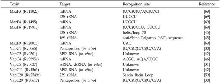
Table 2. Target and recognition site of M. tuberculosis H37Rv RNase toxins
Toxin Target Recognition site Reference
MazF3 (Rv1102c)
MazF4 (Rv1495) MazF6 (Rv1991c)
MazF9 (Rv2801c) VapC1 (Rv0065) VapC2 (Rv0301) VapC4 (Rv0595c) VapC5 (Rv0627) VapC11 (Rv1561) VapC20 (Rv2549c) VapC29 (Rv0617)
mRNA 23S rRNA mRNA mRNA 23S rRNA 16S rRNA mRNA
Pentaprobes (in vitro) MS2 RNA (in vitro) mRNA
ssRNA, dsRNA (in vitro) MS2 RNA (in vitro) 23S rRNA
Pentaprobes (in vitro)
(U/C)U(U/A)C(U/C) UUCCU
UCGCU
(U/C)UCCU, CUCCU helix/loop 70
anti-Shine-Dalgarno (aSD) sequence UAC
(G/C)G(G/C)(G/C/A) Unknown
ACGC, AC(A/U)GC Unknown
Unknown Sarcin Ricin Loop (G/C)G(G/C)(G/C/A)
[69]
[69]
[69]
[69]
[45]
[45]
[69]
[30]
[42]
[46]
[31]
[42]
[59]
[30]
로 확인되었다(Table 2). E. coli MazF toxin의 경우, 리보솜 비의존적으로 mRNA의 ACA 서열을 특이적으로 절단한다 [66]. 결과적으로 이러한 활성은 대부분의 세포 내 mRNA를 절단하게 되고 이를 이용하여 최근에 ACA-less 유전자로부터 단일 단백질을 발현하는 시스템이 개발되었다[49]. 그리고
Staphylococcus aureus의 MazF toxin은 mRNA 상의 UACAU서열을 인식하여 절단하는데, UACAU는 S. aureus의 병원성 관련 유전자들에서 공통적으로 흔하게 나타나는 서열로서 S.
aureus의 MazF-MazE system과 병원성 사이에 가능한 조절
메커니즘이 존재할 것으로 보인다[47]. 반면 Bacillus subtilis의 MazF toxin도 UACAU 서열을 절단하지만, UACAU는 B. sub-
tilis의 2차 대사산물의 생합성과 관련된 유전자들에서 많이나타나는 서열로 B. subtilis의 MazF-MazE system은 2차 대사 산물의 생산 조절과 관련이 있을 것으로 추측된다[39]. 한편
Myxococcus xanthus의 MazF toxin의 경우, GUUGC 서열을인식하여 절단하는 활성을 가진다[35]. 이들은 일반적인 Type II TA 모듈과는 다르게 MazF toxin 유전자 가까이에 MazE antitoxin이 존재하지 않으며, 세포 내 존재하는 전사조절자인 MrpC가 MazF의 antitoxin으로서 기능하는 것이 확인되었다 [35]. MrpC는 mazF의 전사 활성자이며, mrpC와 mazF의 전사 는 Ser/Thr kinase cascade에 의한 MrpC의 인산화를 통해 음 성적으로 조절된다[35]. 그리고 Escherichia coli의 PemK toxin 은 mRNA의 UAH (H is C, A or U), Staphylococcus aureus PemK toxin은 mRNA의 UAUU, Xylella fastidiosa의 PemK toxin은 total RNA의 UACH (H is U or G)를 인식하여 절단하 는 것으로 확인되었다. MqsR toxin은 GCU, Kid toxin은 UAK (K is A or C), ChpBK toxin은 ACY (Y is U, A or G) 부위를 절단한다. 앞서 언급한 일부 toxin들은 rRNA 제한효소 활성을 가지는데 대표적인 것이 E. coli의 MazF toxin으로 이들은 16S rRNA의 3’ 말단을 인식하여 43nt를 제거하며, 특정 mRNA의 개시코돈 상부서열에 존재하는 ACA 서열을 인식하여 절단함
으로써 RBS가 없는 leaderless mRNA를 생산한다. 제거된 부 위는 번역개시와 관련된 anti-RBS 서열을 포함하기 때문에 일 반적인 RBS를 가지는 mRNA들이 결합하는 것을 방해한다.
그러나 이렇게 변형된 리보솜은 MazF에 의하여 발생한 lead- erless mRNA들만을 선택적으로 인식하여 번역이 가능하다 [52]. 그리고 mRNA의 UUCCU 서열을 인식하는 Mycobacte-
rium tuberculosis의 MazF toxin은 M. tuberculosis 23S rRNA의ribosomal A site의 UUCCU 서열을 인식, 절단하여 번역을 억제한다[44].
VapC toxin의 경우, mRNA 절단 부위가 다양하게 나타나 고 23S rRNA 또는 tRNA
fMet를 추가로 절단하기도 한다.
Salmonella enterica와 Shigella flexneri에 존재하는 VapC toxin
의 경우 tRNA
fMet의 anticodon stem-loop를 특이적으로 절단 하는 활성을 가진다[60]. 독특하게도 HicA toxin은 mRNA뿐 만 아니라 tmRNA의 AAAC 부위를 선호적으로 절단하여 번 역속도를 전반적으로 감소시키는 특징이 있다[21]. Kid, MazF toxin과 구조적인 유사성을 가지는 Type III ToxI-ToxN sys- tem의 ToxN toxin 또한 RNase 활성을 가지는 것으로 확인되 었지만 아직 어떠한 RNA를 타겟으로 하는지는 밝혀지지 않 았다[9].
인간질병 치료의 대체 전략
의학의 발달로 인간의 수명이 길어짐에 따라 암이라는 질병
은 인류의 건강을 위협하고 있고 아직도 암의 완전한 정복은
이루어지지 않고 있다. 그뿐만 아니라 human immun-
odeficiency virus (HIV-1), hepatitis C virus (HCV)의 경우,
인간에게 각각 acquired immunodeficiency syndrome (AIDS),
hepatitis C와 같이 심각한 바이러스성 질환을 일으키는 RNA
virus로 좀 더 효과적인 치료법의 개발이 요구되고 있다. 이에
있어서 TA system은 인간의 질병 치료에 있어서 떠오르는
하나의 전략으로 이용될 수 있고, 크게 항암제, 항바이러스제 로 개발할 수 있다.
항암제
Anticancer drug로 이용되기 위해서는 암세포에만 선택적 으로 작용하며 독성을 가지는 것이 이상적이다. 하지만 현재 사용되고 있는 항암제 대부분이 충분한 선택성을 만족하지 못하기 때문에 결과적으로 정상 세포를 손상시켜 부작용을 유발한다. 이러한 문제를 해결하기 위한 대안 중의 하나가 독 성의 조절이 가능한 TA system의 특성을 이용하는 것이다.
TA system은 원핵세포에서만 특이적으로 나타나는 유전적 모듈이지만 이들은 진핵세포에서도 이용될 수 있다. 특히나 리보핵산 가수분해 활성을 갖는 toxin은 진핵세포에서도 활성 을 띠어 apoptosis를 유발하는데, 이를 이용하여 암세포를 공 격하고 정상 세포에서는 toxin의 inhibitor인 antitoxin의 발현 을 유도하면 피해를 최소화할 수 있다. 실제로 ParD (Kid-Kis) TA system을 이용한 selective killing system은 많은 인간 암 세포에서 불활성화되는 전사조절자인 p53 (mutant p53)을 이 용하여 고안되었다. Wild-type p53과 mutant p53에 의해 조절 되는 진핵세포의 프로모터를 이용함으로써 toxin과 antitoxin 의 발현이 각각 독립적으로 조절되도록 하여 toxin과 antitoxin 사이에 상대적인 양적 조절이 가능하도록 하는 것이다. 간단 히 말해서, wild-type p53에 의해 활성화되고 mutant p53에 의해 저해되는 프로모터의 조절 하에 kid toxin의 발현이 이루 어지도록 하고, mutant p53에 의해 활성화되고 wild-type p53 에 의해 저해되는 프로모터의 조절 하에 kis antitoxin의 발현 이 이루어지게 하는 것이다. 이러한 조절 하에 많은 암세포에 서 공통적으로 나타나는 mutant p53은 각각의 프로모터에 결 합하여 Kid toxin의 발현을 유도하고 Kis antitoxin의 발현은 저해하며, 정상 세포에 존재하는 wild-type p53은 Kis anti- toxin의 발현을 유도하고 Kid toxin의 발현은 저해하여 암세포 만 선택적으로 손상된다[7].
항바이러스제
감염성 바이러스 질환의 치료에서도 바이러스에 감염된 세 포만을 선택적으로 공격하는 전략이 필요하다. 이러한 전략은 바이러스 단백질에 의해 특이적으로 인식되는 서열을 이용하 거나 바이러스 단백질 자체를 이용하여 toxin의 발현을 유도 하게 된다.
최근에 HIV-1에 감염된 세포를 공격하기 위한 전략으로 HIV-1 단백질(Tat)의 인식서열(LTR 프로모터)에 의존하여 E.
coli mazF toxin을 발현하는 retroviral vector가 개발되었다.
HIV-1 감염 초기에 생산되는 Tat 단백질의 경우, LTR 프로모 터에 결합하여 다른 HIV-1 단백질들의 발현을 유도한다. 그러 므로 LTR 프로모터 조절 하에 mazF를 발현시키면 HIV-1에 감염된 세포들에서만 발현된 MazF toxin은 세포 내 mRNA와
바이러스 RNA의 ACA 서열을 인식하여 절단함으로써 HIV-1 의 복제를 막는다[36].
많은 RNA 바이러스들이 가지는 protease들은 그들의 복제 를 위해 필수적인 역할을 한다. 그런 의미에서 HIV-1의 HIV PR protease, HCV의 NS3 protease는 특이적 절단서열을 가지 는 viral protease로써 감염 세포를 표적화하기 위해 이용될 수 있다. 각각의 특이적 절단서열을 포함하는 linker로 이어진 MazF-MazEp (MazF-linker-MazE fragment) 형태의 polypep- tide는 정상 세포에서는 TA complex를 이룬 형태로 존재한다.
그러나 HIV-1 혹은 HCV 감염 세포에서 이들이 생산하는 HIV PR protease, NS3 protease에 의해 linker가 절단되면 MazF toxin이 활성화되어 감염 세포를 공격하는 것이다[40].
결 론
박테리아는 평균적으로 TA system을 염색체에 3.8개, 플라 스미드 상에 0.4개 정도 가지고 있으며[14], 현재 추정되는 TA system의 개수만 해도 10,000여 가지를 넘어선다[50]. 주목할 점은, 다른 생물체에 기생하거나 공생하여 살아가는 생물체들 은 공통적으로 TA system을 상대적으로 적게 가지거나 전혀 갖지 않는다[38]. 따라서 독립적으로 살아가는 자유 생활 생물 체들은 비교적 적은 스트레스 환경과 마주하는 기생성 생물체 보다 많은 수의 TA system을 가지며, 다양한 스트레스에 대응 하기 위하여 이러한 TA system을 진화적으로 보존해 온 것이 다. 이렇듯 흔한 TA system의 toxin들은 그 개수만큼이나 다 양한 활성을 나타내지만 공통적으로 toxin이 활성화되면 박테 리아의 성장을 저해하거나 사멸을 유도한다. 박테리아의 성장 을 저해하는 경우, 생육하기 좋은 조건이 되면 antitoxin을 이 용하여 toxin의 활성을 억제함에 따라 다시 성장을 시작한다.
박테리아의 사멸을 유도하는 경우에도 역시나 군집의 지속을
위한 자살 전략이라 볼 수 있다. TA system은 대부분 스트레
스 반응이 주요한 기능이라 할 수 있는데, 각각의 TA system
이 가지는 구체적인 생리학적 기능은 매우 다양하게 나타난
다. E. coli R1 플라스미드에 존재하는 Hok-Sok system,
Kid-Kis system, F 플라스미드에 존재하는 CcdA-CcdB sys-
tem은 플라스미드 유지기능을 하는 TA system의 대표적인
예이다[16]. 플라스미드는 박테리아가 생존하기 위한 필수적
인 요소는 아니지만 생육하기 힘든 환경에서 살아남기 위하여
이러한 TA system을 보존하고 있는 것으로 보인다. 특히
Enterococcus faecalis의 pAD1 플라스미드에 존재하는 Par(Fst-RNA II) system은 독성 플라스미드인 pAD1을 유지하는
기능을 가지며 이는 E. faecalis의 병원성과 연관되어 있을 것으
로 보인다[57]. 그리고 TA system은 persister cell의 형성에도
관여하며, E. coli의 relE, higB, mazF, yafQ, yoeB toxin은 per-
sister cell 내에서 강하게 유도된다. 또한, E. coli MqsR-MqsA
system은 바이오 필름의 형성에 관여하는 대표적인 TA sys-
tem이다. 이 밖에도 박테리오 파지의 감염에 대한 방어 기작으 로서 기능하거나[9], 리보핵산 가수분해 활성을 갖는 toxin은 mRNA의 특정 서열을 인식하여 절단함에 따라 유전자 발현조 절이 이루어지기도 한다[1]. 하지만 아직 특성이 밝혀지지 않 은 TA system의 수가 더 많다는 점을 고려해 볼 때, 이와 같은 TA system의 역할들 외에도 또 다른 기능을 수행하는 TA sys- tem이 존재할 가능성이 있다.
따라서 각종 박테리아와 고세균에서 TA system을 특성화 하는 것은 그들의 생리학적 특성 규명과 동시에 이들을 생물 공학적, 의학적 분야에 응용함으로써 유용한 기술 혹은 치료 제를 개발하는데 한 발짝 다가설 수 있을 것이다.
감사의 글
이 논문은 부산대학교 기본연구지원사업(2년)에 의하여 연 구되었음.
References
1. Bertram, R. and Schuster, C. F. 2014. Post-transcriptional regulation of gene expression in bacterial pathogens by tox- in-antitoxin systems. Front. Cell. Infect. Microbiol. 4, 6.
2. Bi, E., Dai, K., Subbarao, S., Beall, B. and Lutkenhaus, J.
1991. FtsZ and cell division. Res. Microbiol. 142, 249-252.
3. Brown, J. M. and Shaw, K. J. 2003. A novel family of Escherichia coli toxin-antitoxin gene pairs. J. Bacteriol. 185, 6600-6608.
4. Brzozowska, I. and Zielenkiewicz, U. 2013. Regulation of toxin-antitoxin systems by proteolysis. Plasmid 70, 33-41.
5. Bukowski, M., Lyzen, R., Helbin, W. M., Bonar, E., Szalew- ska-Palasz, A., Wegrzyn, G., Dubin, G., Dubin, A. and Wladyka, B. 2013. A regulatory role for Staphylococcus aur- eus toxin-antitoxin system PemIKSa. Nat. Commun. 4, 2012.
6. Cook, G. M., Robson, J. R., Frampton, R. A., McKenzie, J., Przybilski, R., Fineran, P. C. and Arcus, V. L. 2013.
Ribonucleases in bacterial toxin-antitoxin systems. Biochim.
Biophys. Acta 1829, 523-531.
7. de la Cueva-Mendez, G., Mills, A. D., Clay-Farrace, L., Diaz- Orejas, R. and Laskey, R. A. 2003. Regulatable killing of eu- karyotic cells by the prokaryotic proteins Kid and Kis.
EMBO J. 22, 246-251.
8. Dy, R. L., Richter, C., Salmond, G. P. C. and Fineran, P.
C. 2014. Remarkable Mechanisms in Microbes to Resist Phage Infections. Annu. Rev. Virol. 1, 307-331.
9. Fineran, P. C., Blower, T. R., Foulds, I. J., Humphreys, D.
P., Lilley, K. S. and Salmond, G. P. 2009. The phage abortive infection system, ToxIN, functions as a protein-RNA tox- in-antitoxin pair. Proc. Natl. Acad. Sci. USA 106, 894-899.
10. Fozo, E. M., Hemm, M. R. and Storz, G. 2008. Small toxic proteins and the antisense RNAs that repress them.
Microbiol. Mol. Biol. Rev. 72, 579-589.
11. Gerdes, K., Christensen, S. K. and Lobner-Olesen, A. 2005.
Prokaryotic toxin-antitoxin stress response loci. Nat. Rev.
Microbiol. 3, 371-382.
12. Gerdes, K., Thisted, T. and Martinussen, J. 1990. Mechanism of post-segregational killing by the hok/sok system of plas- mid R1: sok antisense RNA regulates formation of a hok mRNA species correlated with killing of plasmid-free cells.
Mol. Microbiol. 4, 1807-1818.
13. Germain, E., Castro-Roa, D., Zenkin, N. and Gerdes, K. 2013.
Molecular mechanism of bacterial persistence by HipA. Mol.
Cell 52, 248-254.
14. Goeders, N. and Van Melderen, L. 2014. Toxin-antitoxin sys- tems as multilevel interaction systems. Toxins (Basel) 6, 304- 324.
15. Hallez, R., Geeraerts, D., Sterckx, Y., Mine, N., Loris, R. and Van Melderen, L. 2010. New toxins homologous to ParE be- longing to three-component toxin-antitoxin systems in Escherichia coli O157:H7. Mol. Microbiol. 76, 719-732.
16. Hayes, F. 2003. Toxins-antitoxins: plasmid maintenance, programmed cell death, and cell cycle arrest. Science 301, 1496-1499.
17. Hurley, J. M. and Woychik, N. A. 2009. Bacterial toxin HigB associates with ribosomes and mediates translation-depend- ent mRNA cleavage at A-rich sites. J. Biol. Chem. 284, 18605- 18613.
18. Jaffe, A., Ogura, T. and Hiraga, S. 1985. Effects of the ccd function of the F plasmid on bacterial growth. J. Bacteriol.
163, 841-849.
19. Jiang, Y., Pogliano, J., Helinski, D. R. and Konieczny, I. 2002.
ParE toxin encoded by the broad-host-range plasmid RK2 is an inhibitor of Escherichia coli gyrase. Mol. Microbiol. 44, 971-979.
20. Jones, L. J., Carballido-Lopez, R. and Errington, J. 2001.
Control of cell shape in bacteria: helical, actin-like filaments in Bacillus subtilis. Cell 104, 913-922.
21. Jorgensen, M. G., Pandey, D. P., Jaskolska, M. and Gerdes, K. 2009. HicA of Escherichia coli defines a novel family of translation-independent mRNA interferases in bacteria and archaea. J. Bacteriol. 191, 1191-1199.
22. Kawano, M., Aravind, L. and Storz, G. 2007. An antisense RNA controls synthesis of an SOS-induced toxin evolved from an antitoxin. Mol. Microbiol. 64, 738-754.
23. Lee, M. W., Rogers, E. E. and Stenger, D. C. 2012. Xylella fastidiosa plasmid-encoded PemK toxin is an endor- ibonuclease. Phytopathology 102, 32-40.
24. Leplae, R., Geeraerts, D., Hallez, R., Guglielmini, J., Dreze, P. and Van Melderen, L. 2011. Diversity of bacterial type II toxin-antitoxin systems: a comprehensive search and func- tional analysis of novel families. Nucleic Acids Res. 39, 5513- 5525.
25. Lioy, V. S., Rey, O., Balsa, D., Pellicer, T. and Alonso, J.
C. 2010. A toxin-antitoxin module as a target for anti- microbial development. Plasmid 63, 31-39.
26. Liu, M., Zhang, Y., Inouye, M. and Woychik, N. A. 2008.
Bacterial addiction module toxin Doc inhibits translation elongation through its association with the 30S ribosomal subunit. Proc. Natl. Acad. Sci. USA 105, 5885-5890.
27. Lopes, A. P., Lopes, L. M., Fraga, T. R., Chura-Chambi, R.
M., Sanson, A. L., Cheng, E., Nakajima, E., Morganti, L. and Martins, E. A. 2014. VapC from the leptospiral VapBC tox- in-antitoxin module displays ribonuclease activity on the in- itiator tRNA. PLoS One 9, e101678.
28. Maezato, Y., Daugherty, A., Dana, K., Soo, E., Cooper, C., Tachdjian, S., Kelly, R. M. and Blum, P. 2011. VapC6, a ribo- nucleolytic toxin regulates thermophilicity in the cren- archaeote Sulfolobus solfataricus. RNA 17, 1381-1392.
29. Masuda, H., Tan, Q., Awano, N., Yamaguchi, Y. and Inouye, M. 2012. A novel membrane-bound toxin for cell division, CptA (YgfX), inhibits polymerization of cytoskeleton pro- teins, FtsZ and MreB, in Escherichia coli. FEMS Microbiol.
Lett. 328, 174-181.
30. McKenzie, J. L., Duyvestyn, J. M., Smith, T., Bendak, K., Mackay, J., Cursons, R., Cook, G. M. and Arcus, V. L. 2012.
Determination of ribonuclease sequence-specificity using Pentaprobes and mass spectrometry. RNA 18, 1267-1278.
31. Miallau, L., Faller, M., Chiang, J., Arbing, M., Guo, F., Cascio, D. and Eisenberg, D. 2009. Structure and proposed activity of a member of the VapBC family of toxin-antitoxin systems. VapBC-5 from Mycobacterium tuberculosis. J. Biol.
Chem. 284, 276-283.
32. Miki, T., Park, J. A., Nagao, K., Murayama, N. and Horiuchi, T. 1992. Control of segregation of chromosomal DNA by sex factor F in Escherichia coli. Mutants of DNA gyrase sub- unit A suppress letD (ccdB) product growth inhibition. J.
Mol. Biol. 225, 39-52.
33. Munoz-Gomez, A. J., Lemonnier, M., Santos-Sierra, S., Berzal- Herranz, A. and Diaz-Orejas, R. 2005. RNase/anti-RNase ac- tivities of the bacterial parD toxin-antitoxin system. J.
Bacteriol. 187, 3151-3157.
34. Mutschler, H. and Meinhart, A. 2011. Epsilon/zeta systems:
their role in resistance, virulence, and their potential for an- tibiotic development. J. Mol. Med. (Berl) 89, 1183-1194.
35. Nariya, H. and Inouye, M. 2008. MazF, an mRNA interfer- ase, mediates programmed cell death during multicellular Myxococcus development. Cell 132, 55-66.
36. Okamoto, M., Chono, H., Kawano, Y., Saito, N., Tsuda, H., Inoue, K., Kato, I., Mineno, J. and Baba, M. 2013. Sustained inhibition of HIV-1 replication by conditional expression of the E. coli-derived endoribonuclease MazF in CD4+ T cells.
Hum. Gene Ther. Methods 24, 94-103.
37. Overgaard, M., Borch, J., Jorgensen, M. G. and Gerdes, K.
2008. Messenger RNA interferase RelE controls relBE tran- scription by conditional cooperativity. Mol. Microbiol. 69, 841-857.
38. Pandey, D. P. and Gerdes, K. 2005. Toxin-antitoxin loci are highly abundant in free-living but lost from host-associated prokaryotes. Nucleic Acids Res. 33, 966-976.
39. Park, J. H., Yamaguchi, Y. and Inouye, M. 2011. Bacillus subtilis MazF-bs (EndoA) is a UACAU-specific mRNA interferase. FEBS Lett. 585, 2526-2532.
40. Park, J. H., Yamaguchi, Y. and Inouye, M. 2012. Intramolec- ular regulation of the sequence-specific mRNA interferase activity of MazF fused to a MazE fragment with a linker
cleavable by specific proteases. Appl. Environ. Microbiol. 78, 3794-3799.
41. Park, S. J., Son, W. S. and Lee, B. J. 2013. Structural overview of toxin-antitoxin systems in infectious bacteria: a target for developing antimicrobial agents. Biochim. Biophys. Acta 1834, 1155-1167.
42. Ramage, H. R., Connolly, L. E. and Cox, J. S. 2009.
Comprehensive functional analysis of Mycobacterium tu- berculosis toxin-antitoxin systems: implications for patho- genesis, stress responses, and evolution. PLoS Genet. 5, e1000767.
43. Sala, A., Bordes, P. and Genevaux, P. 2014. Multiple tox- in-antitoxin systems in Mycobacterium tuberculosis. Toxins (Basel) 6, 1002-1020.
44. Schifano, J. M., Edifor, R., Sharp, J. D., Ouyang, M., Konkimalla, A., Husson, R. N. and Woychik, N. A. 2013.
Mycobacterial toxin MazF-mt6 inhibits translation through cleavage of 23S rRNA at the ribosomal A site. Proc. Natl.
Acad. Sci. USA 110, 8501-8506.
45. Schifano, J. M., Vvedenskaya, I. O., Knoblauch, J. G., Ouyang, M., Nickels, B. E. and Woychik, N. A. 2014. An RNA-seq method for defining endoribonuclease cleavage specificity identifies dual rRNA substrates for toxin MazF- mt3. Nat. Commun. 5, 3538.
46. Sharp, J. D., Cruz, J. W., Raman, S., Inouye, M., Husson, R. N. and Woychik, N. A. 2012. Growth and translation in- hibition through sequence-specific RNA binding by Mycobacterium tuberculosis VapC toxin. J. Biol. Chem. 287, 12835-12847.
47. Siboo, I. R., Chambers, H. F. and Sullam, P. M. 2005. Role of SraP, a Serine-rich surface protein of staphylococcus aur- eus, in binding to human platelets. Infect. Immun. 73, 2273- 2280.
48. Stieber, D., Gabant, P. and Szpirer, C. 2008. The art of se- lective killing: plasmid toxin/antitoxin systems and their technological applications. BioTechniques 45, 344-346.
49. Suzuki, M., Zhang, J., Liu, M., Woychik, N. A. and Inouye, M. 2005. Single protein production in living cells facilitated by an mRNA interferase. Mol. Cell 18, 253-261.
50. Unterholzner, S. J., Poppenberger, B. and Rozhon, W. 2013.
Toxin-antitoxin systems: Biology, identification, and appli- cation. Mob. Genet. Elements 3, e26219.
51. Van Melderen, L., Bernard, P. and Couturier, M. 1994. Lon- dependent proteolysis of CcdA is the key control for activa- tion of CcdB in plasmid-free segregant bacteria. Mol.
Microbiol. 11, 1151-1157.
52. Vesper, O., Amitai, S., Belitsky, M., Byrgazov, K., Kaberdina, A. C., Engelberg-Kulka, H. and Moll, I. 2011. Selective trans- lation of leaderless mRNAs by specialized ribosomes gen- erated by MazF in Escherichia coli. Cell 147, 147-157.
53. Vogel, J., Argaman, L., Wagner, E. G. and Altuvia, S. 2004.
The small RNA IstR inhibits synthesis of an SOS-induced toxic peptide. Curr. Biol. 14, 2271-2276.
54. Wang, X., Lord, D. M., Cheng, H. Y., Osbourne, D. O., Hong, S. H., Sanchez-Torres, V., Quiroga, C., Zheng, K., Herrmann, T., Peti, W., Benedik, M. J., Page, R. and Wood,
초록:박테리아의 toxin-antitoxin system과 생명공학기술 응용
김윤지․황지환*
(부산대학교 생명시스템학과)
Toxin-antitoxin (TA) system은 박테리아와 고세균에서 진화적으로 보존되어 흔히 발견되는 유전적 모듈이다.
기본적으로 이 시스템은 세포 내 toxin과 그들의 억제자로 작용하는 antitoxin으로 구성되어있으며, 현재 총 다섯 가지 유형으로 구분된다. 공통적으로 toxin은 스트레스 조건에서 활성화됨으로써 세포 내 다양한 과정을 억제하는 활성을 가지는데 이는 결과적으로 세포 사멸 혹은 가역적인 생장 저해를 일으킨다. Toxin의 이러한 효과들은 유전 자 발현의 조절, 성장 조절, programmed cell arrest, programmed cell death, persister cell의 형성, 박테리오파지 방어기작, 가동성 유전인자의 안정화, 플라스미드 유지 기작 등 다양한 생리학적 역할을 나타낸다. 그러므로 TA system은 일반적인 스트레스 반응모듈로서 여겨진다. 하지만 이를 역이용한다면 TA system으로부터 toxin을 활 성화 시키는 인자를 개발하여 새로운 항균 물질로 이용할 수 있다. 그뿐만 아니라 TA system은 toxin의 세포 사멸 효과를 이용하여 원하는 타겟 유전자가 존재하는 세포만 선택적으로 살아남도록 하는 효율적인 클로닝 전략에 이용될 수 있다. 또한, toxin의 서열 특이적 리보핵산 가수분해효소 활성을 이용하여 타겟 단백질 이외의 단백질 합성을 막아 효과적인 단일 단백질 대량 생산을 위해서도 이용할 수 있다. 더 나아가 일부 TA system의 toxin은 진핵 세포에서도 세포 독성을 나타내기 때문에 암세포, 바이러스 감염 세포에서 toxin의 발현을 유도하여 세포 사멸을 일으킴으로써 인간의 질병 치료로 이어질 수 있다.
T. K. 2012. A new type V toxin-antitoxin system where mRNA for toxin GhoT is cleaved by antitoxin GhoS. Nat.
Chem. Biol. 8, 855-861.
55. Wang, X., Lord, D. M., Hong, S. H., Peti, W., Benedik, M.
J., Page, R. and Wood, T. K. 2013. Type II toxin/antitoxin MqsR/MqsA controls type V toxin/antitoxin GhoT/GhoS.
Environ. Microbiol. 15, 1734-1744.
56. Weaver, K. E., Ehli, E. A., Nelson, J. S. and Patel, S. 2004.
Antisense RNA regulation by stable complex formation in the Enterococcus faecalis plasmid pAD1 par addiction system. J. Bacteriol. 186, 6400-6408.
57. Weaver, K. E., Weaver, D. M., Wells, C. L., Waters, C. M., Gardner, M. E. and Ehli, E. A. 2003. Enterococcus faecalis plasmid pAD1-encoded Fst toxin affects membrane perme- ability and alters cellular responses to lantibiotics. J.
Bacteriol. 185, 2169-2177.
58. Williams, J. J. and Hergenrother, P. J. 2012. Artificial activa- tion of toxin-antitoxin systems as an antibacterial strategy.
Trends Microbiol. 20, 291-298.
59. Winther, K. S., Brodersen, D. E., Brown, A. K. and Gerdes, K. 2013. VapC20 of Mycobacterium tuberculosis cleaves the sarcin-ricin loop of 23S rRNA. Nat. Commun. 4, 2796.
60. Winther, K. S. and Gerdes, K. 2011. Enteric virulence asso- ciated protein VapC inhibits translation by cleavage of ini- tiator tRNA. Proc. Natl. Acad. Sci. USA 108, 7403-7407.
61. Yamaguchi, Y. and Inouye, M. 2009. mRNA interferases, se- quence-specific endoribonucleases from the toxin-antitoxin systems. Prog. Mol. Biol. Transl. Sci. 85, 467-500.
62. Yamaguchi, Y., Park, J. H. and Inouye, M. 2009. MqsR, a
crucial regulator for quorum sensing and biofilm formation, is a GCU-specific mRNA interferase in Escherichia coli. J.
Biol. Chem. 284, 28746-28753.
63. Yamaguchi, Y., Park, J. H. and Inouye, M. 2011. Toxin-anti- toxin systems in bacteria and archaea. Annu. Rev. Genet. 45, 61-79.
64. Zhang, J., Zhang, Y., Zhu, L., Suzuki, M. and Inouye, M.
2004. Interference of mRNA function by sequence-specific endoribonuclease PemK. J. Biol. Chem. 279, 20678-20684.
65. Zhang, Y. and Inouye, M. 2011. RatA (YfjG), an Escherichia coli toxin, inhibits 70S ribosome association to block trans- lation initiation. Mol. Microbiol. 79, 1418-1429.
66. Zhang, Y., Zhang, J., Hoeflich, K. P., Ikura, M., Qing, G.
and Inouye, M. 2003. MazF cleaves cellular mRNAs specifi- cally at ACA to block protein synthesis in Escherichia coli.
Mol. Cell 12, 913-923.
67. Zhang, Y., Zhu, L., Zhang, J. and Inouye, M. 2005. Characte- rization of ChpBK, an mRNA interferase from Escherichia coli. J. Biol. Chem. 280, 26080-26088.
68. Zhu, L., Inoue, K., Yoshizumi, S., Kobayashi, H., Zhang, Y., Ouyang, M., Kato, F., Sugai, M. and Inouye, M. 2009.
Staphylococcus aureus MazF specifically cleaves a pentad sequence, UACAU, which is unusually abundant in the mRNA for pathogenic adhesive factor SraP. J. Bacteriol. 191, 3248-3255.
69. Zhu, L., Zhang, Y., Teh, J. S., Zhang, J., Connell, N., Rubin, H. and Inouye, M. 2006. Characterization of mRNA interfer- ases from Mycobacterium tuberculosis. J. Biol. Chem. 281, 18638-18643.