심전도 동기 심장 CT가 이용되면서 각종 심장 기형에 대한 비침습적인 검사가 가능해졌다. 특히 심장 초음파로 관찰하기 에 제한이 있었던 상종격동의 혈관과 구조물의 이상을 파악할 수 있다. 저자들은 심전도 동기 심장 CT로 심장 초음파 검사 에서는 종괴로 오인되었던 우심실 앞을 지나는 변이 정맥을 발 견할 수 있었는데, 지금까지 문헌에 보고된 적이 없는 심장 전 방을 주행하는 지속성 좌측 상대 정맥으로 생각된다.
증례 보고
44세 남자 환자의 오른 손 양성 낭종 제거 수술을 위한 수술 전 흉부방사선촬영에서 심장 비대가 발견되었고, 이어서 시행 한 경흉부 심장 초음파 검사에서 우심실을 압박하는 종괴가 의 심되었다(Fig. 1). 심장초음파에서는 종괴 외의 소견으로 중 도의 편심성 삼첨판 역류와 경도의 폐동맥 고혈압이 있었고 우 심방과 우심실이 커져 있었다. 심전도 검사에서 부정맥은 관찰 되지 않았고 우심방 확장과 우심실 비대 소견이 있었다. 환자 는 과거력에 특이소견이 없었고 혈압도 110/80 mmHg로서 정상이었다. 운동에 의해 유발되는 호흡곤란은 없었다.
종괴로 의심되는 병변을 확인하고자 심전도 동기 심장 CT를 시행하였다. CT는 8 채널 다중검출기 CT(General Electric LightSpeed Ultra; General Electric Medical Systems, Milwaukee, WI, U.S.A.)에서 120 Kv, 300 mA의 매개변수 를 이용하여 시행하였다. 우측 상지 정맥에 있는 카테터를 통 해 120 mL의 조영제를 초당 4 mL의 속도로 주입하여 조영증 강을 한 후 영상을 얻었다. 영상은 심전도 신호를 바탕으로 60% RR 간격에서 후향적 동기화 방법으로 만들었다(절편두 께, 1.25 mm; 절편간격, 1 mm). 심전도 동기 심장 CT에서
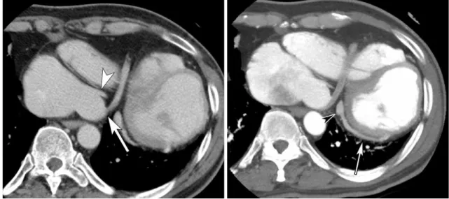
좌측 내경정맥과 좌측 쇄골하정맥이 합류하여 만들어진 정맥 은 일반적인 좌측 완두정맥이나 지속성 좌측 상대정맥과 달리 전방으로 주행하여 흉벽 하에서 늑골연골접합부하로 진행하여 우심실 전방에서 하방으로 주행하다가 우측 후방으로 주행하 여 관상정맥동(coronary sinus)입구의 후방에서 관상정맥동 과 별도로 우심방으로 연결되었다. 이 정맥은 관상정맥동이나 다른 심장의 정맥들과 연결이 없었다(Fig. 2, 3). 이 정맥의 경로에 접한 우심실이 눌린 듯한 변형을 보였다. 좌측 내유정 맥은 이 정맥과는 별도로 정상적으로 있었다. 우측 상대정맥으 로 유입되는 좌측 완두정맥(무명정맥)은 없었으며 좌측 하 갑 상선 정맥은 우측 하 갑상선정맥과 합류하여 우측 상대정맥으 로 연결되었다(Fig. 2A). 우측 상대정맥은 좌측완두정맥이 연 결되지 않는다는 것 외에는 이상소견이 없었다. 우심방 비대와
─ 369 ─ 대한영상의학회지 2008;59:369-371
우심실 전방으로 주행하는 지속성 좌측 상대정맥: 증례 보고1
이유경∙이 활∙박은아∙강미진∙정진욱∙박재형
44세 남자의 심장초음파에 심장 전방의 종괴로 의심되는 병변을 확인하고자 심전도 동기 심 장 CT를 실시하였다. 심장 CT에서 좌측 내경정맥과 쇄골하정맥이 합류하여 우심실 전방으로 내려오는 정맥을 확인할 수 있었고, 이 정맥은 심장 하연에서 관상정맥동과 별도로 우심방으로 연결되었다. 이 변이 정맥은 이전에 보고된 적이 없는 우심실 전방으로 주행하는 지속성 좌측 상대정맥으로 생각된다.
1서울대학교병원 영상의학과
이 논문은 2008년 6월 8일 접수하여 2008년 9월 4일에 채택되었음.
Fig. 1. A transthoracic echocardiography shows the lesion (ar- row) anterior to the right ventricle.
우심실 비대가 있었고, 혈관조영술은 시행하지 않았다. 우심방 으로 연결되는 이 정맥은 해부학적 변이로서 우심실 전방으로 주행하는 지속성 좌측 상대정맥으로 생각되며 환자는 별다른 처치 없이 추적관찰 중이다.
고 찰
이번 증례에서 이 변이 정맥은 좌완과 좌측 두경부의 혈관과
연결되므로 좌측 전주정맥(anterior cardinal vein)에서 유래 한 구조물이다. 좌측 전주정맥에서 형성되는 구조물로 좌측 완 두정맥 혹은 지속성 좌측 상대정맥을 생각할 수 있는데, 이 변 이 정맥은 우측 상대정맥으로 연결되지 않으므로 좌측 완두 정 맥으로 보기는 어렵고 지속성 좌측 상대정맥으로 생각된다.
태생기 때 심장의 양측에서 각각 전주정맥(anterior cardinal vein)과 후주정맥(posterior cardinal vein)이 합해 져서 총주정맥(common cardinal vein 또는 duct of Cuvier)을 형성하고 정맥동(sinus venosus)으로 연결된다.
그리고 양측 전주정맥(anterior cardinal vein) 사이에 연결 이 생겨서 좌측 완두정맥이 되고, 그 하방의 좌측 전주정맥은 폐쇄된다. 폐쇄된 부분보다 하방에서 좌측 총주정맥은 좌심방
─ 370 ─
이유경 외: 우심실 전방으로 주행하는 지속성 좌측 상대정맥
A B
Fig. 2. Three-dimensional volume-rendered images show the anomalous vein (white arrows in Figure 2A, 2B) which is an exten- sion of the left subclavian vein and left internal jugular vein courses anterior to the right ventricle, draining into the right atrium. A common trunk of both inferior thyroidal veins (arrowhead in Figure 2A) connects to the right superior vena cava (asterisk in Figure 2A). Note compressed segment of the coronary sinus (black arrowheads in Figure 2B). The great cardiac vein (white arrowhead in Figure 2B) connects to opening of the coronary sinus.
A B
Fig. 3. A. An axial image at the level of the right atrium at venous phase. The drainage of the anomalous vein (arrow) to the right atrium is separate from that of the coronary sinus (arrowhead).
B. A maximum-intensity-projection image at arterial phase. The great cardiac vein connects (arrowhead) to opening of the coronary sinus, and the posterior vein of left ventricle (arrow) connects to the coronary sinus.
의 경사 정맥(the oblique vein of the left atrium of Marshall)이 되고 정맥동의 좌측 각(left horn of sinus venosus)은 관상정맥동이 된다. 대부분은 이런 과정을 거치나 드물게 좌측 주정맥이 폐쇄되지 않으면, 좌측 주정맥이 지속성 좌측 상대정맥이 되어 관상정맥동을 통해 우심방으로 연결된 다(1). 지속성 좌측 상대정맥은 92%에서는 전술한 대로 우심 방으로 연결되나 나머지에서는 좌심방으로 유입된다(2). 선천 성 심장기형이 있을 때의 지속성 좌측 상대정맥의 빈도는 보고 에 따라 2.8%에서 4.3%에 이르는데, 일반 인구에서의 빈도인 0.3%보다 높다(2).
발생학을 바탕으로 추론해 봤을 때 이번 증례에서는 전술한 지속성 좌측 상대정맥이 만들어지는 기전에 더하여 좌측 전주 정맥과 후주정맥이 합해져서 총주정맥을 형성하는 단계부터 이상이 있었던 것으로 생각된다. 좌측 전주정맥이 좌측 후주정 맥과 합류하여 총주정맥(common cardinal vein)을 형성하여 정맥동의 좌측 각으로 연결되어야 하는데 비정상적으로 좌측 전주정맥이 심장관보다 전방으로 주행하여 정맥동에 합류한 것으로 설명할 수 있다. 그 이후에 좌측 전주정맥은 폐쇄되지 않고 지속성 좌측 상대정맥이 되고 좌측 완두정맥은 만들어지 지 않은 것으로 생각된다.
혹은 좌측 전주정맥이 비정상적인 혈관과 연결되어 우심방으 로 연결될 수 있겠다. 좌측 완두 정맥이 만들어지는 시기에 여
러 개의 상하좌우 연결로 이루어진 정맥총이 양측 전주정맥 사 이에 만들어졌다가 좌측 완두정맥만 남기고 사라진다는 가설을 제시한 보고가 있다(3). 이러한 정맥총 중 원시 대동맥 폐동맥 계의 전방을 지나서 정맥동으로 연결된 경로가 좌측 전주정맥 과 연결되어 이 변이 정맥이 생성되었을 가능성이 있겠다.
이번 증례처럼 지속성 좌측 상대정맥으로 생각되는 혈관이 우심실 전방으로 주행하여 관상정맥동 입구 전방에서 관상정 맥동과 별도로 우심방으로 연결되는 경우는 이전에 보고된 적 이 없다. 하지만, 이 환자에서 이 정맥은 좌측 내경정맥과 좌측 쇄골하 정맥이 합류하여 만들어지는 유일한 정맥으로서 지속 성 좌측 상대정맥에 상응하는 구조물로 생각된다.
참 고 문 헌
1. Campbell M, Deuchar DC. The left-sided superior vena cava. Br Heart J 1954;16:423-439
2. Biffi M, Boriani G, Frabetti L, Bronzetti G, Branzi A. Left superior vena cava persistence in patients undergoing pacemaker or car- dioverter-defibrillator implantation: a 10-year experience. Chest 2001;120:139-144
3. Takada Y, Narimatsu A, Kohno A, Kawai C, Hara H, Harasawa A et al. Anomalous left brachiocephalic vein: CT findings. J Comput Assist Tomogr 1992;16:893-896
─ 371 ─ 대한영상의학회지 2008;59:369-371
J Korean Radiol Soc 2008;59:369-371
Address reprint requests to : Whal Lee, M.D., Department of Radiology, Seoul National University Hospital, 28 Yongon-dong, Chongno-gu, Seoul 110-744, Korea
Tel. 82-2-2072-2584 Fax. 82-2-743-6385 E-mail: [email protected]
Multislice CT of a Persistent Left Superior Vena Cava Coursing Anterior to the Right Ventricle: A Case Report1
Youkyung Lee, M.D., Whal Lee, M.D., Eun-Ah Park, M.D., Mi-jin Kang, M.D., Jin Wook Chung, M.D., Jae Hyung Park, M.D.
1Department of Radiology, Seoul National University College of Medicine, Institute of Radiation Medicine, Seoul National University Medical Research Center
Clinical Research Institute, Seoul National University Hospital, Korea
EKG-gated cardiac CT revealed a variant vein in a 44-year-old man that was misinterpreted as a mass on echocardiography. The variant vein was an extension of the confluence of the left internal jugular vein and left subclavian vein and coursed anterior to the right ventricle. It connected to the right atrium directly at the infe- rior surface of the heart. The variant vein was likely a persistent left superior vena cava, a variant that has nev- er been reported.
Index words :Superior vena cava
X-ray computed tomography Heart