213
Copyrights © 2014 The Korean Society of Radiology
INTRODUCTION
Azygos vein has many variations in their venous tributaries.
Among them, the absence of the azygos vein is a very rare form of the congenital venous anatomic variation, and only a few cas- es have been reported in the literature up to recently (1-3). We report the radiologic findings of the congenital absence of the azygos vein with hemiazygos vein, which drained into the left superior vena cava via superior intercostal vein (SICV).
CASE REPORT
A 65-year-old man diagnosed as rectal cancer visited our colorectal cancer clinic. He did not show any chest symptoms.
He underwent preoperative chest posteroanterior radiograph and postoperative contrast-enhanced chest computed tomogra- phy (CT) to monitor metastasis in the thorax. On the chest ra- diograph, azygos arch shadow was not visible in its usual loca- tion, and there was an aortic nipple, a focal bulge adjacent to the aortic arch, representing the left SICV (Fig. 1A). The chest CT
showed no azygos vein at its usual location and the left SVC was present (Fig. 1B-D). Hemiazygos vein was slightly dilated and drained into the left SVC via prominent left SICV (Fig. 1D, E).
DISCUSSION
Because of the complexity of its developmental stages, the car- dinal vein system could undergo a variety number of congenital anomalies during the embryonic period (4). While in the devel- opmental period, the cardinal system consists of anterior and posterior cardinal veins. By the eighth week, right and left ante- rior cardinal veins become connected by an anastomotic duct, which later become the superior vena cava on right side (from right anterior cardinal vein and right common cardinal vein) and left brachiocephalic vein on left side (from left anterior car- dinal vein) (3, 4). However, when the left common cardinal vein fails to be obliterated, it becomes the left SVC (4).
The second most important system of the great thoracic veins is the subcardinal veins that form the azygos and the hemiazy- gos veins (4). Azygos vein is corresponded by the right subcar-
Case Report
pISSN 1738-2637 / eISSN 2288-2928 J Korean Soc Radiol 2014;70(3):213-215 http://dx.doi.org/10.3348/jksr.2014.70.3.213
Received November 10, 2013; Accepted February 5, 2014 Corresponding author: Jeong Geun Yi, MD Department of Radiology, Konkuk University School of Medicine, 120-1 Neungdong-ro, Gwangjin-gu, Seoul 143-729, Korea.
Tel. 82-2-2030-5576 Fax. 82-2-2030-5549 E-mail: [email protected]
This is an Open Access article distributed under the terms of the Creative Commons Attribution Non-Commercial License (http://creativecommons.org/licenses/by-nc/3.0) which permits unrestricted non-commercial use, distri- bution, and reproduction in any medium, provided the original work is properly cited.
Absence of the azygos vein is a very rare variant of venous tributary arrangement which has been reported only in few cases so far. We hereby introduce the chest ra- diographic and computed tomographic findings of the congenital absence of the azygos vein with bilateral superior vena cava, incidentally detected during a follow- up for rectal cancer. The hemiazygos vein is drained into persistent left superior vena cava via left superior intercostal vein, so called the “aortic nipple”.
Index terms Azygos Vein
Bilateral Superior Vena Cava Hemiazygos Vein
Superior Intercostal Vein Computed Tomography
Congenital Absence of the Azygos Vein with Persistent Left Superior Vena Cava: A Case Report
좌측 상대정맥을 동반한 선천성 기정맥 결손: 증례 보고
Younghee Yim, MD, Jeong Geun Yi, MD, Inyoung Song, MD, Jeong Hee Park, MD
Department of Radiology, Konkuk University School of Medicine, Seoul, Korea
Congenital Absence of the Azygos Vein with Persistent Left Superior Vena Cava
214
J Korean Soc Radiol 2014;70(3):213-215 jksronline.orgThere have been several case reports about the radiologic find- ings of the absence of azygos vein (1-3, 5-7). All of these cases showed no azygos vein at the normal anatomic position and the
“aortic nipple” in the dependent drainage side (i.e., left SVC, left innominate vein) on the chest radiograph. Chest CT showed the
“aortic nipple” and the agenesis of the azygos vein. It also showed some cases with the right SVC only (1) or the left SVC only (2, 3, 5), and the patients with bilateral SVC (6, 7) like in this case.
Absence of the azygos vein is a very rare congenital venous anomaly, but we should take this anomaly into account when the chest radiograph fails to show azygos arch shadow on its usual location and shows the “aortic nipple” on the lateral side of aortic arch. The chest CT is the best way to confirm the agenesis of the azygos vein and the associated thoracic venous anomalies based on the chest radiograph.
REFERENCES
1. Arslan G, Cubuk M, Ozkaynak C, Sindel T, Lüleci E. Absence dinal vein and hemiazygos vein by the left subcardinal vein. The
connection of the right and left subcardinal veins usually forms at the level of the sixth or seventh thoracic vertebra (5). The left subcardinal vein undergoes obliteration cranial to the anasto- motic site, or it may persist as the accessory hemiazygos vein.
The accessory hemiazygos vein is connected to the left SICV medial to the distal aortic arch (6). The left SICV is developed from the embryonic posterior cardinal veins. The left SICV then courses anteriorly beside the aortic arch to meet the left brachio- cephalic vein posteriorly (6).
A persistent left SVC is considered to be the most common anomalous systemic vein-to-cardiac connection which is seen in 0.3–0.5% of general population (7). Up to 90% of the people with persistent left SVC present with the right SVC as well. About 65% of the people with persistent left SVC have no left brachio- cephalic vein or atrophic one. About 20% of them show left su- perior intercostal vein forming a communication between hemi- azygos vein and the left SVC, producing a left hemiazygos arch like the current case (7).
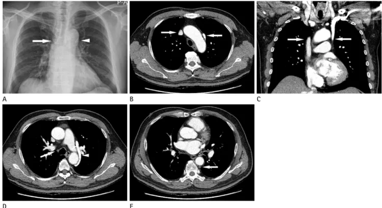
Fig. 1. A 65-year-old man with rectal cancer.
A. Chest radiograph shows no azygos arch shadow in its usual site (arrow) and shows focal bulge of aortic arch (aortic nipple) (arrowhead).
B, C. Contrast enhanced axial (B) and coronal (C) CT images show bilateral SVC (arrows).
D. Contrast enhanced axial CT scan also shows the superior intercostal vein, the “aortic nipple” (arrowhead), beside the aortic arch and the ab- sence of the azygos vein at its usual site (arrow).
E. Contrast enhanced axial scan shows the slightly dilated hemiazygos vein (arrow).
E B
D
A C
Younghee Yim, et al
215
jksronline.org J Korean Soc Radiol 2014;70(3):213-215
5. Kullnig P, Melzer G, Hausegger K, Einspieler R. Computed tomographic diagnosis of left superior vena cava and ab- sence of the azygos vein: case report. Cardiovasc Intervent Radiol 1990;13:47-49
6. Arslan G, Ozkaynak C, Cubuk M, Sindel T, Lüleci E. Absence of the azygos vein associated with double superior vena cava--a case report. Angiology 1999;50:81-84
7. Yazdi HR, Sotoudeh H, Taslimi R. Absence of azygos vein in an adult patient with polysplenia syndrome. Eur J Radiol Extra 2006;59:89-92
of the azygos vein. Clin Imaging 2000;24:157-158
2. Arslan G, Cubuk M, Ozkaynak C. Absence of the azygos vein associated with left superior vena cava. Eur J Radiol Extra 2005;54:15-17
3. Hatfield MK, Vyborny CJ, MacMahon H, Chessare JW. Con- genital absence of the azygos vein: a cause for “aortic nipple” enlargement. AJR Am J Roentgenol 1987;149:273- 274
4. Cardiovascular system. In Moore KL, Persaud TVN, Torchia MG. The Developing Human: clinically oriented embryolo- gy, 9th ed. Philadelphia: Saunders, 2013:289-342
좌측 상대정맥을 동반한 선천성 기정맥 결손: 증례 보고
임영희 · 이정근 · 송인영 · 박정희
기정맥의 선천적 결손은 매우 드문 혈관 기형이다. 우리는 대장암 환자에서 기정맥이 없고, 한 편으로 반기정맥이 대동맥 궁 측부의 좌측 상부늑간정맥, 이른바 “대동맥 유두”를 이루어 좌측 상대정맥(좌우 상대정맥이 있음)으로 혈류가 유입된 소견을 흉부 X-선 사진 및 흉부 전산화단층촬영 영상으로 관찰하였기에 보고한다.
건국대학교 의학전문대학원 영상의학교실