J Korean Soc Radiol 2016;74(6):394-398 http://dx.doi.org/10.3348/jksr.2016.74.6.394
INTRODUCTION
Congenital intrahepatic inferior vena cava (IVC) interruption with azygos/hemiazygos continuation is a rare congenital anom- aly (1, 2) and hemiazygos vein continuation draining into the right atrium via a persistent left superior vena cava (SVC) is even rarer (3). Furthermore, a single coronary artery (SCA) ostium, in the absence of other cardiac disease, is an extremely rare coro- nary artery anomaly (4). We presented a very rare case of inci- dentally found interrupted IVC with hemiazygos vein continua-
tion combined with a left SCA diagnosed by multi-detector computed tomography (MDCT). The dilated hemiazygos vein drained directly into the persistent left SVC.
CASE REPORT
A 50-year-old woman was referred to our institution for medi- cal screening due to an incidental finding on abdominal ultraso- nography. Ultrasonography was unable to demonstrate the he- patic segment of the IVC. She was scheduled for chest, abdomen
Interrupted Inferior Vena Cava with Hemiazygos Continuation
in an Adult with a Persistent Left Superior Vena Cava and Left Single Coronary Artery: A Case Report
성인에서 발견된 하대정맥 단절, 반홀정맥 연속, 좌상대정맥 존속과 좌측 단일 관상동맥 동반: 증례 보고
Yeo Jin Kim, MD
1, Se Hwan Kwon, MD
1*, Sung-Eun Ahn, MD
1, Soo-Joong Kim, MD
2, Jong Soo Shin, MD
3, Joo Hyeong Oh, MD
1Departments of 1Radiology, 2Cardiology, College of Medicine, Kyung Hee University, Seoul, Korea
3Department of Radiology, Kyung Hee University Hospital at Gangdong, Seoul, Korea
A 50-year-old woman was referred to our institution for medical screening due to an incidental finding on abdominal ultrasonography. She underwent chest, abdo- men and cardiac multi-detector computed tomography (MDCT). Her MDCT revealed absence of the hepatic segment of the inferior vena cava (IVC), with hemiazygos continuation and a left single coronary artery. The dilated hemiazygos vein drained directly into the persistent left superior vena cava (SVC). Herein, we reported a very rare case combining an incidentally found interrupted IVC with hemiazygos vein continuation, persistent left SVC and a left single coronary artery diagnosed by MDCT.
Index terms
Vena Cava, Superior/Abnormalities Vena Cava, Inferior/Abnormalities Coronary Vessel Anomalies Vascular Malformations Azygos Vein/Abnormalities
Tomography, X-Ray Computed/Methods Congenital
Received September 12, 2015 Revised November 30, 2015 Accepted December 17, 2015
*Corresponding author: Se Hwan Kwon, MD Department of Radiology, College of Medicine, Kyung Hee University, 23 Kyungheedae-ro, Dongdaemun-gu, Seoul 02447, Korea.
Tel. 82-2-958-8622 Fax. 82-2-968-0787 E-mail: [email protected]
This is an Open Access article distributed under the terms of the Creative Commons Attribution Non-Commercial License (http://creativecommons.org/licenses/by-nc/3.0) which permits unrestricted non-commercial use, distri- bution, and reproduction in any medium, provided the original work is properly cited.
and cardiac MDCT because she wanted whole body evaluation.
On the day of admission, the patient’s physical examination and electrocardiogram (ECG) were unremarkable. She reported mild chest discomfort with no other abdominal or respiratory symp- toms. Her history included hypertension (diagnosed 5 years pre- viously), for which she had been taking antihypertensive drugs for the past 5 years. There was no history of trauma, diabetes, or surgery. The patient had no connective tissue disorders or other systemic anomalies, and no significant family history of disease.
A chest X-ray revealed clear lungs and a normal-sized heart.
Transthoracic echocardiography revealed a dilated coronary si- nus, but no other abnormalities were noted.
Initially, cardiac CT with retrospective ECG gating was per- formed with a 128-slice MDCT scanner (Ingenuity core 128;
Philips Medical Systems, Best, the Netherlands). A 70-second delayed phase chest and abdomen CT was subsequently per- formed. CT scan demonstrated absence of the hepatic segment of the IVC with hemiazygos continuation; the dilated hemiazy-
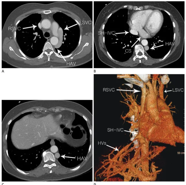
Fig. 1. Axial (A-C) and three-dimensional computed tomography (D) images showing an absent hepatic segment of the inferior vena cava (IVC) with hemiazygos continuation. The dilated hemiazygos vein (HAV) drains into the persistent left superior vena cava (LSVC). The LSVC drains into the right atrium through the dilated coronary sinus (CS). Hepatic veins (HVs) form a confluence and drain directly into the right atrium via supra- hepatic IVC (SH-IVC).
RSVC = right superior vena cava A
C
B
D
gos vein drained into the persistent left SVC (Figs. 1, 2, Supple- mentary Movie 1 in the online-only Data Supplement). Hepatic veins drained directly into the supra-hepatic part of the IVC and right atrium. The right SVC normally drained into the right atri- um and the left SVC also drained into the right atrium through the dilated coronary sinus. The patient had no left brachioce- phalic vein. A three-dimensional volume-rendering cardiac CT image showed the SCA arising from the left coronary sinus of Valsalva, and an absent right coronary artery (Fig. 3, Supplemen- tary Movie 2 in the online-only Data Supplement). The SCA pro- duced the anterior descending branch in the usual pattern, which then continued in the atrioventricular groove as the left circum- flex branch; it traveled beyond the crux into the right atrioven- tricular groove, where it provided branches to the right ventricle and atrium. There were no significant atherosclerotic lesions in the coronary arteries, and the aorta and other major vascular structures were normal. MDCT contributed additional diagnos- tic information and confirmed the diagnosis, which remained equivocal on routine abdominal ultrasound. The patient was dis- charged in good condition without any medical treatment.
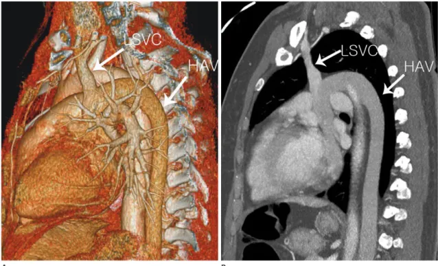
Fig. 2. Three-dimensional volume-rendering (A) and oblique sagittal maximum intensity (B) images showing the dilated hemiazygos vein (HAV) draining into the LSVC.
LSVC = left superior vena cava
A B
Fig. 3. Three-dimensional volume-rendering image showing the single coronary artery (SCA) arising from the left coronary sinus of Valsalva, and absence of the right coronary artery.
LAD = left anterior descending artery
DISCUSSION
Interruption of the IVC with azygos/hemiazygos continuation is a rare, but well-known, anomaly (1, 2) with a prevalence of 0.6-2.0% in patients with congenital heart disease and < 0.3%
among otherwise-normal patients (2). The condition is typically described as a congenital heart malformation associated with heterotaxy (cardiosplenic) syndrome, particularly in the form of left isomerism or polysplenia (5, 6). However, an isolated occur- rence without heart disease may go unnoticed during early life and may be detected incidentally during a radiological examina- tion or vascular interventions. Recent advances in MDCT tech- nology have led to incidental diagnoses of IVC or other vascular anomalies.
Hemiazygos continuation draining into the right atrium via a persistent left SVC is rare (3); SCA is extremely rare, with an inci- dence of 0.024-0.066% in the general population undergoing coronary angiography (4, 7, 8). SCA may be associated with oth- er severe congenital cardiac malformations, such as persistent truncus arteriosus, tetralogy of Fallot, and pulmonary atresia. In our case, the hemiazygos vein continued and connected to the persistent left SVC posteriorly, finally entering the right atrium via the enlarged coronary sinus. MDCT also revealed an anoma- lous SCA arising from the left sinus of Valsalva, together with ab- sence of the right coronary artery. According to the classification by Lipton et al. (7) and Yamanaka and Hobbs (8), our present case can be classified as group I (L-1 subtype). According to the definition, the ‘L-1’ pattern implies that the right coronary artery is congenitally absent and the left circumflex artery is markedly dominant. Fortunately, there were no significant symptoms or other associated cardiac or vascular anomalies. To our knowl- edge, no reported case has completely matched our findings.
Knowledge of the presence of an interrupted IVC, persistent left SVC, or coronary artery anomalies is important for both sur- geons and radiologists to prevent problems when planning an endovascular intervention or surgery, such as coronary angiogra- phy, right heart catheterization, electrophysiological studies, car- diopulmonary bypass surgery, cardiac transplantation, femoral vein catheter advancement, IVC filter placement, or temporary pacing through the transfemoral route (2, 6, 9).
In summary, we reported a very rare case of an incidentally de- tected interrupted IVC with hemiazygos continuation combined
with a left SCA. The dilated hemiazygos vein drained directly into the persistent left SVC.
SUPPLEMENTARY MOVIE LEGENDS
Movie 1. Chest CT axial images.
Movie 2. Cardiac CT axial images during mid-diastole.
Supplementary Materials
The online-only Data Supplement is available with this article at http://dx.doi.org/10.3348/jksr.2016.74.6.394.
REFERENCES
1. Yilmaz E, Gulcu A, Sal S, Obuz F. Interruption of the inferior vena cava with azygos/hemiazygos continuation accompa- nied by distinct renal vein anomalies: MRA and CT assess- ment. Abdom Imaging 2003;28:392-394
2. Vijayvergiya R, Bhat MN, Kumar RM, Vivekanand SG, Gro- ver A. Azygos continuation of interrupted inferior vena cava in association with sick sinus syndrome. Heart 2005;91:e26 3. Yildiz AE, Cayci FS, Genc S, Cakar N, Fitoz S. Right nut- cracker syndrome associated with left-sided inferior vena cava, hemiazygos continuation and persistant left superior vena cava: a rare combination. Clin Imaging 2014;38:340-345 4. Desmet W, Vanhaecke J, Vrolix M, Van de Werf F, Piessens J,
Willems J, et al. Isolated single coronary artery: a review of 50,000 consecutive coronary angiographies. Eur Heart J 1992;
13:1637-1640
5. Tubau A, Grau J, Filgueira A, Juan M, Estremera A, Ferrer MI, et al. Prenatal and postnatal imaging in isolated inter- ruption of the inferior vena cava with azygos continuation.
Prenat Diagn 2006;26:872-874
6. Minniti S, Visentini S, Procacci C. Congenital anomalies of the venae cavae: embryological origin, imaging features and report of three new variants. Eur Radiol 2002;12:2040-2055 7. Lipton MJ, Barry WH, Obrez I, Silverman JF, Wexler L. Iso- lated single coronary artery: diagnosis, angiographic classi- fication, and clinical significance. Radiology 1979;130:39-47 8. Yamanaka O, Hobbs RE. Coronary artery anomalies in
126,595 patients undergoing coronary arteriography. Cathet Cardiovasc Diagn 1990;21:28-40
성인에서 발견된 하대정맥 단절, 반홀정맥 연속, 좌상대정맥 존속과 좌측 단일 관상동맥 동반: 증례 보고
김여진
1· 권세환
1* · 안성은
1· 김수중
2· 신종수
3· 오주형
150세 여성이 복부 초음파에서 우연히 발견된 이상소견을 주소로 건강검진을 위해 본원에 내원하여 흉부, 복부 및 심장 컴 퓨터단층촬영을 시행하게 되었다. 컴퓨터단층촬영에서 하대정맥의 간 분절이 없고, 반홀정맥이 연속되며 좌측 단일 관상 동맥이 발견되었고 늘어난 반홀정맥은 존속된 좌상대정맥으로 직접 배출되었다. 우리는 다중검출 컴퓨터단층촬영으로 하 대정맥 단절, 반홀정맥 연속, 좌상대정맥 존속과 좌측 단일 관상동맥의 매우 드문 혈관기형들이 복합되어 있는 증례를 발 견하여 이를 보고한다.
경희대학교 의과대학 1영상의학과, 2심장내과, 3강동경희대학교병원 영상의학과
9. Imamura M, Abraham B, Garcia X, Knecht KR, Frazier E, Shinkawa T. New transplant technique with hemiazygos
continuation and interrupted inferior vena cava. Ann Tho- rac Surg 2013;96:1882-1884