and Public Awareness
Metabolic Syndrome Is Associated With Venous Thromboembolism in the Korean Population
Moon Ju Jang, Won-il Choi, Soo-Mee Bang, Taeseung Lee, Yeo-Kyeoung Kim, Walter Ageno, Doyeun Oh
Background—The metabolic syndrome (MS) is a known risk factor for arterial thromboembolism. Preliminary reports have also suggested the association between MS and venous thromboembolism (VTE).
Methods and Results—In this case– control study, we investigated the association between MS and VTE in Korean patients. Patients with objectively diagnosed VTE and healthy control subjects underwent clinical assessment for the presence of MS according to the National Cholesterol Education Adult Treatment Panel III criteria modified with body mass index (WHO Asian Pacific Perspective, 2000). The presence of known risk factors for VTE was ascertained.
Patients with VTE secondary to cancer were excluded. The prevalence of MS was compared between VTE group and controls. Two hundred eight VTE patients and 300 controls were assessed. VTE was idiopathic in 91 patients and secondary to a known risk factor in 117. The prevalence of MS was significantly higher in VTE patients (47.6%) than in controls (37.7%) (OR: 1.50; 95% CI: 1.05 to 2.15, P⫽0.026). After adjusting for age, sex, and smoking status, metabolic MS remained independently associated with VTE (OR: 1.56; 95% CI: 1.07 to 2.27, P⫽0.020). In the subgroup analysis, MS was also independently associated with idiopathic VTE (OR: 1.71; 95% CI: 1.04 to 2.81, P⫽0.033), but not with secondary VTE (OR: 1.43; 95% CI: 0.91 to 2.99, P⫽0.121). Multivariate analysis demonstrated that high BMI (OR: 1.70, 95% CI: 1.01 to 2.87), decreased HDL cholesterol (OR: 1.99, 95% CI: 1.17 to 3.39), and elevated fasting glucose levels (OR: 2.31; 95% CI: 1.35 to 3.94) were associated with idiopathic VTE.
Conclusion—MS is associated with VTE and in particular with idiopathic VTE in the Korean population. (Arterioscler Thromb Vasc Biol. 2009;29:311-315.)
Key Words: venous thromboembolism metabolic syndrome
Venous thromboembolism (VTE) and arterial thrombosis are usually considered as distinct disease entities be- cause of their different anatomic location, risk factors, clini- cal presentation, and modalities for prevention and treatment.
However, several recent studies have elucidated the associa- tion between VTE and atherosclerosis. The most important basis for this association is that VTE and atherosclerosis may share common risk factors including obesity, diabetes, hyper- tension, and hyperlipidemia.1,2 A number of studies have consistently demonstrated that idiopathic VTE is associated with an increased risk of subsequent cardiovascular events3,4 and that patients with idiopathic VTE have an increased prevalence of asymptomatic atherosclerotic lesions.5
The metabolic syndrome is a cluster of risk factors for atherosclerosis, including abdominal obesity, hypertension,
insulin resistance, dyslipidemia with high triglycerides, and low high-density lipoprotein (HDL) cholesterol. A few recent studies have suggested that the metabolic syndrome may also be a risk factor for VTE in whites.6 – 8
The incidence of VTE in the Asian population has been generally found to be lower than in whites, but it appears to be rapidly increasing,9 –11possibly because of the widespread adop- tion of a westernized lifestyle which also results in an increasing prevalence of the metabolic syndrome. Although the metabolic syndrome has become an important health concern in Asian countries, as much as it is in Western countries, the association between the metabolic syndrome and VTE has never been studied in Asian populations. To address this issue, we carried out a case– control study to investigate the association between the metabolic syndrome and VTE in a Korean population.
Received January 7, 2009; revision accepted January 15, 2009.
From the Department of Internal Medicine (M.J.J., D.O.), College of Medicine, Pochon CHA University, Sungnam, Korea; the Department of Internal Medicine (W.C.), College of Medicine, Keymyung University, Daegu, Korea; the Department of Internal Medicine (S.-M.), Seoul National University Bundang Hospital, Sungnam, Korea; the Department of Vascular Surgery (T.L.), Seoul National University Bundang Hospital, Sungnam, Korea; the Department of Internal Medicine (Y.-K.K.), College of Medicine, Chonnam National University, Kwangju, Korea; and the Department of Clinical Medicine (W.A.), University of Insubria, Varese, Italy.
Correspondence to Doyeun Oh, Department of Internal Medicine, College of Medicine, Pochon CHA University, Sungnam, Korea. E-mail [email protected] or Walter Ageno, U.O. Medicinal Ospedale di Circolo, Vale Borri 57, 21100 Varese, Italy. E-mail [email protected]
© 2009 American Heart Association, Inc.
Arterioscler Thromb Vasc Biol is available at http://atvb.ahajournals.org DOI: 10.1161/ATVBAHA.109.184085 311
Patients and Methods Study Population
The study was conducted between January 2006 and May 2008 by the Korean Deep Vein Thrombosis Working Party (KDVTWP).
During this period, the 4 participating centers in KDVTWP enrolled consecutive Korean patients with recent (6 months) objective diag- nosis of deep vein thrombosis (DVT) or pulmonary embolism (PE).
VTE was defined as idiopathic or secondary depending on the presence or absence of any of the following risk factors: recent surgery (⬍3 months), recent trauma/fracture (⬍3 months), immobi- lization (⬎7 days), severe medical disease, pregnancy, use of oral contraceptives, and known hypercoagulable disease. VTE was clas- sified as secondary if there was at least 1 of these risk factors.
Patients with VTE secondary to known cancer were excluded from the study. The control group was selected among subjects visiting the Bundang CHA Health Promotion Center for periodic health exami- nation. These were individuals who had no medical history of VTE or malignancy. The Institutional Review Board of Bundang CHA Hospital approved the research protocol and written informed consent was obtained from all participating individuals.
Methods
Data were recorded on computer-based case report form at each participating hospital and were submitted to a centralized coordinating center through a Korean DVT registry website (http://kdvt.chamc.
co.kr). Patients’ identities remain confidential because they are identified by unique numbers assigned by the study coordinating center. At regular intervals, data quality was monitored and docu- mented electronically to detect inconsistencies or errors. The computer-based case report form comprised the following data of patients: age, sex, weight, height, body mass index (BMI), waist circumference, systolic and diastolic blood pressure, history of smoking, hypertension, diabetes mellitus, hyperlipidemia, current use of antihypertensive, antidiabetic drug and antilipidemic drugs, and laboratory results (serum fasting glucose, triglyceride, and HDL cholesterol).
The time interval between VTE event and physical measurement and blood collection had to be at least 6 months. Body weight was measured in light underwear by a precision scale to the nearest 0.5 kg, and body height was measured by a precision meter to the nearest 0.01 meter. BMI was calculated as body weight (kg) divided by the square of the height (m). The circumference of the waist was measured with a retractable steel tape, with the subject in the
standing position, as described by Ashwell et al.12 The waist measurement to be recorded was the smallest girth between the rib cage and the iliac crest. Blood pressure was measured in the right arm, with subject in the supine position after 10-minute rest by using a mercury sphygmomanometer of appropriate cuff size. Venous blood was drawn from an antecubital vein with plastic syringes after an overnight fast and was collected in polystyrene tubes and then glucose, HDL cholesterol, and triglycerides were determined in fresh plasma.
The presence of the metabolic syndrome in patients and controls was defined in accordance with the National Cholesterol Education Program (NCEP) Adults Treatment Panel III (ATP III) criteria13and modified with Asia–Pacific criteria for obesity based on BMI (ⱖ25 kg/m2) or waist circumference (ⱖ90 cm for men, ⱖ80 cm for women).14BMI was used for participants with missing data on waist circumference to diagnose metabolic syndrome. In the presence of 3 or more of the following risk factors the metabolic syndrome was diagnosed: (1) elevated waist circumference:ⱖ90 cm in men, ⱖ80 cm in women, or BMI: ⱖ25 kg/m2 in both sexes; (2) elevated triglycerides:ⱖ150 mg/dL or ongoing drug treatment for elevated triglycerides; (3) reduced HDL cholesterol:⬍40 mg/dL in men, ⬍50 mg/dL in women or ongoing antilypidemic treatment; (4) elevated blood pressure:ⱖ130 mm Hg systolic blood pressure, ⱖ85 mm Hg diastolic blood pressure, or ongoing antihypertensive treatment; (5) elevated fasting glucose: ⱖ110 mg/dL or ongoing antidiabetic treatment.
Statistical Analysis
Statistical analyses were conducted by using SPSS 13.0. Differences between the group of VTE patients and controls were assessed using the Student t test. Categorical variables were compared using the chi-squared test. Logistic regression analyses were performed to select significant risk factor for VTE among components of meta- bolic syndrome. Multivariate analysis was performed to select independent risk factors for VTE among those clinical variables and components of metabolic syndrome using logistic regression analy- sis. Odds ratios and corresponding 95% confidence intervals were calculated. Statistical significance was determined to be P⬍0.05.
Results Baseline Characteristics
Baseline characteristics of patients with VTE and controls are shown in Table 1. A total of 208 patients with VTE and 300 Table 1. Baseline Characteristics
Total VTE (n⫽208) P Values* Idiopathic VTE (n⫽91) P Values* Secondary VTE (n⫽117) P Values* Controls (n⫽300)
Mean age, years (SD) 58.1 (16.2) 0.900 56.5 (15.7) 0.332 59.3 (16.5) 0.542 58.3 (13.0)
Male sex, n (%) 91 (43.8) 0.649 44 (48.4) 0.278 47 (40.2) 0.825 125 (41.7)
Family history of VTE, n (%) 4 (1.9) 0.028 1 (1.1) 0.233 3 (2.6) 0.022 0 (0.0)
Smokers, n (%) 57 (27.4) 0.088 26 (28.6) 0.117 31 (26.5) 0.238 62 (20.7)
Mean systolic BP, mm Hg (SD) 129.8 (20.5) 0.002 129.5 (19.0) 0.023 130.1 (21.7) 0.013 124.5 (16.5) Mean diastolic BP, mm Hg (SD) 79.7 (12.1) 0.330 79.5 (11.8) 0.354 79.9 (12.4) 0.506 80.7 (10.9) Mean TG, mg/dL⫺1(SD) 140.8 (78.4) 0.955 153.5 (85.7) 0.281 130.9 (71.1) 0.297 141.2 (97.6)
Mean HDL, mg/dL⫺1(SD) 47.3 (15.3) 0.001 46.2 (14.0) 0.001 48.2 (16.3) 0.002 53.5 (13.2)
Mean glucose, mg/dL⫺1(SD) 124.8 (73.3) 0.001 118.9 (38.9) 0.001 129.3 (91.4) 0.001 103.8 (24.6)
Mean BMI, kg/m⫺2(SD) 24.4 (5.1) 0.035 24.4 (3.6) 0.048 24.5 (6.0) 0.131 23.6 (3.2)
DM (%) 33 (15.9) 0.114 8 (8.8) 0.567 25 (21.4) 0.012 34 (11.3)
HTN (%) 82 (39.4) 0.005 31 (34.1) 0.235 51 (43.6) 0.002 82 (27.3)
Antilipidemic therapy with
statins (%) 24 (11.5) 0.161 11 (12.1) 0.204 13 (11.1) 0.252 23 (7.7)
Antihypertensive therapy (%) 59 (28.4) 0.147 22 (24.2) 0.777 37 (31.6) 0.061 68 (22.7)
*P value as the total VTE group, idiopathic VTE group, and secondary VTE group compared with control group.
BMI indicates body mass index; DVT, deep vein thrombosis; HDL, high-density lipoprotein; SD, standard deviation.
controls were enrolled in the study. Of VTE patients, 74 (35.6%), 66 (31.7%), and 68 (32.7%) patients were diagnosed as having isolated DVT, DVT with PE, and isolated PE, respectively. VTE was idiopathic in 91 patients and second- ary to a known risk factor in 117. Among components of the metabolic syndrome, systolic blood pressure and serum glucose levels were significantly higher in patients with VTE and also in the subgroups of patients with idiopathic or secondary VTE than in controls. Also, mean HDL cholesterol levels were significantly lower in all VTE populations than in controls. Conversely, diastolic blood pressure and triglycer- ide levels were not statistically different among the groups.
BMI was significantly higher both in patients with all VTE and idiopathic VTE than that in controls. At the time of study enrollment, 24 (11.5%) VTE patients and 23 (7.7%) controls were receiving therapy with statins, and 59 (28.4%) VTE patients and 68 (22.7%) controls were receiving therapy with antihypertensive drugs.
Prevalence of the Metabolic Syndrome
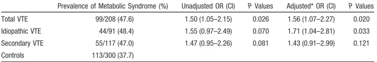
The prevalence of the metabolic syndrome was 47.6% in patients with VTE, 48.4% in patients with idiopathic VTE, 47.0% in patients with secondary VTE, and 37.7% in controls (Table 2). In multivariate analysis, after adjusting for age, sex, and smoking status in the comparison between idiopathic and secondary VTE and controls, the prevalence of the metabolic syndrome was significantly higher in VTE patients than in controls (OR: 1.56; 95% CI: 1.07 to 2.27) and in the subgroup of patients with idiopathic VTE than in controls (OR: 1.71; 95% CI: 1.04 to 2.81). There was no statistically significant difference between patients with secondary VTE and controls (OR: 1.43; 95% CI: 0.91 to 2.99).
Association Between the Individual Components of the Metabolic Syndrome and VTE
In univariate analysis, the prevalences of decreased HDL cholesterol levels and increased glucose levels were signifi-
cantly more frequent in the population of VTE patients (OR:
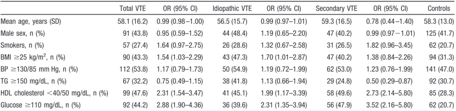
2.30; 95% CI: 1.59 to 3.33, and OR: 3.04; 95% CI: 2.06 to 4.50), and in the populations of patients with idiopathic VTE (OR: 2.07; 95% CI: 1.28 to 3.36, and OR: 2.51; 95% CI: 1.52 to 4.17) or secondary VTE (OR: 2.49; 95% CI: 1.60 to 3.86, and OR: 3.52; 95% CI: 2.23 to 5.57) than in controls. The prevalence of high BMI was significantly more frequent in the population of patients with VTE (OR: 1.67; 95% CI: 1.16 to 2.41) than in controls. In multivariate analysis, after adjusting for components of the metabolic syndrome, age, sex, and smoking status, decreased HDL cholesterol levels, and increased glucose levels remained independently associ- ated with total VTE (OR: 2.31; 95% CI: 1.54 to 3.47, and OR:
2.88; 95% CI:1.90 to 4.36, respectively), idiopathic VTE (OR: 1.99; 95% CI: 1.17 to 3.39, and OR: 2.31; 95% CI: 1.35 to 3.94, respectively), and secondary VTE (OR: 2.73; 95%
CI: 2.14 to 5.80, and OR: 3.52; 95% CI: 2.16 to 5.80, respectively), whereas increased BMI was independently associated with total VTE (OR: 1.54; 95% CI: 1.03 to 2.29) and idiopathic VTE (OR: 1.70; 95% CI: 1.01 to 2.87), but not with secondary VTE (OR: 1.38; 95% CI: 0.84 to 2.26).
Discussion
This is the first study to demonstrate an association between the metabolic syndrome and VTE in an Asian population. In particular, patients with idiopathic VTE, but not with second- ary VTE had a significantly higher prevalence of the meta- bolic syndrome than healthy control subjects. Among the components of metabolic syndrome, high BMI, decreased HDL cholesterol levels, and increased glucose levels were independently associated with idiopathic VTE.
The results of the present study are consistent with the results of previous studies conducted in white populations.
The association between the metabolic syndrome and VTE was suggested for the first time by Ageno et al.8 Two subsequent case– control studies6,7 showed similar findings.
Ageno et al observed a significantly greater prevalence of the Table 2. Association Between the Metabolic Syndrome and VTE
Prevalence of Metabolic Syndrome (%) Unadjusted OR (CI) P Values Adjusted* OR (CI) P Values
Total VTE 99/208 (47.6) 1.50 (1.05–2.15) 0.026 1.56 (1.07–2.27) 0.020
Idiopathic VTE 44/91 (48.4) 1.55 (0.97–2.49) 0.070 1.71 (1.04–2.81) 0.033
Secondary VTE 55/117 (47.0) 1.47 (0.95–2.26) 0.081 1.43 (0.91–2.99) 0.121
Controls 113/300 (37.7)
*Adjusted by age, sex and smoke.
OR indicates odds ratio; CI, confidence interval; VTE, venous thromboembolism.
Table 3. Univariate Analysis Examining the Components of the Metabolic Syndrome Total VTE
(n⫽208) OR (95% CI)
Idiopathic VTE
(n⫽91) OR (95% CI)
Secondary VTE
(n⫽117) OR (95% CI)
Controls (n⫽300) BMIⱖ25 kg/m2, n (%) 90 (43.3) 1.67 (1.16 –2.41) 43 (47.3) 1.44 (0.90 –2.31) 47 (40.2) 1.47 (0.95–2.29) 94 (31.3) BPⱖ130/85 mm Hg, n (%) 112 (53.8) 1.34 (0.94–1.91) 50 (54.9) 1.38 (0.86–2.20) 62 (53.0) 1.27 (0.83–1.95) 141 (47.0) TGⱖ150 mg/dL, n (%) 67 (32.2) 1.07 (0.73–1.57) 38 (41.8) 1.62 (1.00–2.63) 29 (24.8) 0.75 (0.46–1.21) 92 (30.7) HDL cholesterol⬍40/50 mg/dL, n (%) 99 (47.6) 2.30 (1.59–3.33) 41 (45.1) 2.07 (1.28–3.36) 58 (49.6) 2.49 (1.60–3.86) 85 (28.3) Glucoseⱖ110 mg/dL, n (%) 92 (44.2) 3.04 (2.06–4.50) 36 (39.6) 2.51 (1.52–4.17) 56 (47.9) 3.52 (2.23–5.57) 62 (20.7) OR indicates odds ratio; CI, confidence interval; VTE, venous thromboembolism; BMI, body mass index; BP, blood pressure; TG, triglycerides; HDL, high-density lipoprotein.
metabolic syndrome in patients with idiopathic DVT than in those with secondary DVT and in matched controls. The prevalence of the metabolic syndrome in our study and in the Italian study are quite similar: 48.4% and 50.5%, respec- tively, in the population of patients with idiopathic VTE and 37.7% and 34.6%, respectively, in controls. Conversely, the prevalence of the metabolic syndrome in patients with sec- ondary VTE was higher in our study (47.0%) than in the Italian study (27%). In the study by Ay et al, the prevalence of the metabolic syndrome was lower both in cases and controls as compared to our study, but the association between the metabolic syndrome and recurrent VTE was statistically significant.6 These discrepant prevalences of metabolic syndrome among studies are attributed to variable sample sizes and different definition of metabolic syndrome among studies. Finally, Ambrosetti et al have shown in an observational case– control study that the presence of the metabolic syndrome significantly increased the risk of VTE after acute cardiac events.7The multiple adjusted odds ratio of the 4 case– control studies were quite similar, being 1.94 (95% CI: 1.04 to 3.63), 2.20 (95% CI: 1.10 to 4.30), and 2.38 (95% CI: 1.64 to 3.12), respectively in previous studies and 1.71 (95% CI: 1.04 to 2.81) in our study (Table 5). This is interesting given the different ethnicities of the studied populations. All previous case– control studies were con- ducted in whites, whereas our study was conducted in Asian patients only. The metabolic syndrome is an increasingly common disease both in Western Europe and in South Korea.
On the other hand, it is usually conceived that the incidence of VTE is lower in the Asian population than in whites, although recent studies carried out in Asian patients under- going major orthopedic surgery have shown that VTE rates at least for what concerns postsurgical VTE are quite similar.15
Indeed, the prevalence of some risk factors such as inherited thrombophilia is clearly different between whites and Asians.16,17The metabolic syndrome may be a common risk factor for VTE in Asian and in whites and may at least partially explain the observed increase in the rate of VTE in Asian patients.9,18 Among components of the metabolic syndrome, we found high BMI (OR 1.70; 95% CI: 1.01 to 2.87), decreased HDL cholesterol levels (OR: 1.99; 95% CI:
1.17 to 3.39), and increased glucose levels (OR: 2.31; 95% CI 1.35 to 3.94) to be independently associated with idiopathic VTE. Conversely, Ageno et al reported that waist circumfer- ence (OR 4.62; 95% CI: 2.06 to 10.38) and triglycerides (OR:
2.59; 95% CI: 1.27 to 5.29) were independently associated with idiopathic DVT.8 Further research needs to elucidate potential ethnic differences in the role of other cardiovascular risk factors.
Recently, two similar population-based cohort studies aimed to clarify the nature of the association between atherosclerosis and VTE.19,20 In these studies, the authors demonstrated that subclinical atherosclerosis itself is not a risk factor for VTE. Taken together, we may consider atherosclerosis as an additive factor rather than an indepen- dent risk factor for VTE possibly because of underlying biological links between atherosclerosis and VTE.
The results of our study corroborate the evidence that patients with VTE, especially patients with idiopathic VTE, should also be carefully assessed for their risk of atheroscle- rosis. Whether the detection of cardiovascular risk factors and the application of appropriate lifestyle changes and medica- tions will reduce the risk of cardiovascular disease and recurrent VTE in this patient population will need to be carefully addressed in future studies.
Table 4. Multivariate Analysis Examining the Components of the Metabolic Syndrome
Total VTE OR (95% CI) Idiopathic VTE OR (95% CI) Secondary VTE OR (95% CI) Controls Mean age, years (SD) 58.1 (16.2) 0.99 (0.98 –1.00) 56.5 (15.7) 0.99 (0.97–1.01) 59.3 (16.5) 0.78 (0.44 –1.40) 58.3 (13.0) Male sex, n (%) 91 (43.8) 0.95 (0.59–1.52) 44 (48.4) 1.19 (0.65–2.20) 47 (40.2) 0.99 (0.97⫺1.01) 125 (41.7) Smokers, n (%) 57 (27.4) 1.64 (0.97–2.75) 26 (28.6) 1.32 (0.67–2.58) 31 (26.5) 1.82 (0.96–3.45) 62 (20.7) BMIⱖ25 kg/m2, n (%) 90 (43.3) 1.54 (1.03–2.29) 43 (47.3) 1.70 (1.01–2.87) 47 (40.2) 1.38 (0.84–2.26) 94 (31.3) BPⱖ130/85 mm Hg, n (%) 112 (53.8) 1.17 (0.79–1.73) 50 (54.9) 1.19 (0.72–1.99) 62 (53.0) 1.23 (0.76–1.99) 141 (47.0) TGⱖ150 mg/dL, n (%) 67 (32.2) 0.75 (0.49–1.15) 38 (41.8) 1.13 (0.66–1.94) 29 (24.8) 0.50 (0.29–0.87) 92 (30.7) HDL cholesterol⬍40/50 mg/dL, n (%) 99 (47.6) 2.31 (1.54–3.47) 41 (45.1) 1.99 (1.17–3.39) 58 (49.6) 2.73 (2.14–5.80) 85 (28.3) Glucoseⱖ110 mg/dL, n (%) 92 (44.2) 2.88 (1.90–4.36) 36 (39.6) 2.31 (1.35–3.94) 56 (47.9) 3.52 (2.16–5.80) 62 (20.7) OR indicates odds ratio; CI, confidence interval; VTE, venous thromboembolism; BMI, body mass index; BP, blood pressure; TG, triglycerides; HDL, high-density lipoprotein.
Table 5. Summary of Clinical Studies About Association Between Metabolic Syndrome and VTE
Investigators VTE Patients (No.)
Prevalence of Metabolic Syndrome (%)
Adjusted OR (95% CI)
Patients Controls
Ageno et al8 93 47/93 (50.5) 37/107 (34.6) 1.94 (1.04 –3.63)
Ay et al6 116 40/116 (35) 26/129 (20) 2.20 (1.10–4.30)
Ambrosetti et al7 86 44/86 (51) 29/95 (30) 2.38 (1.64–3.12)
Present study 208 99/208 (47.6) 113/300 (37.7) 1.56 (1.07–2.27)
MS indicates metabolic syndrome; OR, odds ratio; CI, confidence interval; VTE, venous thromboembolism.
This study has several limitations. First of all, control subjects were selected from a group of asymptomatic healthy individuals in whom the diagnosis of VTE was not excluded using objective methods. There is a possibility that a few patients with asymptomatic VTE were selected to represent control subjects. However, the likelihood of missing a num- ber of asymptomatic VTE events to sufficiently interfere with our findings is extremely low. Second, we defined the metabolic syndrome in accordance with the NCEP ATP III criteria modified with the Asia–Pacific criteria for obesity based on BMI (ⱖ25 kg/m2) or waist circumference (ⱖ90 cm for men, ⱖ80 cm for women). Therefore, we should use caution when comparing our results with the results of previous studies that used the unmodified NCEP definition.
Third, patients with idiopathic VTE were not completely investigated for the presence of occult malignancy in our study. Therefore we may have misclassified some patients with secondary risk factor for VTE as patients with idiopathic VTE. However, the influence on our results is likely low.
In conclusion, patients with idiopathic VTE have a signif- icantly higher prevalence of the metabolic syndrome than healthy controls in the Korean population. The metabolic syndrome might play a pathogenetic role in idiopathic VTE.
Acknowledgments
This work was supported by the research fund from the Korean Ministry of Science and Technology (R01-2006-000-10654-0).
Disclosures
None.
References
1. Ageno W, Becattini C, Brighton T, Selby R, Kamphuisen PW. Cardio- vascular risk factors and venous thromboembolism: a meta-analysis.
Circulation. 2008;117:93–102.
2. Cushman M. Epidemiology and risk factors for venous thrombosis. Semin Hematol. 2007;44:62– 69.
3. Prandoni P, Ghirarduzzi A, Prins MH, Pengo V, Davidson BL, Sorensen H, Pesavento R, Iotti M, Casiglia E, Iliceto S, Pagnan A, Lensing AW.
Venous thromboembolism and the risk of subsequent symptomatic ath- erosclerosis. J Thromb Haemost. 2006;4:1891–1896.
4. Becattini C, Agnelli G, Prandoni P, Silingardi M, Salvi R, Taliani MR, Poggio R, Imberti D, Ageno W, Pogliani E, Porro F, Casazza F. A prospective study on cardiovascular events after acute pulmonary embolism. Eur Heart J. 2005;26:77– 83.
5. Prandoni P, Bilora F, Marchiori A, Bernardi E, Petrobelli F, Lensing AW, Prins MH, Girolami A. An association between atherosclerosis and venous thrombosis. N Engl J Med. 2003;348:1435–1441.
6. Ay C, Tengler T, Vormittag R, Simanek R, Dorda W, Vukovich T, Pabinger I. Venous thromboembolism–a manifestation of the metabolic syndrome. Haematologica. 2007;92:374 –380.
7. Ambrosetti M, Ageno W, Salerno M, Pedretti RF, Salerno-Uriarte JA.
Metabolic syndrome as a risk factor for deep vein thrombosis after acute cardiac conditions. Thromb Res. 2007;120:815– 818.
8. Ageno W, Prandoni P, Romualdi E, Ghirarduzzi A, Dentali F, Pesavento R, Crowther M, Venco A. The metabolic syndrome and the risk of venous thrombosis: a case-control study. J Thromb Haemost. 2006;4:1914 –1918.
9. Sakon M, Maehara Y, Yoshikawa H, Akaza H. Incidence of venous thromboembolism following major abdominal surgery: a multi-center, prospective epidemiological study in Japan. J Thromb Haemost. 2006;4:
581–586.
10. Lee LH, Gu KQ, Heng D. Deep vein thrombosis is not rare in Asia–the Singapore General Hospital experience. Ann Acad Med Singapore. 2002;
31:761–764.
11. Kobayashi T, Nakamura M, Sakuma M, Yamada N, Sakon M, Fujita S, Seo N. Incidence of pulmonary thromboembolism (PTE) and new guidelines for PTE prophylaxis in Japan. Clin Hemorheol Microcirc.
2006;35:257–259.
12. Ashwell M, Chinn S, Stalley S, Garrow JS. Female fat distribution-a simple classification based on two circumference measurements. Int J Obes. 1982;6:143–152.
13. Executive Summary of The Third Report of The National Cholesterol Education Program (NCEP) Expert Panel on Detection, Evaluation, And Treatment of High Blood Cholesterol In Adults (Adult Treatment Panel III). JAMA. 2001;285:2486 –2497.
14. Steering Committee of the WHO Western Pacific Region, IASO &
IOTF. The Asia–Pacific perspective: redefining obesity and its treatment, Australia, 2000.
15. Piovella F, Wang CJ, Lu H, Lee K, Lee LH, Lee WC, Turpie AG, Gallus AS, Planes A, Passera R, Rouillon A. Deep-vein thrombosis rates after major orthopedic surgery in Asia. An epidemiological study based on postoperative screening with centrally adjudicated bilateral venography.
J Thromb Haemost. 2005;3:2664 –2670.
16. Miyata T, Kimura R, Kokubo Y, Sakata T. Genetic risk factors for deep vein thrombosis among Japanese: importance of protein S K196E mutation. Int J Hematol. 2006;83:217–223.
17. Jun ZJ, Ping T, Lei Y, Li L, Ming SY, Jing W. Prevalence of factor V Leiden and prothrombin G20210A mutations in Chinese patients with deep venous thrombosis and pulmonary embolism. Clin Lab Haematol.
2006;28:111–116.
18. Liew NC, Moissinac K, Gul Y. Postoperative venous thromboembolism in Asia: a critical appraisal of its incidence. Asian J Surg. 2003;26:154 –158.
19. Reich LM, Folsom AR, Key NS, Boland LL, Heckbert SR, Rosamond WD, Cushman M. Prospective study of subclinical atherosclerosis as a risk factor for venous thromboembolism. J Thromb Haemost. 2006;4:1909 –1913.
20. van der Hagen PB, Folsom AR, Jenny NS, Heckbert SR, O’Meara ES, Reich LM, Rosendaal FR, Cushman M. Subclinical atherosclerosis and the risk of future venous thrombosis in the Cardiovascular Health Study.
J Thromb Haemost. 2006;4:1903–1908.
and Doyeun Oh
Moon Ju Jang, Won-il Choi, Soo-Mee Bang, Taeseung Lee, Yeo-Kyeoung Kim, Walter Ageno
Print ISSN: 1079-5642. Online ISSN: 1524-4636
Copyright © 2009 American Heart Association, Inc. All rights reserved.
Greenville Avenue, Dallas, TX 75231
is published by the American Heart Association, 7272 Arteriosclerosis, Thrombosis, and Vascular Biology
doi: 10.1161/ATVBAHA.109.184085 2009;29:311-315 Arterioscler Thromb Vasc Biol.
http://atvb.ahajournals.org/content/29/3/311
World Wide Web at:
The online version of this article, along with updated information and services, is located on the
http://atvb.ahajournals.org//subscriptions/
at:
is online Arteriosclerosis, Thrombosis, and Vascular Biology
Information about subscribing to Subscriptions:
http://www.lww.com/reprints
Information about reprints can be found online at:
Reprints:
document.
Question and Answer
Permissions and Rights page under Services. Further information about this process is available in the
which permission is being requested is located, click Request Permissions in the middle column of the Web Copyright Clearance Center, not the Editorial Office. Once the online version of the published article for
can be obtained via RightsLink, a service of the Arteriosclerosis, Thrombosis, and Vascular Biology
in
Requests for permissions to reproduce figures, tables, or portions of articles originally published Permissions: