Korean Guidelines for the Prevention of Venous Thromboembolism
This guideline focuses on the primary prevention of venous thromboembolism (VTE) in Korea. The guidelines should be individualized and aim at patients scheduled for major surgery, as well as patients with a history of trauma, high-risk pregnancy, cancer, or other severe medical illnesses. Currently, no nation-wide data on the incidence of VTE exist, and randomized controlled trials aiming at the prevention of VTE in Korea have yielded few results. Therefore, these guidelines were based on the second edition of the Japanese Guidelines for the Prevention of VTE and the eighth edition of the American College of Chest Physicians (ACCP) Evidenced-Based Clinical Practice Guidelines. These guidelines establish low-, moderate-, and high-risk groups, and recommend appropriate thromboprophylaxis for each group.
Key Words: Guideline; Prevention; Venous Thromboembolism Soo-Mee Bang1,*, Moon Ju Jang2,*,
Doyeun Oh2, Yeo-Kyeoung Kim3, In Ho Kim4, Sung-Soo Yoon4,
Hwi-Joong Yoon5, Chul-Soo Kim6, and Seonyang Park4 on behalf of Korean Society of Thrombosis and Hemostasis
*Soo-Mee Bang and Moon Ju Jang contributed equally to this work.
Department of Internal Medicine1, Seoul National University Bundang Hospital, Seongnam;
Department of Internal Medicine2, School of Medicine, CHA University, Seongnam; Department of Internal Medicine3, Chonnam National University Medical School, Gwangju; Department of Internal Medicine4, Seoul National University, Seoul;
Department of Hematology-Oncology5, School of Medicine, Kyung Hee University, Seoul; Department of Internal Medicine6, Inha University College of Medicine, Incheon, Korea
Received: 23 February 2010 Accepted: 5 July 2010 Address for Correspondence:
Doyeun Oh, M.D.
Department of Internal Medicine, School of Medicine, CHA University, 222 Yatap-dong, Bundang-gu, Seongnam 463-836, Korea
Tel: +82.31-780-5217, Fax: +82.31-780-5208 E-mail: [email protected]
DOI: 10.3346/jkms.2010.25.11.1553 • J Korean Med Sci 2010; 25: 1553-1559
INTRODUCTION
Venous thromboembolism (VTE) is a thrombotic disorder of the venous system, which includes deep vein thrombosis (DVT) and pulmonary embolism (PE). Approximately half of all un- treated DVT cases are complicated by PE, and conversely, 50%
to 80% of all untreated PE cases are associated with DVT (1, 2).
VTE is a well-recognized, public health issue in developed countries. In the United States of America, 200,000 new cases of PE occur each year, and 50,000 of these result in death. PE is the third most common fatal vascular disorder following coronary artery disease (CAD) and cerebrovascular accident (CVA) (3).
Thromboprophylaxis has been recommended for the following reasons: the high incidence of VTE in hospitalized patients; the difficulty of early diagnosis due to vague symptomatology; the cost-effectiveness of medical prophylaxis; and, the high mortal- ity of PE without early diagnosis and adequate treatment. Health authorities in developed countries have established guidelines
based on the available evidence and recommended thrombo- prophylaxis to medical societies in order to improve health stan- dards and reduce health costs (4, 5).
The incidence of VTE in the Korean population has known to be lower than in the Caucasian population; however, it appears to be rapidly increasing in large part from the widespread adop- tion of the Western lifestyle. Additionally, the elderly comprise the largest proportion of the Korean population, and advanced age has been recognized as a risk factor for VTE. Despite the rise in the incidence of VTE, many physicians in Korea still are not aware of the significance of VTE and the risk of sudden death associated with inappropriate management of this condition.
Recently, we published a guidline which was a revised version of the Japanese Guidelines for the Prevention of VTE; however, it was cumbersome for physicians to read (6). The following guidelines represent a simplified, practical version of the afore- mentioned guidelines, complete with risk stratification (low, moderate, and high) and thromboprophylaxis recommenda-
tions for each group.
GENERAL GUIDELINES Risk stratification
Accurate and prompt stratification of thrombotic risk should be undertaken for every patient based on available evidence. The method for risk evaluation should be simple, efficient, and cost- effective. Most hospitalized patients have at least one risk factor for VTE, and decisions regarding the risk of VTE should include considerations of current and future risks (7). Accepted risk fac- tors for the development of VTE include previous VTE; major surgery; pelvis or femur fracture; major trauma; cancer; preg- nancy and the postpartum period; long-term immobilization;
comorbidities such as congestive heart failure (CHF), chronic obstructive pulmonary disease (COPD) requiring mechanical ventilation, or CVA; spinal cord injury (SCI), old age, obesity, estrogen replacement therapy, varicose veins, and other acquired or hereditary thrombophilic conditions.
Selection of the appropriate thromboprophylaxis depends upon the level of thrombotic risk taken on by each patient (8- 10). The general consensus establishes obesity, exogenous es- trogen, and varicose veins as low-risk conditions; advanced age, prolonged immobilization, CHF, COPD, central venous catheter placement, chemotherapy, and sepsis as moderate-risk condi- tions; and, previous VTE, hereditary thrombophilia, and lower extremity paralysis as high-risk conditions.
The American College of Chest Physicians (ACCP) Evidenced- Based Clinical Practice Guidelines (8th Edition) for the Preven- tion of VTE uses three thrombotic risk groups (low, moderate, and high) based on clinical evidence in the Caucasian popula- tion. Thromboprophylaxis is recommended for all patients with a moderate or high risk for VTE (11). However, the Japanese Guideline recommends that active prophylaxis should be initi- ated at one level higher than the ACCP guidelines because the Japanese population is less susceptible to VTE than the Cauca- sian population. The Korean Society on Thrombosis and He- mostasis (KSTH) adopted the Japanese risk stratification guide- lines due to the ethnic similarity between the Korean and Japa- nese populations.
Thromboprophylaxis
Screening compression ultrasonography (CUS) or pulmonary computed tomography (CT) angiography can be used to detect VTE in some patients, like only in high-risk patients who can not receive thrombophylaxis; however, these modalities are ex- pensive and unreliable at times. These are not routinely recom- mended for detection of VTE instead of pharmacoprophylaxsis.
Non-pharmacologic methods are applicable in patients with a high bleeding risk. However, supportive evidence is limited, and they are not cost-effective. Pharmacologic methods are reason-
able and cost-effective, and are therefore recommended as the initial form of prophylaxis in most patients without a high bleed- ing risk.
Non-pharmacologic methods
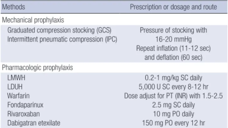
Exercise increases venous blood flow and reduces venous sta- sis, and thus, helps prevent VTE. However, no standardized ex- ercise has been implemented for the prevention of VTE in hos- pitalized patients. The application of graduated compression stockings (GCS) at the time of admission can prevent VTE in at- risk patients. Use of intermittent pneumatic compression (IPC) can be helpful in at-risk surgical patients (Table 1) (12). Place- ment of an inferior vena cava (IVC) filter is only recommended for patients at high-risk of VTE when pharmacologic thrombo- prophylaxis is contraindicated (13-16).
Pharmacologic methods
Pharmacologic methods for thromboprophylaxis include low- molecular weight heparin (LMWH), low-dose unfractionated heparin (LDUH), fondaparinux, warfarin, dabigatran, and riva- roxaban. The recommended daily doses are LMWH 20-100 U/
kg (0.2-1 mg/kg) subcutaneously (SC) daily; LDUH 5,000 U ev- ery 8 to 12 hr SC, fondaparinux 2.5 mg SC daily; dabigatran etex- ilate 150 mg every 12 hr orally; and, rivaroxaban 10 mg orally each day. Warfarin should be dosed daily to maintain an inter- national normalized ratio (INR) in the range 1.5 to 2.5 (Table 1).
The duration of thromboprophylaxis depends upon the per- ceived benefits of anticoagulation versus the risks of bleeding and overall cost.
Stratification of VTE risk in hospitalized patients
Based on clinical evidence, the risk of VTE in surgical patients can be stratified from low to high (Table 2). High-risk situations include major surgery in patients with previous VTE or a hyper- coagulable state, major orthopedic surgery, CVA, major trauma, or SCI.
The Korean guidelines are not based upon clinical evidence,
Table 1. Methods of thromboprophylaxis
Methods Prescription or dosage and route
Mechanical prophylaxis
Graduated compression stocking (GCS)
Intermittent pneumatic compression (IPC) Pressure of stocking with 16-20 mmHg Repeat inflation (11-12 sec)
and deflation (60 sec) Pharmacologic prophylaxis
LMWH LDUH Warfarin Fondaparinux Rivaroxaban Dabigatran etexilate
0.2-1 mg/kg SC daily 5,000 U SC every 8-12 hr Dose adjust for PT (INR) with 1.5-2.5
2.5 mg SC daily 10 mg PO daily 150 mg PO every 12 hr LMWH, low-molecular-weight heparin; LDUH, low-dose unfractionated heparin; SC, subcutaneously; PT, prothrombin time; INR, international normalized ratio; PO, per os.
but rather upon a consensus in experts panel of Korean Society of Thrombosis and Hemostasis (KSTH). Therefore, these guide- lines represent the most current opinions for good clinical prac- tices for the prevention of VTE. The ultimate decision regarding thromboprophylaxis should be individualized by case and de- termined by the attending physician.
GENERAL SURGERY
The principles of risk assessment for general surgery are based on the type of surgery (minor or major), age (<40 yr, 40-60 yr, and ≥60 yr), and the presence of additional risk factors, such as cancer or previous VTE (11). According to these principles, cas- es were classified into three risk groups (Table 3). Early and fre- quent ambulation was recommended for low-risk patients.
Mechanical methods (GCS and/or IPC) or pharmacologic meth- ods (LMWH, LDUH, or fondaparinux) are recommended for moderate-risk patients. LMWH, fondaparinux, or warfarin are recommended for high-risk patients scheduled for major can- cer surgery with additional risk factors for VTE and for high-risk patients with a previous VTE or thrombophilia. When antico-
agulation is contraindicated in a high-risk patient, IPC is rec- ommended. For patients undergoing entirely laparoscopic pro- cedures who do not have additional thromboembolic risk fac- tors, routine use of thromboprophylaxis (other than early and frequent ambulation) is discouraged (17).
ORTHOPEDIC SURGERY
Patients scheduled for major orthopedic surgery, including to- tal hip replacement (THR), total knee replacement (TKR), and hip fracture surgery (HFS), represent a group at particularly high risk of VTE. In the West, the incidence of VTE among pa- tients that undergo major orthopedic surgery ranges from 41%
to 60%, but the epidemiological data on the incidence of VTE in the Asian population varies (11). A recent Korean study found the incidence of postoperative VTE in TKR, THR, and HFS, to be 40.4%, 8.7%, and 16.4%, respectively, using CT pulmonary angiography and indirect CT venography (18).
For patients scheduled to undergo TKR, THR, or HFS, LMWH, warfarin or fondaparinux is recommended for thrombopro- phylaxis (Table 4) (19, 20). GCS and/or IPC are recommended in patients with a risk of bleeding. The current recommended duration for anticoagulation is 7 to 10 days, but extended use through 35 days has been proposed in the 8th ACCP guideline for the prevention of VTE after discharge from the hospital (4).
Patients scheduled for vertebral surgery have a moderate risk for VTE and should wear GCS and/or utilize IPC. Patients sched- uled for surgery of the upper extremity or lower leg (distal to the knee) has a low risk for VTE and do not require prophylaxis. Rou- tine CUS screening is not helpful in these patients.
NEUROSURGERY
The incidence of DVT and subsequent PE in neurosurgery pa- tients has been reported as high as 25%, and the PE mortality rate has been reported from 9% to 50% (21, 22). The risk factors that contribute to the high frequency of VTE in neurosurgical patients include a prior VTE; type of surgery (cranial, spinal, or vascular); duration of surgery; malignancy; infection; immobi- lization; venous stasis; chronic lower extremity swelling; lower Table 2. VTE risk-stratification and recommended prophylactic methods for each risk
group
Risk Patients or Surgery Thromboprophylaxis
Low Minor surgery in mobile patients,
Medical patients who are fully mobile Not necessary and Early ambulation Moderate General open gynecological surgery,
General open urologic surgery, Medical patients on bed rest or sick
GCS, IPC, LMWH, LDUH or Fondaparinux
High THR, TKR, HFS, Major trauma, Spinal cord injury, Major surgery
in patients with previous VTE or thrombophilia
LMWH, Warfarin or Fondaparinux; IPC*
*Recommended for patients with a risk of bleeding; consider switching to anticoa- gulants when the bleeding risk abates.
VTE, venous thromboembolism; GCS, graduated compression stockings; IPC, inter- mittent pneumatic compression; LMWH, low-molecular-weight heparin; LDUH, low- dose unfractionated heparin; THR, total hip replacement; TKR, total knee replacement;
HFS, hip fracture surgery.
Table 3. Levels of VTE risk and recommendations for general surgery
Risk group Procedures Thromboprophylaxis
Low Major surgery in age <40 yr, Minor surgery in age <60 yr
Early ambulation
Moderate Major surgery in age >40 yr or with risk factor, Non-major surgery in age >60 yr
or with risk factor
GCS, IPC, LMWH, LDUH, or Fondaparinux
High Major cancer surgery with additional risk factor, Major surgery in patients with
previous VTE or thrombophilia
LMWH, Warfarin, or Fondaparinux; IPC*
*Recommended in patients with a risk of bleeding; consider switching to anticoagulants when the bleeding risk abates.
VTE, venous thromboembolism; GCS, graduated compression stockings; IPC, inter- mittent pneumatic compression; LMWH, low-molecular-weight heparin; LDUH, low- dose unfractionated heparin; VTE, venous thromboembolism.
Table 4. Levels of VTE risk and recommendations for orthopedic surgery
Risk group Procedures Thromboprophylaxis
Low Surgery of upper extremity,
Surgery of fracture distal to the knee Early ambulation
Moderate Vertebral surgery GCS or IPC
High Total hip replacement, Total knee
replacement, Hip fracture surgery LMWH, Warfarin, or Fondaparinux; IPC*
*Recommended in patients with a risk of bleeding; consider switching to anticoagulants when the bleeding risk abates.
VTE, venous thromboembolism; GCS, graduated compression stockings; IPC, inter- mittent pneumatic compression; LMWH, low-molecular-weight heparin; LDUH, low- dose unfractionated heparin.
extremity trauma; advanced age; CHF; obesity; and, sleep apnea.
Early and frequent ambulation for low-risk neurosurgery pa- tients who have undergone surgery (not craniotomy) is recom- mended. For moderate-risk patients scheduled for craniotomy (for reasons other than brain tumor), GCS and/or IPC is recom- mended. IPC, LDUH or LMWH are suggested for patients sched- uled for craniotomy for a brain tumor. For high-risk patients scheduled for craniotomy with a concomitant history of VTE or thrombophilia, a combined thromboprophylactic approach with a mechanical method (GCS and/or IPC) and a pharmacologic method (LMWH or LDUH) is recommended.
UROLOGIC SURGERY
Risk factors for VTE in urologic surgery patients include an age
≥40 yr, obesity, malignancy, recent surgery, previous VTE, an open (vs transurethral) procedure, general anesthesia, and a long operation time. Major urologic surgery may present higher risks of PE and VTE than general surgery.
The risk of VTE has been associated with operation time (<45 min and ≥45 min). For patients scheduled for a low-risk proce- dure, early ambulation and exercise without any other form of thromboprophylaxis is recommended. On the other hand, for patients scheduled for major surgery, thromboprophylaxis with GCS or IPC; LMWH, LDUH, or fondaparinux; or a combination of mechanical and pharmacologic thromboprophylaxis is indi- cated. For abdominal (open) surgery on the kidney or retroper- itoneum, the aforementioned principles outlined for general surgery should be followed. Nephrectomy presents the same level of risk as radical prostatectomy.
OBSTETRIC AND GYNECOLOGIC SURGERY
The incidence of VTE is high in pregnancy and may occur at any stage during pregnancy or in the weeks following delivery. The risk factors for VTE in pregnant women include previous VTE, a family history of VTE, presence of anti-phospholipid antibody, age ≥40 yr, prolonged bed rest, placenta previa, Caesarian sec- tion, and lower extremities varicosities. For women with a his- tory of abortion, intrauterine death, toxemia, placenta previa, and intrauterine growth retardation, monitoring for thrombo-
philia and evaluating for thrombosis must be continued through- out the pregnancy.
Giant uterine myoma, previous surgery for an ovarian tumor, ovarian cancer, uterine or cervical cancer, severe intrapelvic ad- hesions, ovarian hyperstimulation syndrome, hormonal thera- py, and particularly, protracted lymph node dissection requir- ing transfusion are considered important risk factors for VTE (23, 24). Estrogen or progesterone should be administered with caution in postmenopausal women with a high risk of VTE.
For patients with risk factors, general thromboprophylaxis, such as, lower extremity exercise on a bed, GCS, IPC, and ade- quate hydration postpartum are recommended. Early ambula- tion should be encouraged even after a normal delivery in low- risk patients. In addition, pharmacologic thromboprophylaxis with LMWH or warfarin should be considered in patients with risk factors other than Caesarian section. For high-risk pregnan- cies with documented thrombophilia, such as, positive anti-phos- pholipid antibody or previous VTE, pharmacologic thrombopro- phylaxis with LMWH is recommended (25). Warfarin is contra- indicated during pregnancy (category X) (26). However, warfa- rin can replace LMWH after delivery and be used for 6 weeks to 3 months for continued postpartum thromboprophylaxis (25).
For patients with gynecologic disease, management strategies should follow those outlined for general surgery patients.
MEDICAL CONDITIONS
Acutely ill medical patients represent a clinically heterogeneous group and demonstrate differing risks of VTE. Despite extensive studies in medical patients, the morbidity and mortality of VTE remains significant. Without prophylaxis, the incidence of DVT and PE in general medical patients has been reported to range from 10% to 30% (5, 27, 28).
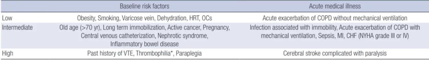
The risk of VTE was determined by assessing the probability of VTE in acutely ill medical patients according to predisposing risk factors (age >70 yr, obesity, long-term immobility, tobacco use, varicosities, dehydration, estrogens, cancer, previous DVT, paraplegia, congenital or acquired thrombophilia, and inflam- matory bowel disease [IBD]) and acute medical illnesses cur- rently under treatment (COPD exacerbation, mechanical venti- lator therapy, infection, CHF, and CVA) (Table 5). Acute ischemic
Table 5. Risk factors of VTE in general medical patients
Baseline risk factors Acute medical illness
Low Obesity, Smoking, Varicose vein, Dehydration, HRT, OCs Acute exacerbation of COPD without mechanical ventilation Intermediate Old age (>70 yr), Long term immobilization, Active cancer, Pregnancy,
Central venous catheterization, Nephrotic syndrome, Inflammatory bowel disease
Infection associated with immobility, Acute exacerbation of COPD with mechanical ventilation, Sepsis, MI, CHF (NYHA grade III or IV)
High Past history of VTE, Thrombophilia*, Paraplegia Cerebral stroke complicated with paralysis
*congenital thrombophilia such as antithrombin III deficiency or protein C or S deficiency, or acquired thrombophilia like antiphospholipid antibody syndrome.
VTE, venous thromboembolism; HRT, hormone replacement therapy; OCs, oral contraceptives; COPD, chronic obstructive pulmonary disease; MI, myocardial infarction; CHF, congestive heart failure; NYHA, New York Heart Association, VTE, venous thromboembolism.
stroke patients with restricted mobility should have LDUH or LMWH administrated 24 hr after thrombolytic therapy. If a pa- tient has an acute intracranial hemorrhage or a contraindica- tion to anticoagulation, GCS and/or IPC can be used. However, GCS and IPC can increase venous return and should be used carefully in cases of fluid overload, such as severe CHF. For am- bulatory cancer patients with cyclic chemotherapy, routine thromboprophylaxis is not recommended; however, cancer pa- tients with restricted mobility due to other acute medical illness- es are considered high-risk and should have thromboprophy- laxis administered (4).
Patients with CHF, COPD, sepsis, or IBD also have a higher prevalence of VTE, and thromboprophylaxis using GCS, IPC, LMWH, LDUH, or fondaparinux is recommended (29, 30). The majority of patients admitted to the intensive care unit (ICU) have multiple risk factors for VTE. These patients should be rou- tinely assessed and offered thromboprophylaxis with LDUH or LMWH. GCS and/or IPC can be used when there is a contrain- dication to anticoagulation.
MAJOR TRAUMA AND SPINAL CORD INJURY
Patients with major trauma are classified as high-risk. Major trauma includes multiple serious injuries, head trauma with mental status changes, severe pelvic fracture, and multiple frac- tures of a lower extremity. VTE can cause significant morbidity in patients experiencing major trauma and occurs in up to 50%
of patients without thromboprophylaxis (31). Furthermore, PE represents the third-leading cause of death among those who survive beyond 24 hr.
Routine thromboprophylaxis is recommended for all major trauma patients. Those patients with a high bleeding risk should receive LMWH or LDUH after primary hemostasis. On the other hand, if pharmacologic thromboprophylaxis is contraindicated due to active bleeding or a sustained high risk of bleeding, me- chanical (GCS or IPC) thromboprophylaxis should be imple- mented.
All patients with acute SCI should receive routine thrombo- prophylaxis. Initially, GCS and/or IPC and careful observation for bleeding is recommended, followed by LMWH thrombo- prophylaxis when primary hemostasis is complete. For patients with a high bleeding risk, such as those with intra-abdominal bleeding, traumatic subarachnoid hemorrhage, or spinal hema- toma, pharmacologic thromboprophylaxis should be delayed until the risk of further bleeding has diminished.
NEURAXIAL ANESTHESIA
Neuraxial anesthesia is a comprehensive term used for spinal, epidural, and caudal blocks. The risk for the development of spinal or epidural hematoma may be elevated by the concomi-
tant use of anticoagulants and antiplatelet agents (32, 33). The established risk factors for spinal or epidural hematoma after neuraxial blockade include an underlying hemostatic disorder, an anatomically-deformed vertebral column, traumatic inser- tion of a needle or catheter, repeated insertion attempts, con- comitant anticoagulation, continuous use of epidural catheters, and old age (11, 34).
To improve the safety of neuraxial blockade in patients receiv- ing or scheduled to receive anticoagulant prophylaxis, several guidelines have been established. Neuraxial anesthesia/anal- gesia should generally be avoided in patients with a bleeding disorder and in situations when preoperative hemostasis is im- paired by antithrombotic drugs. An epidural catheter should be removed when the anticoagulant effect is minimal (usually just before the next scheduled SC injection). Anticoagulation pro- phylaxis should be delayed for at least 2 hr after the removal of a spinal needle or epidural catheter. If warfarin is required, con- tinuous epidural analgesia should not be used for longer than 1 or 2 days. Finally, monitoring for cord compression syndrome is required when patients are receiving anticoagulation medi- cation (32, 35).
SUMMARY
These guidelines emphasize the primary prevention of VTE with thromboprophylaxis for Korean patients experiencing surgery, obstetric delivery, trauma, cancer, and severe medical illness.
Based on VTE risk factors (age, immobility, history of VTE, co- morbid illness, and type of surgery or trauma), patients can be stratified into low-, moderate-, and high-risk groups. For high- risk patients (major orthopedic surgery, major trauma, SCI, and major surgery with a history of VTE or thrombophilia), throm- boprophylaxis with LMWH, warfarin, or fondaparinux is recom- mended. Mechanical methods of thromboprophylaxis should be used primarily in patients with a high bleeding risk. For mod- erate-risk patients (general open gynecological surgery, general open urologic surgery, and medical patients on bed rest), throm- boprophylaxis with either a mechanical method (GCS and/or IPC) or a pharmacologic method (LMWH, LDUH, or fondapa- rinux) can be utilized. For low-risk patients (minor surgery in mobile patients and medical patients who are fully mobile), early and frequent ambulation is the only recommended throm- boprophylaxis. In conclusion, this article outlines the first Kore- an guidelines issued for primary VTE prevention and provides a useful reference for clinicians. These guidelines need to be updated based on results from well controlled studies conduct- ed in Korea.
REFERENCES
1. Huisman MV, Buller HR, ten Cate JW, van Royen EA, Vreeken J, Kersten
MJ, Bakx B. Unexpected high prevalence of silent pulmonary embolism in patients with deep venous thrombosis. Chest 1989; 95: 498-502.
2. Moser KM, Fedullo PF, LitteJohn JK, Crawford R. Frequent asymptom- atic pulmonary embolism in patients with deep venous thrombosis.
JAMA 1994; 271: 223-5.
3. Anderson FA Jr, Wheeler HB, Goldberg RJ, Hosmer DW, Patwardhan NA, Jovanovic B, Forcier A, Dalen JE. A population-based perspective of the hospital incidence and case-fatality rates of deep vein thrombosis and pulmonary embolism. The Worcester DVT Study. Arch Intern Med 1991; 151: 933-8.
4. Geerts WH, Bergqvist D, Pineo GF, Heit JA, Samama CM, Lassen MR, Colwell CW; American College of Chest Physicians. Prevention of ve- nous thromboembolism: American College of Chest Physicians Evidence- Based Clinical Practice Guidelines (8th Edition). Chest 2008; 133 (6 Sup- pl): 381S-453S.
5. Nicolaides AN, Breddin HK, Fareed J, Goldhaber S, Haas S, Hull R, Kalo- diki E, Myers K, Samama M, Sasahara A; Cardiovascular Disease Edu- cational and Research Trust and the International Union of Angiology.
Prevention of venous thromboembolism. International Consensus State- ment. Guidelines compiled in accordance with the scientific evidence. Int Angiol 2001; 20: 1-37.
6. The Korea Society on Thrombosis and Hemostasis. Japanese guideline for prevention of venous thromboembolism. Korean J Thromb Hemost 2009; 16: Supplement 1.
7. Anderson FA Jr, Wheeler HB, Goldberg RJ, Hosmer DW, Forcier A. The prevalence of risk factors for venous thromboembolism among hospital patients. Arch Intern Med 1992; 152: 1660-4.
8. Zhan C, Miller MR. Excess length of stay, charges, and mortality attribut- able to medical injuries during hospitalization. JAMA 2003; 290: 1868-74.
9. Samama MM, Dahl OE, Quinlan DJ, Mismetti P, Rosencher N. Quanti- fication of risk factors for venous thromboembolism: a preliminary study for the development of a risk assessment tool. Haematologica 2003; 88:
1410-21.
10. Heit JA, O’Fallon WM, Petterson TM, Lohse CM, Silverstein MD, Mohr DN, Melton LJ 3rd. Relative impact of risk factors for deep vein throm- bosis and pulmonary embolism: a population-based study. Arch Intern Med 2002; 162: 1245-8.
11. Geerts WH, Pineo GF, Heit JA, Bergqvist D, Lassen MR, Colwell CW, Ray JG. Prevention of venous thromboembolism: the Seventh ACCP Con- ference on Antithrombotic and Thrombolytic Therapy. Chest 2004; 126 (3 Suppl): 338S-400S.
12. Scurr JH, Coleridge-Smith PD, Hasty JH. Regimen for improved effective- ness of intermittent pneumatic compression in deep venous thrombosis prophylaxis. Surgery 1987; 102: 816-20.
13. Rodriguez JL, Lopez JM, Proctor MC, Conley JL, Gerndt SJ, Marx MV, Taheri PA, Greenfield LJ. Early placement of prophylactic vena caval fil- ters in injured patients at high risk for pulmonary embolism. J Trauma 1996; 40: 797-802.
14. Carlin AM, Tyburski JG, Wilson RF, Steffes C. Prophylactic and thera- peutic inferior vena cava filters to prevent pulmonary emboli in trauma patients. Arch Surg 2002; 137: 521-5.
15. Rogers FB, Shackford SR, Ricci MA, Wilson JT, Parsons S. Routine pro- phylactic vena cava filter insertion in severely injured trauma patients decreases the incidence of pulmonary embolism. J Am Coll Surg 1995;
180: 641-7.
16. Giannoudis PV, Pountos I, Pape HC, Patel JV. Safety and efficacy of vena cava filters in trauma patients. Injury 2007; 38: 7-18.
17. Lee KW, Bang SM, Kim S, Lee HJ, Shin DY, Koh Y, Lee YG, Cha Y, Kim YJ, Kim JH, Park DJ, Kim HH, Oh D, Lee JS. The incidence, risk factors and prognostic implications of venous thromboembolism in patients with gastric cancer. J Thromb Haemost 2010; 8: 540-7.
18. Cha SI, Lee SY, Kim CH, Park JY, Jung TH, Yi JH, Lee J, Huh S, Lee HJ, Kim SY. Venous thromboembolism in Korean patients undergoing ma- jor orthopedic surgery: a prospective observational study using comput- ed tomographic (CT) pulmonary angiography and indirect CT venogra- phy. J Korean Med Sci 2010; 25: 28-34.
19. Warwick D. New concepts in orthopaedic thromboprophylaxis. J Bone Joint Surg Br 2004; 86: 788-92.
20. Lieberman JR, Hsu WK. Prevention of venous thromboembolic disease after total hip and knee arthroplasty. J Bone Joint Surg Am 2005; 87:
2097-112.
21. Agnelli G, Piovella F, Buoncristiani P, Severi P, Pini M, D’Angelo A, Bel- trametti C, Damiani M, Andrioli GC, Pugliese R, Iorio A, Brambilla G.
Enoxaparin plus compression stockings compared with compression stockings alone in the prevention of venous thromboembolism after elec- tive neurosurgery. N Engl J Med 1998; 339: 80-5.
22. Black PM, Baker MF, Snook CP. Experience with external pneumatic calf compression in neurology and neurosurgery. Neurosurgery 1986; 18:
440-4.
23. Schorge JO, Goldhaber SZ, Duska LR, Goodman A, Feldman S. Clini- cally significant venous thromboembolism after gynecologic surgery. J Reprod Med 1999; 44: 669-73.
24. Ageno W, Manfredi E, Dentali F, Silingardi M, Ghezzi F, Camporese G, Bolis P, Venco A. The incidence of venous thromboembolism following gynecologic laparoscopy: a multicenter, prospective cohort study. J Thromb Haemost 2007; 5: 503-6.
25. Leonhardt G, Gaul C, Nietsch HH, Buerke M, Schleussner E. Thrombo- lytic therapy in pregnancy. J Thromb Thrombolysis 2006; 21: 271-6.
26. Ageno W, Crotti S, Turpie AG. The safety of antithrombotic therapy dur- ing pregnancy. Expert Opin Drug Saf 2004; 3: 113-8.
27. Caprini JA, Arcelus JI, Reyna JJ. Effective risk stratification of surgical and nonsurgical patients for venous thromboembolic disease. Semin Hematol 2001; 38 (2 Suppl 5): 12-9.
28. Samama MM, Cohen AT, Darmon JY, Desjardins L, Eldor A, Janbon C, Leizorovicz A, Nguyen H, Olsson CG, Turpie AG, Weisslinger N. A com- parison of enoxaparin with placebo for the prevention of venous throm- boembolism in acutely ill medical patients. Prophylaxis in Medical Pa- tients with Enoxaparin Study Group. N Engl J Med 1999; 341: 793-800.
29. Kniffin WD Jr, Baron JA, Barrett J, Birkmeyer JD, Anderson FA Jr. The ep- idemiology of diagnosed pulmonary embolism and deep venous throm- bosis in the elderly. Arch Intern Med 1994; 154: 861-6.
30. Morpurgo M, Schmid C, Mandelli V. Factors influencing the clinical di- agnosis of pulmonary embolism: analysis of 229 postmortem cases. Int J Cardiol 1998; 65 Suppl 1: S79-82.
31. Geerts WH, Code KI, Jay RM, Chen E, Szalai JP. A prospective study of venous thromboembolism after major trauma. N Engl J Med 1994; 331:
1601-6.
32. Horlocker TT, Wedel DJ, Benzon H, Brown DL, Enneking FK, Heit JA, Mulroy MF, Rosenquist RW, Rowlingson J, Tryba M, Yuan CS. Regional anesthesia in the anticoagulated patient: defining the risks (the second
ASRA Consensus Conference on Neuraxial Anesthesia and Anticoagula- tion). Reg Anesth Pain Med 2003; 28: 172-97.
33. Moen V, Dahlgren N, Irestedt L. Severe neurological complications after central neuraxial blockades in Sweden 1990-1999. Anesthesiology 2004;
101: 950-9.
34. Vandermeulen EP, Van Aken H, Vermylen J. Anticoagulants and spinal-
epidural anesthesia. Anesth Analg 1994; 79: 1165-77.
35. Rowlingson JC, Hanson PB. Neuraxial anesthesia and low-molecular- weight heparin prophylaxis in major orthopedic surgery in the wake of the latest American Society of Regional Anesthesia guidelines. Anesth Analg 2005; 100: 1482-8.