Head & Neck 환자의 방사선 치료시 Metal Artifact의 감소를 위한 Gantry Tilt Scan의 유용성 평가
서울아산병원 방사선종양학과
이충환ㆍ윤인하ㆍ홍동기ㆍ백금문ㆍ권경태
목 적: 두경부 환자의 CT simulation시 metal artifact가 발생하여 영상의 질 저하와 선량계산의 오차를 유발 할 수 있어 metal artifact를 줄이기 위한 gantry tilt scan의 유용성을 평가하고자 한다.
대상 및 방법: Metal artifact를 줄이기 위하여 20o gantry tilt scan하여 0o로 재구성한 이미지를 획득하였다. AAPM CT performance Phantom을 이용하여 CT number를 비교 분석하고, 아크릴 팬텀을 이용하여 체적의 일치여부를 확인하였다.
Metal artifact에 의한 영향을 평가하기 위해 Intensity volume Histogram (IVH)을 통한 CT number의 균질성 및 Dose Volume histogram (DVH)을 통한 선량평가를 시행하였다.
결 과: CT number와 체적의 비교에서는 차이가 0.5% 이하로 나타났다. IVH를 비교 분석결과 gantry tilt scan에서 artifact에 의한 영향이 감소되어 CT number의 균질도가 향상되고, DVH 비교결과는 양쪽 이하선의 0.2∼6%의 평균선량 오차를 감소시 켰다.
결 론: Head & Neck 방사선치료 시 metal artifact 때문에 체표윤곽의 구별이 어렵고 선량의 오차가 발생한다. Gantry tilt scan 을 이용하면 정확한 조직의 묘사가 가능하고, CT number 균질도가 향상되어 선량의 오차를 줄일 수 있었다. Gantry tilt scan 은 Head & Neck 방사선치료 시 정확한 방사선치료에 매우 유용함을 확인하였다.
핵심용어: gantry tilt scan, metal artifact, CT number, IVH, DVH
이 논문은 2010년 4월 5일 접수하여 2010년 7월 19일 채택되었음.
책임저자:이충환, 서울아산병원 방사선종양학과 Tel: 02)3010-2781, Fax: 02)3010-6950 E-mail: [email protected]
서 론
방사선 치료분야에서 전산화 단층 촬영(CT, Computed Tomoghraphy)의 활용은 이제는 없어서는 안 될 중요한 부분 이 되었다. CT영상을 입체적으로 재구성할 수 있는 기술의 발달과 함께 CT 모의 치료기가 사용되면서 종양의 위치나 모양을 3차원적으로 재구성하여 Beam's eye view를 통해 여 러 각도에서 종양을 좀 더 정확히 구분하고 종양 주변의 방 사선에 민감한 정상 조직들을 제외할 수 있게 되었다.
Radiation Therapy Planning System (RTP)의 적용으로 방 사선의 선량 분포를 입체적으로 확인할 수 있게 되면서 이를 Dose Volume Histogram (DVH)이라는 지표로 구현하여 실제 치료 시행 전 종양과 장기에 조사되는 방사선량의 분포를 입 체적으로 미리 확인하고 방사선의 방향이나 조사야를 변경 하여 가장 적절한 선량을 얻을 수 있다. 이러한 과정들을 통 하여 Tumor Control probability (TCP, 종양 제어 확률)와
Normal Tissue Complication Probability (NTCP, 정상조직 합 병증 확률)를 예측하는 것이 가능하게 되었다.1)
전산화 치료계획시 정상조직의 선량과 치료부위 선량의 분포는 매우 중요하다. 치료 부위의 균일한 선량분포를 얻기 위하여 여러 가지 방법을 이용하고 있다. 특히 두경부(Head
& Neck)종양의 방사선 치료 시 체표윤곽의 변화가 심하여 이에 따른 선량 불 균일에 대한 보정이 필요하다.2) 현재 방 사선 치료의 불 균질에 관한 보정은 CT number와 전자 밀도 의해 결정되며, CT 영상에서 metal이 삽입되어 있는 경우 metal artifact가 발생하여 CT number와 전자 밀도에 변화가 발생할 수 있어 이러한 값의 변화는 선량 계산에 오류를 일 으키는 요인이 되기도 한다.3-9)
Metal artifact가 CT영상에 미치는 영향은 크게 다음의 3가 지로 나눌 수 있는데 첫째는 다양한 파장으로 구성된 X선속 이 금속을 통과할 때에 파장에 따라서 감쇠되는 정도가 다르 기 때문에 발생하는 선속 경화 현상, 둘째는 금속을 통과하 는 광자의 수가 급격히 감소함으로 해서 발생하는 광자 고갈 현상, 셋째는 금속과 인접하는 해부학적 구조들 간의 좋은 대조도의 차이로 인하여 움직임에 의한 간섭현상이 증대되
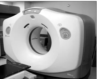
Fig. 1. LightSpeed RT 16 (GE. USA).
Fig. 2. Comparison of original image (A) and gantry tilt image (B).
는 것이다.10)
이러한 metal artifact의 영향을 줄이기 위한 노력들은 하 드웨어와 소프트웨어적으로 많은 연구와 논문들이 보고되었
다.11-14) 그중에 한 가지 방법으로 두경부 환자의 경우 Gantry
tilt scan을 이용하여 metal artifact를 줄이는 방법이 있다.15,16) 진단을 목적으로 하는 두경부 CT검사에서는 metal artifact가 발생하는 경우 gantry tilt scan을 이용하여 artifact가 생기는 방향을 바꿔, 필요한 영역을 정확하게 묘사하는 방법이 이용 된다. 그러나 gantry tilt scan image는 방사선 치료계획에 사 용되는 것이 불가능하기 때문에 참고영상으로만 사용되었으 며, 이를 보완하기 위하여 Multi Planar Reconstruction (MPR)기법을 이용하여 original image와 같은 0o의 recon- struction image를 획득하였다.
방사선 치료계획시 gantry tilt scan image를 적용하여 metal artifact를 감소시켜 실제 종양과 주변의 정상조직의 선명한
영상을 획득하고, CT number값의 오류를 줄여 선량계산의 정확도를 향상 시킬 수 있다. 이에 본 논문에서는 Gantry tilt scan의 유용성을 평가하여 방사선 치료계획 시 영상의 질 향 상과 정확한 선량계산을 적용하는 것이 본 논문의 목적이다.
대상 및 방법 1. 실험재료
- LightSpeed RT 16 (GE. USA)
- AAPM CT performance Phantom (CIRS. USA) - MATLAB 7.10.0 (R2010a)
- CERR (The Computational Environment for Radiotherapy Research)
- Rando phantom (The Phantom Laboratory. USA) - Acrylic phantom (hand-made)
- External beam planning system (EclipseTM V.8.9 Varian USA)
2. 실험방법 1) Gantry tilt scan
본 실험에서는 LightSpeed RT 16( GE. USA)을 이용하여 gantry tilt scan의 유용성을 평가하였다(Fig. 1). CT simu- lation시 metal artifact에 의한 영향을 줄이기 위해서 하위 (inferior) 방향으로 20o tilt 한 후 CT scan하여 tilt image를 획득하고 일반적인 gantry 0o의 original scan과 비교하였다.
Original image와 gantry tilt scan image를 비교하면(Fig. 2), original image는 metal artifact가 dental 바로 아래 나타나 두 경부 환자의 경우 종양과 정상조직에 영상의 화질 저하와 CT number의 변화가 생긴다. Gantry tilt scan image는 arti- fact를 위쪽으로 이동시켜 필요한 영역에서 artifact를 감소시
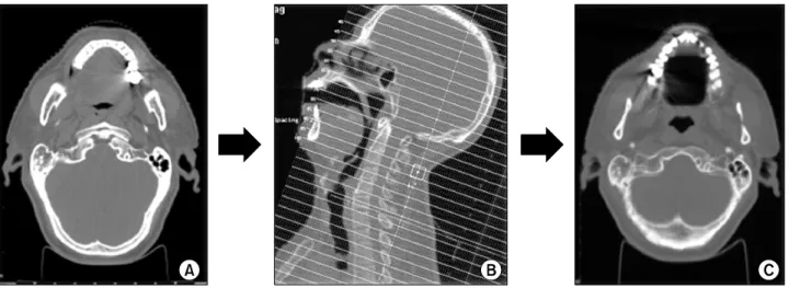
Fig. 3. Process of reconstruction by gantry tilt scan: 20o gantry tilt scan (A), multi planar reconstruction (B), reconstruction image (C).
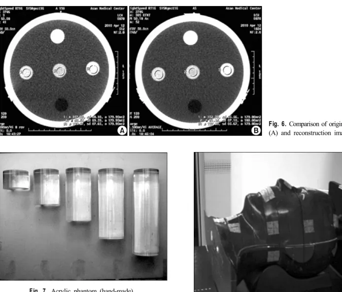
Fig. 5. AAPM CT performance phantom (CIRS. USA).
Fig. 4. Relationship between the relative electron and CT number.
켜 종양과 정상 조직의 정확한 구별이 가능하다 이렇게 획득 한 image를 Multi Planar Reconstruction (MPR)기법을 이용 하여서 original image와 같은 0o의 reconstruction image를 획 득하고 DICOM file의 X, Y, Z축 기준점을 MATLAB으로 재 설정하여 방사선치료 계획에 적용함으로서 gantry tilt scan의 유용성을 평가 하고자 한다(Fig. 3).
2) CT number 재현성의 평가
CT에 의한 영상의 재구성 알고리즘은 CT값이라는 것을 산출하며 이 값들은 감약 계수와 관련이 있다. CT값은 물의 경우를 0으로 하고 공기의 경우 −1,000에서 뼈의 +1,000까 지의 범위이다. 이런 한 방법으로 규정화된 CT값은 Houns- field값(H)이라고 한다. Hounsfield값(H)은 다음 값으로 나타 낼 수 있다.
× (1)
μ는 선감약계수이다. CT값은 감약 계수와 직선 관계를 나타낸다. CT number는 전자밀도 상관관계를 가지고 있다 (Fig. 4).17,18) 이러한 상관관계는 방사선 치료에 사용되는 photon beam에서 중요하며 방사선 치료에 사용되는 방사선 에너지의 대부분은 Compton scattering이 발생한다. Compton scattering에서 조직에 대한 photon의 흡수와 산란은 조직의 전자밀도의 주된 영향을 받는다. 따라서 CT number는 전자 밀도의 추론이 가능하므로 현재 선량계산 알고리즘에서 불 균질 조직의 보정을 위한 적절한 요소로 사용되고 있다. 이 렇게 획득한 전자 밀도는 방사선 치료의 선량계산에 있어서 필수적이며 대부분 방사선 치료계획에 CT영상이 이용되고
Fig. 7. Acrylic phantom (hand-made).
Fig. 6. Comparison of original image (A) and reconstruction image (B).
Fig. 8. Rando phantom (The Phantom Laboratory. USA).
있다.19-24)
Gantry tilt scan에 의한 CT number의 변화를 확인하기 위 해 재현성 실험을 하였다. Orignal image (0o)와 Gantry tilt scan (20o)후 reconstruction image (0o)의 CT number 값의 차 이를 알아보기 위하여 AAPM performance phantom을 이용 하였다(Fig. 5). 125 kVp, 200 mA, slice thickness 2.5 mm의 조건으로 10회씩 측정하여 같은 지점에서 5가지 물질의 CT number calibration block을 이용하여 각각의 CT number 값 을 비교하고 분석하였다(Fig. 6).
3) 체적(volume) 재현성의 평가
Gantry tilt scan후 Multi Planar Reconstruction (MPR)기법 에 의한 reconstruction과정에서 volume의 변화로 영상의 왜 곡과 선량계산의 오차가 발생할 수 있기 때문에 original im- age와 reconstruction image의 volume에 대한 재현성 평가를 실험하였다. 125 kVp, 200 mA, slice thickness 2.5 mm의 조 건으로 자체 제작한 지름이 3 cm이고 높이가 각각 2, 4, 6, 8,
10 cm의 원기둥 아크릴 phantom을 각각 10회씩 scan하고 두 image의 아크릴 phantom을 external beam planning system인 Eclipse를 이용하여 각각 contouring하여 volume을 차이를 비 교 분석 하였다(Fig. 7).
4) Metal artifact 영향의 평가
Metal artifact의 영향에 따른 CT number와 선량계산에서 차이를 알아보기 위하여 rando phantom을 이용하여 비교분 석하였다(Fig. 8). (1) artifact가 없는 normal dental영상, (2) rando phantom에 metal을 삽입하여 artifact가 있는 dental filling 영상, (3) dental filling상태에서 tilt scan하여 artifact 를 감소시킨 reconstruction image 등의 3가지 image에 동일 한 이하선영역을 좌우 한 쌍을 설정하였다(Fig 9). Intensity Volume Histogram (IVH)을 이용하여 각각의 CT number의
Fig. 10. Patient's image with metal artifact.
Fig. 9. Comparison of three images using the rando phantom (A: normal dental, B: dental filling, C: tilt image after dental filling).
분포를 분석 하였다. IVH는 DVH와 비슷한 개념으로 선량대 신에 CT number값을 대신하여 Volume과 CT number 상관관 계를 나타낸 것으로 본 논문에서는 CT number의 균질도를 판단하기 위하여 사용하였다. IVH의 기울기가 급할수록 균 질도가 좋은 것이고, 기울기가 완만할수록 균질도가 떨어지 는 것을 의미한다.
Metal artifact의 영향에 따른 이하선의 선량변화를 실험하 기 위해 RT, RAO, ANT, LAO, LT방향에서 45o 간격으로 5 개의 beam을 선택하여 전산화 치료계획을 시행하였으며, 3 가지 image의 이하선의 평균선량(mean dose)을 DVH (Dose Volume Histogram)로 비교 분석 하였다.
5) Gantry tilt scan의 임상적용
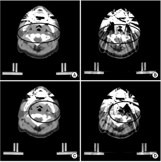
Metal artifact가 있는 두경부 암 환자의 경우 영상의 질 저 하로 종양과 정상조직에 구분이 어렵다(Fig. 10). 본원에 내 원한 metal artifact가 있는 10명의 두경부 암 환자를 대상으 로 gantry 각도가 0o인 original scan과 20o인 gantry tilt scan
의 방법으로 두 가지 image를 획득하여 비교 분석 하였다.
Artifact에 의한 영상의 질 저하로 종양과 정상조직의 구별이 어려워 조직 묘사의 개인차가 발생할 수 있기 때문에 정확한 비교를 위해 gantry tilt scan을 이용하여 artifact가 감소된 re- construction image를 이용하여 종양과 정상조직을 설정하여 artifact가 있는 original image에 동일한 volume을 설정하였 다. 즉 동일한 structure에 다른 두 영상을 사용하여 volume차 이에 의한 영향을 최소화하여 이하선의 CT number와 평균 선량을 비교분석 하였다. 이하선을 설정한 이유는 개인 간의 volume의 차이가 적고, metal artifact의 영향을 가장 잘 받는 정상조직으로 artifact에 의해서 차이를 볼 수 있는 장기이다.
IVH를 통해 조직 내 CT number의 균질도를 비교분석하였 고, 전산화 치료계획에 적용했을 때 선량의 차이를 알아보기 위하여 DVH를 이용하여 두 영상의 평균선량을 비교분석 하 였다.
현재 전산화 치료계획 장치에서에서는 gantry tilt scan이
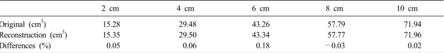
2 cm 4 cm 6 cm 8 cm 10 cm
Original (cm3) 15.28 29.48 43.26 57.79 71.94
Reconstruction (cm3) 15.35 29.50 43.34 57.77 71.96
Differences (%) 0.05 0.06 0.18 −0.03 0.02
Table 2. Comparison of volume using acrylic phantom
Fig. 11. Comparison of IVH of three images using the rando phantom.
Acrylic Nylon Polycarbonate Polystyrene Polyethylene
Original (HU) 124.82 93.78 103.79 −23.22 −89.55
Reconstruction (HU) 124.90 94.08 103.54 −23.27 −89.81
Difference (%) 0.06 0.31 −0.24 0.02 0.29
Table 1. Comparison of CT number using AAPM CT performance phantom 적용되지 않기 때문에 DICOM file을 MATLAB을 이용하여 좌
표의 기준점을 재설정하여서 전산화 치료계획에 적용하였다.
결 과
CT number 재현성을 AAPM CT performance Phantom을 이용하여 평가한 결과는 5가지 물질의 CT number calibra- tion 블록의 CT number값이 Table 1과 같은 결과를 얻었다.
분석 결과 original image와 reconstruction image는 5가지 물 질의 CT number가 모두 기준 값(각 핀의 CT 감약 계수
±10HU)에 포함되어 정확한 방사선 선량계산이 가능하였다.
두 영상의 CT number의 차이는 −0.24∼+0.31%로 gantry tilt scan에 대한 변화는 없었으며reconstruction image를 전산 화 치료계획에 적용할 때 전자밀도의 변화도 없었다.
Original image와 reconstruction image의 volume에 대한 재현성의 평가 결과는 5가지의 아크릴 팬텀의 체적은 Table
2와 같은 결과를 얻었다. Volume의 차이는 −0.03∼+0.18%
로 gantry tilt scan에 의한 reconstruction image에서 volume의 왜곡은 나타나지 않기 때문에 정확한 방사선 치료계획의 적 용이 가능하였다.
Metal artifact의 영향에 관한 결과는 Rando phantom을 이 용한 CT number의 균질도에 대한 IVH평가에서 artifact가 없 는 normal dental영상에서는 경사도가 급하여 균질한 CT number를 가지고 있는 영상임을 확인하였다. Dental filling상 태에서 tilt scan하여 reconstructed image한 영상에서는 경사 도가 normal dental영상과 비슷한 기울기를 나타내었으며 ar- tifact의 감소로 인한 균질도의 향상을 확인하였다. 앞의 두 영상과는 다르게 artifact가 있는 dental filling 영상에서는 완 만한 기울기를 나타내어 균질도가 낮았다(Fig. 11).
Fig. 12는 세 가지 영상의 DVH를 나타내는 것으로 metal 이 삽입된 dental filling영상은 artifact의 영향에 의해 다른 두 영상과는 다른 분포를 나타냈다. 평균선량의 비교에서도
RT parotid gland (%)
Difference (%)
LT parotid gland (%)
Difference (%)
Artifact O 48.5 3.6 50.2 3.6
Artifact X 44.9 48.6
0.3 0.4
Gantry tilt 44.6 40.2
Table 3. Comparison of the mean dose of three kinds images
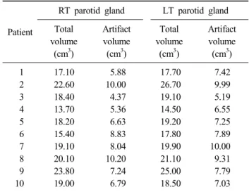
Patient
RT parotid gland LT parotid gland Total
volume (cm3)
Artifact volume
(cm3)
Total volume (cm3)
Artifact volume
(cm3)
1 17.10 5.88 17.70 7.42
2 22.60 10.00 26.70 9.99
3 18.40 4.37 19.10 5.19
4 13.70 5.36 14.50 6.55
5 18.20 6.63 19.20 7.25
6 15.40 8.83 17.80 7.89
7 19.10 8.04 19.90 10.00
8 20.10 10.20 21.10 9.31
9 23.80 7.24 25.00 7.79
10 19.00 6.79 18.50 7.03
Table 4. The comparison of the volume receiving the influence of the metal artifact and whole parotid gland volume
Fig. 12. Comparison of DVH of three images using the rando phantom.
Fig. 13. Comparison of IVH of original image and reconstruction image for head and neck patient.
artifact가 없는 normal dental영상을 기준으로 artifact가 있는 dental filling영상에서는 양측 이하선에서 평균 3.6%의 선량 의 차이를 나타냈다. Artifact를 감소시킨 gantry tilt scan에서 는 0.3%, 0.4%의 적은 평균선량의 차이를 보였다(Table 3).
10명의 두경부 환자를 gantry tilt scan에 적용한 결과는 양
측 이하선의 전체 체적은 좌측 이하선: 18.79 cm3, 우측 이하 선: 20.00 cm3로 나타났다. 이중에서 artifact의 영향을 받는 부분은 좌측 이하선: 7.33 cm3, 우측 이하선: 7.84 cm3로 양측 모두 약 39% 정도의 영역에서 artifact의 영향을 받았다 (Table 4).
Fig. 13은 양측 이하선의 CT number값과 volume의 관계를 IVH로 나타낸 것이다. Original image는 metal artifact의 영향 으로 IVH상에서 기울기는 완만하게 나타났으며, 완만한 경 사도는 artifact의 영향으로 CT number값이 원래 값이 아닌 다른 값으로 인식한다는 것을 의미한다. Artifact의 영향을 감소시킨 gantry tilt scan에서는 IVH상에서 기울기는 급하게 나타나 이하선의 CT number 균질도가 향상되었다. Gantry tilt scan을 적용하면 정확한 CT number를 얻을 수 있다.
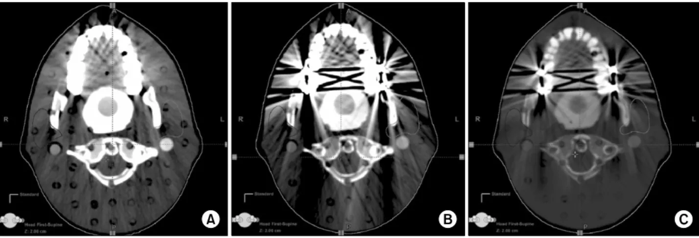
Fig. 14. Comparison of the metal artifacts of the original image and reconstruction image (A and C is reconstruction image, B and D is original image).
Patient RT. parotid gland (%) LT. parotid gland (%)
1 1.8 2.9
2 0.7 1.1
3 6.0 −1.5
4 0.8 0.4
5 −4.7 0.3
6 0.2 0.2
7 −1.1 −1.1
8 4.4 0.6
9 −0.2 0.5
10 −1.0 −0.3
Table 5. Comparison of the mean dose for head and neck cancer patients
Table 5는 DVH 이용한 metal artifact의 영향으로 인한 평 균선량 차이를 나타낸다. Metal artifact의 정도에 따라 최대 6%, 최소 0.2%의 평균선량의 차이가 발생했다. Artifact가 심 할수록 많은 선량차이를 나타났으며, 전산화 치료계획에서
선량의 오차가 발생할 수 있는 요인이 된다.
Fig. 14는 gantry tilt scan후 reconstruction image로 종양과 정상조직 주변에서 artifact에 의한 영향이 감소되어 정확한 조직 구분이 가능하였고 영상의 질이 향상되어 정확한 방사 선 치료가 가능하다.
고안 및 결론
방사선 치료에서 CT의 적용은 두 가지 면에서 매우 중요 하다. 첫째는 정확한 체표윤곽을 얻을 수 있으며, 종양과 정 상조직의 묘사가 유용하다. 다른 하나는 조직 불 균질 보정 을 위한 CT number형태의 값을 제공한다. 실질적인 견해에 서 첫째 면이 더 중요하다. 체표윤곽, 종양과 정상조직의 정 확한 설정은 전산화 치료계획에 중요할 뿐 아니라 선량 분포 의 계산에서도 중요하다. Metal artifact에 의해서 종양과 정 상조직의 기하하적 외형을 부정확하게 설정할 수 있고, 외형 은 정확한데 CT number의 불 균질에 의해서 부정확한 전자
밀도의 사용하게 되어 결과적으로 정확한 방사선 치료에 선 량오류를 발생할 수 있다.25)
이런 단점을 해결하기 위하여 gantry tilt scan을 이용하여 종양과 정상조직 주변의 영상의 질을 향상 시켜 정확한 조직 의 체적을 설정하고, 조직 내 CT number의 균질도를 향상시 켜 정확한 전자밀도의 획득으로 선량계산의 오차를 감소시 킬 수 있다. Gantry tilt scan의 유용성평가보다 선행된 origi- nal image와 reconstruction image의 재현성 평가에서 CT number와 volume의 차이는 0.5% 미만으로 나타나 재현성이 우수하였다. 이와 같은 결과로 실제 임상에 적용할 수 있는 타당성을 확보하였으며, Rando phantom을 이용한 실험결과 에서 gantry tilt scan경우 metal artifact를 감소시켜 artifact가 없는 경우와 비슷한 CT number 균질도와 평균선량의 결과 를 얻었다.
전산화 치료계획을 임상에 적용한 결과는 이하선의 약 39%정도에서 artifact에 의한 영상왜곡과 선량계산의 오차가 발생하였다. 이러한 영향을 이하선뿐만 아니라 CTV와 PTV 에도 영향을 주어 정확한 방사선치료에 장해 요인이 되었다.
Gantry tilt scan을 이용하여 metal artifact에 의한 오류를 감소시켜 종양과 정상조직의 정확한 설정이 가능한 장점이 있어 보이지 않는 부분을 정확하게 설정할 수 있고, 정확한 방사선 치료가 가능하였다. 또한 IVH상에서 CT number의 균질도가 향상되어 정확한 전자밀도의 계산이 가능하였다.
Metal artifact의 발생에 따라 IVH와 DVH의 변화에 영향 을 주었으며 많은 artifact영상에서 IVH와 DVH가 적은 arti- fact의 영상보다 변화가 심하였다. Artifact의 영향이 큰 경우 는 최대 6% 평균 선량의 차이가 발생하였고, 적은 경우는 0.2%의 평균선량 차이가 나타났다. Artifact에 의한 선량의 차이는 정확한 방사선 치료를 위해서 보정이 필요하다.
Gantry tilt scan을 임상에 적용한다면 정확한 종양과 정상 조직의 설정이 가능하고, 정확한 CT number값을 획득함으로 서 선량계산의 오차를 줄여서 정확한 방사선 치료가 가능하 였다. 본 논문에서 Head & Neck 방사선 치료시 gantry tilt scan의 유용성을 확인하였다.
참고문헌
1. Choi SK: Recent advances in radiotherapy. Korean J Clin Geri 2008;9(2):218-224
2. 백금문, 김대섭, 박광호 등: 두경부 종양에서 Forward IMRT 유용성에 관한 고찰. 대한방사선치료학회지 2003;1:41-52 3. 김경태, 주상규: 두 경부 종양의 CT영상을 이용한 방사선 치
료계획 시 Artifact가 선량 계산에 미치는 영향. 대한방사선치
료학회지 2001;1:109-112
4. 이제희, 김보겸, 박흥득: 방사선 치료계획 시 불 균질 보정에 관한 고찰. 대한방사선치료학회지 2006;2:89-95
5. Webster GJ, Rowbottom CG, Mackay RI: Evaluation of the impact of dental artifacts on intensity-modulated radiotherapy planning for the head and neck. Radiother Oncol 2009;93:
553-558
6. Schneider U, Pedroni E, Lomax A: The calibration of CT Hounsfield units for radiotherapy treatment planning. Phys Med Biol 1996;41:111-124
7. Kim Y, Tome WA, Bal M: The impact of dental artifacts on head and neck IMRT dose distributions. Radiother Oncol 2006;79:198-202
8. Kim Y, Tome WA: On the radiobiological impact of metal artifacts in head and neck IMRT in terms of tumor control probability (TCP) and normal tissue complication probability (NTCP). Med Bio Eng Comput 2007;45:1045-1051
9. Jikun W, George AS, Wen CH et al.: Dosimetric impact of a CT metal artefact suppression algorithm for proton, electron and photon therapies. Phys Med Biol 2006;51:5183-5197 10. Park H, Lee CH, Kim DK, Park CS: The elimination of the
linear artifacts by the metal restorations in the three dimen- sional computed tomographic images using the personal computer and software. Korean J Oral Maxillofac Radiol 2003;33(3):151-159
11. Rinkel J, Dillon WP, Funk T, Gould R, Prevrhal S: Computed tomographic metal artifact reduction for the detection and quantization of small features near large metallic implants. J Comput Assist Tomogr 2008;32:621-629
12. Park WS, Kim KD, Shin HK, Lee SH: Reduction of metal artifact in three-dimensional computed tomography (3DCT) with dental impression materials. Conf Proc IEEE Eng Med Biol Soc 2007;2007:3496-3499
13. Mehran Y, Luc G, Luc B: An adaptive approach to metal artifact reduction in helical computed tomography for radiation therapy treatment planning. Int J Radiat Oncol Biol Phys 2005;62:1224-1231
14. Iskandar K, Hans J, Maarten LP: Implication of artefacts reduction in the planning ct originating from implanted fiducial markers. Medical Dosimetries 2010
15. Yasuo N, Hiroyuki I, Takahiro M et al.: Influence and improvement of metal artifacts in dental structures by CT for radiation treatment planning reconstruction of transverse images using oblique images by gantry tilt scanning. Nippon Hoshasen Gijutsu Gakkai Zasshi 2007;63(3):326-334 16. Yasuo N, Kiyoshi S, Takahiro M et al.: Improvement of metal
artifact in dental structures by X-ray CT: reconstruction of transverse image using oblique image by gantry tilt scanning.
Nippon Hoshasen Gijutsu Gakkai Zasshi 2006;62(60):863-866 17. Hendee WR, Lbbott GS, Hendee EG: Radiation therapy
physics. 3rd ed. Philadelphia: John Wiley and Sons, 2005;
248-251
18. Levitt SH, Potish RA, Khan FM: Technological basis of radiation therapy. 3th ed. Philadelphia: Williams & Wilkins, 1999;104-116
19. Chapman A, Butson M, Quach1 K, et al.: Verification of CT number to density conversion for a simulator-CT attachment.
Australas Phy Eng Sci Med 2002;25:78-80
20. Thomas SJ: Relative electron density calibration of CT scanners for radiotherapy treatment planning. Br J Radiol 1999;72:781-786
21. Coolens C, Childs PJ: Calibration of CT Hounsfield units for radiotherapy treatment planning of patients with metallic hip prostheses: the use of the extended CT-scale. Phys Med Biol 2003;48:1591-1603
22. Huizenga H, Storchi PR: The use of computed tomography numbers in dose calculations for radiation therapy. Acta Radiol Oncol 1985;24:509-519
23. Coolens C, Childs PJ: Calibration of CT Hounsfield units for radiotherapy treatment planning of patients with metallic hip prostheses. Phys Med Biol 2003;1591-1603
24. James CH, Ben N, Robert K: Applications of simulator computed tomography number for photon dose calculations during radiotherapy treatment planning. Radiother Oncol 2000;55:65-73
25. Khan FM: The physics of radiation therapy. 3rd ed.
Philadelphia: Williams & Wilkins, 2003;231-234
Abstract
Evaluation of using Gantry Tilt Scan to Head & Neck of Patients during Radiation Therapy for Reduction of Metal Artifact
Chung Hwan Lee, In Ha Yun, Donggi Hong, Geum Mun Back, Gyeong Tae Kwon Department of Radiation Oncology, Asan Medical Center, Seoul, Korea
Purpose: the degradation of an image quality and error of the beam dose calculation can be caused because the metal artifact is generated during the CT simulation of head and neck patient. The usability of the gantry tilt scan for reducing the metal artifact tries to be appraised.
Materials and Methods: The inferior 20˚ gantry tilt scan was made in order to reduce the metal artifact and 0˚
reconstruction image was acquired. The AAPM CT performance Phantom was used in order to compare the CT number of the reconstructed image and Original image. the difference of volume was compared by using the acrylic phantom. The homogeneity of the CT number was evaluated the Intensity volume Histogram (IVH) as in order to evaluate an influence by the metal artifact. A dose was evaluated as the Dose Volume Histogram (DVH).
Results: in the comparison of the CT number and volume, the difference showed up less than 0.5%. As to the comparison of IVH, in the gantry tilt scan, influence by an artifact was reduced and the homogeneity of the CT number was improved. The comparison of DVH result reduced the mean dose error of the both sides parotid 0.2~
6%.
Conclusion: In the Head & Neck radiation therapy, It is difficult and to distinguish tumor and normal tissue and the error of dose is generated by the metal artifact. The delineation of the exact organization was possible if the Gantry tilt scan was used. The CT number homogeneity was improved and the error of dose could be reduced. The Gantry tilt scan confirmed in the Head & Neck radiation therapy to be very useful in the exact radiation therapy.
Key words: gantry tilt scan, metal artifact, CT number, IVH, DVH