형에서의 수술적교정은 대부분 완전교정이 되지 않는
9
0
0
전체 글
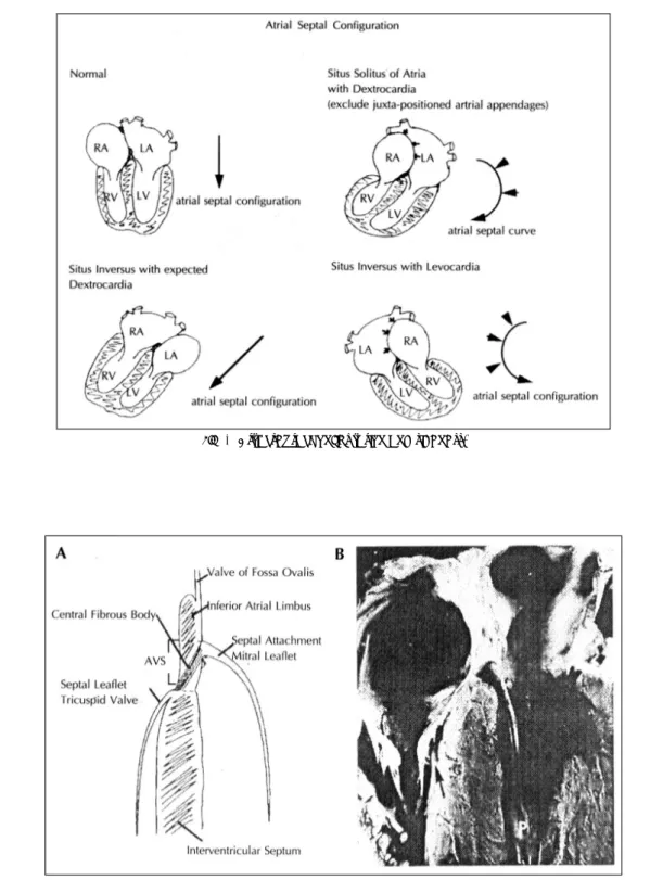
(2) 경계로 반대방향에 위치하게 되나 ambigus의 경우 같. crux를 관찰하는 것이 중요하다(Fig. 4). 좀더 apex. 은 방향에 위치하던가 IVC가 interupt되고 hemiaz-. 로 치우친 atrioventricular valve가 경우가 tricuspid. ygos로 연결되기도 한다.. valve로 해당 심실이 우심실이 된다. 2) 심실의 trabecular pattern을 관찰할 수 있는데 이때에는 moderate. 2) Juxtapostion of atral appendages 좌, 우심방의 appendage가 인접하여 있는 경우를 juxtapostion of atrial appenages라고 하며 두 appendage가 좌측에 있을 수도(left juxtaposed atrial appendages) 혹은 우측에(right juxtaposed atrial appendgae)있을 수도 있다. 심초음파도상으로는 심첨부4방 단층영상에서 특징적인 심방중격모양을 관찰할 수 있 고, 단축영상에서 great artery 하방으로 두 개의 appendage를 관찰할 수 있다. 이 단층영상에서는 통상의 심방중격이 상하방향을 갖는데 비하여 좌우방향을 갖 게 된다(Fig. 2).. band의 존재가 가장 눈에 띄는 소견이다(Fig. 5). 이 외에도 3) AV valve의 leaflet수, 4) papillary muscle 의 수 및 5) chordal insertion의 pattern이 좌우심방 의 구분에 도움이 된다. AV valve의 connection으로 는 chordal insertion에 따라 stradding와 overriding 으로 구분하는데 이는 apical 4 chamber plane에서 가장 잘 평가할 수 있다(Fig. 6). 또한 univentricular connection을 보이는 경우로는 double inlet, single, inlet, common inlet의 양상을 보일 수 있다(Fig. 7).. 4. Ventriculoarterial connection의 평가 ventriculoatrial connection의 평가에서는 두 개의. 3) Dextrocardia, levocardia 심장의 sideness는 levocardia, dextrocardia와 mesocardia로 나누어진다. 심첨부가 왼편으로 향할 경우 levocardia, 오른편으로 향할 경우 dextrocardia라 칭 하게 된다. Situs solutis인 경우 정상적으로는 심첨부. great artery중 어느것이 aorta인지 또 어느것이 pulmonary artery인지를 판별하여야 한다. 심초음파를 통 한 이의 판별은 탐촉자를 기울여 주행방향을 추적하면 pulmonary artery의 경우 bifurcation하게 된다(Fig. 8).. 가 왼편으로 향하는 levocardia의 소견을 보여야 하. 5. Great artery의 configuration. 며 반대로 situs inversus는 심첨부가 오른족으로 향. Great artery들의 configuration으로 두 great artery. 하는 dextrocardai의 소견을 보인다(mirror image de-. 가 cross할 경우 단축영상상 한 circle과 다른 saus-. xtrocardia). 그러나 situs가 solitus이면서 심첨부만 오. age shape의 structure를 관찰할 수 있는데 반하여. 른쪽으로 향하는 경우도 있는데(pivotal type dextroc-. 두 great artery가 pararell할 경우 두 개의 circle로. ardia:dextroversion) 이런 경우 심초음파상으로는. 관찰된다. 또한 이 영상에서 aorta와 pulmonary art-. atrial septum과 ventricular septum의 configuration. ery 사이에 어느것이 앞쪽에 위치하며 두 great artery. 이 정상적인 경우와 다르게 된다(Fig. 3).. 의 좌우관계를 알 수 있다(Fig. 9).. 3. Atrioventricular connection의 평가. 6. Shunt lesion의 존재유무 및 obstructive 혹은 regurgitant lesion의 존재 유무. situs가 결정된 경우 각 심방과 연결되어 있는 ventricle이 우심실인가 좌심실인가를 판별하면 atrioven-. 이는 통상적인 Doppler 심초음파검사법에서 사용하. tricular connection을 평가할 수 있다. 먼저 심실로. 는 방법과 다를 것이 없다. 그러나 많은 경우 이의 확. 규정하기 위하여서는 ventricular inlet이나 AV valve. 인과 정도를 확인하기 어려운 경우를 접하게 된다. 예. fibrous ring의 50% 이상을 수용하여야 한다. 이런조. 를 들어 coarctation of aorta의 경우 소아연령에서는. 건을 만족하지 못할 경우 rudimentary chamber라 하. suprasternal notch view에서 Doppler를 이용하여. 며 rudimentry chamber는 다시 outlet(aorta나 pulm-. 확인할 수 있으나 성인에서는 어려운 경우가 흔하며. inary artery)의 50% 이상을 수용하는 경우에는 outlet. 이러한 경우 subxiphoid view에서 descending aorta. chamber로, 그러하지 못할 경우 trabecular pouch로. 에서의 이완시기 flow turbulence를 관찰할 수 있다. 부르게 된다. 심초음파상 좌우심실을 판별하는데는 1). (Fig. 10).. - 683 -.
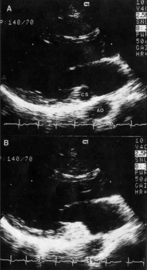
(3) to left shunt는 보이지 않는데 반하여 descending aor-. 7. Contrast echocardiography의 이용. ta가 contrast되는 현상을 관찰할 수 있다(Fig. 13).. 1) Dilated coronary sinus. References. 우심실에 용적과다를 보이는 상황에서 coronary sinus가 확장될 수 있다. 그러나 이러한 상황을 보이지 않은 경우 가장 흔한 원인으로는 persistent left sided SVC가 coronary sinus로 drain되는 경우이다. 이의 확인은 left arm으로부터 contrast echocardiography 를 시행하면 coronary sinus로 drain되는 것을 확인 할 수 있다(Fig. 11, 12).. 2) Right to left shunt의 확인 Contrast echocardiography를 이용하면 right to left shunt를 용이하게 판벼랄 수 있다. 이중 특히 PDA 의 경우 존재자체 조차 놓치는 경우가 있는데 contrast echocardiography를 이용하는 경우 intracardiac right. 1) JK Perloff:The clinical recognition of congenital heart disease. Fourth ed. W.B. Saunders Co., Phladelphia, 1995 2) Seward JB, Tajik AJ, Edward WD, Hagler DJ:Twodimensional echocardiographic atlas. Vol 1. Congential heart disease. Springer-Verlag, New York, 1987 3) Backer AE, Anderson RH:Cardiac pathology. Churchill Livingstone, London, 1983 4) Roberts WC:Adult congential heart disease. F.A. Davis Co.m Philadelphia, 1987 5) Perloff JK, Child JS:Congenital heart disease in adults. Fourth ed. W.B. Saunders Co., Philadelphia, 1991 6) Williams RG, Bierman FZ, Sanders SP:Echocardiographic diagnosis of cardiac malformations. Little, Brown, Boston, 1986. - 684 -.
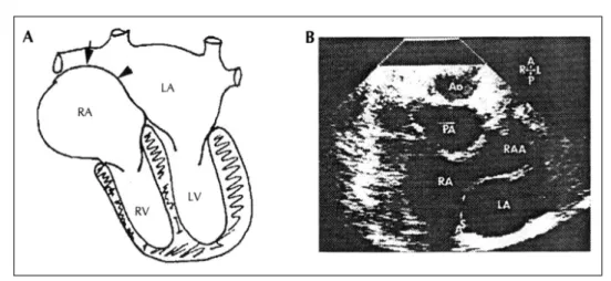
(4) □ 손 대 원 사진부도 (1)□. Fig. 1. Left sided atrium(*) in this figure could be recognized as left atrium by identifying the pulmonary venous drainage(arrow).. Fig. 2. A:Easiest feature ot recognize the juxtaposed atrial appendages is a curvature of the atrial spptum on 4chamber views. B:Most diagnostic observation is the visualization of both atrial appendages posterior to the great arteries. The plane of the atrial septum on short axis is from left to right as opposed to up and down.. - 685 -.
(5) □ 손 대 원 사진부도 (2)□. Fig. 3. Atrial septal configuration and sideness.. Fig. 4. Details of internal cardiac crux. Upper and lower poles of cardiac crux represent inferior atrial limbus and interventricular septum, respectively. Note the more apically displaced septal leaflet of tricuspid valve.. - 686 -.
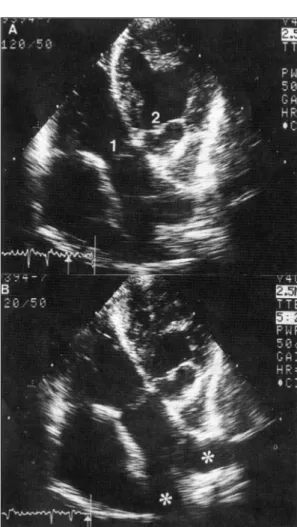
(6) □ 손 대 원 사진부도 (3)□. Fig. 5. Same patient as in figure 1. Valve seen on the right side in this figure is more apically displaced, indicating tricuspid valve. Thus ventricle connected to the tricuspid valve is the right ventricle representing atrioventricular discordance in this patient. Prominent moderate band(arrowhead) also helps recognizing the right ventricle.. Fig. 6. Atriventricular valve connection-straddling and overiding. Two condition may coexist.. - 687 -.
(7) □ 손 대 원 사진부도 (4)□. Fig. 7. Univentricular connection.. Fig. 9. Same patient as in figure 1, 5 and 6. Short axis plane of the great arteries. As the great artery located on the left side in the figure(A) was identified as pulmonary artery, two circle in this figure indicates that pulmonary artery and aorta(B) are running parallel side by side.. Fig. 8. A:Same patient as in figure 1 and 5. Two great arteries are seen(1, 2). B:By tilting the tranducer, great artery located on the left side in the figure(1) bifurcates(*) indicating that this great artery is the pulmonary artery.. - 688 -.
(8) □ 손 대 원 사진부도 (5)□. Fig. 10. In patient with coarctation of aorta, turbulant flow during diastole(arrows) was noticed in the descending aorta.. Fig. 11. A:Diated coronary sinus in the parasternal long axis view. B:contrast injection in the left side arm vein opacifies the coronary sinus indication persistent left sided SVC draining into coronary sinus. CS:coronary sinus, AO:aorta.. - 689 -.
(9) □ 손 대 원 사진부도 (6)□. Fig. 12. A:Coronary sinus could be visualized by slightly tilting the transducer downward at the apical 4-chamber view. B:Contrast injection in the left side arm vein showed the drainage into the coronary sinus. RV:right ventricle, LV:left ventricle, RA:right atrium.. Fig. 13. A:Right ventrcle was dilated and hypertrophied indicating RV pressure overload. B:Descending aorta at the subxyphoid position. C:After contrast injection RV was opacified. No contrast in the left-sided chambers suggesting the abence of intracardiac right to left shunt. D:Descending aorta was opacified. Thus patent ductus arteriosus with Eisenmenger syndrome could be diagnosed.. - 690 -.
(10)
수치

+3

관련 문서
④는 그림 c와 d같이 가장 높은 만족을 주는 무차별곡선이 가격선의 Y절편이나 X절편에서 만나는 경우이다.
Chest X-ray: Posterio-anterior (PA) View - Left Anterior Oblique
• 눈에 보이지 않는 개념은 적당한 비유를 활용해서 나타 내기... 선과 도형으로
ㅇ 미국 등 많은 OECD국가에서 사람들이 사회적 접촉을 꺼리는 현상이 발생하는 것도 사회적 소외와 뒤쳐짐을 악화시키는 요인임.. - 기존 유럽 사회는 교회,
core exercise program showed not only increasing peak torque of the right leg, left leg in Hip Extension of exercise group but also increasing peak torque of the right leg, left
줄자를 이용하여 보이지 않는 두 점간의 거리를 재어보는 체험을 하고 느낀 소감을 기록해보세요.. 호수
소음 공학에서는 소음은 물체와 공기가 부딪히면서 나는 소음이 있고, 기계적 진동에 의해 발생하는 소음이 있다. 소음 공 학은 과거에는 소음을 줄이거나 없애는
센서방식의 경우 일정 각도가 벗어난 경우 태양 추적이 되지 않는 점을 보완하기 위해 프로그램에 의해 시간에 따른 태양의 대강의 위치를 결정 하고 센서로 정확한
