© 2017 The Korean Ophthalmological Society
This is an Open Access article distributed under the terms of the Creative Commons Attribution Non-Commercial License (http://creativecommons.org/licenses /by-nc/3.0/) which permits unrestricted non-commercial use, distribution, and reproduction in any medium, provided the original work is properly cited.
Original Article
Treatment of Bilateral Retinal Angiomatous Proliferation with Anti-vascular Endothelial Growth Factor: 12-Month Outcome
Jae Min Kim
1, Jae Hui Kim
1, Young Suk Chang
2, Jong Woo Kim
1, Chul Gu Kim
1, Dong Won Lee
11
Department of Ophthalmology, Kim’s Eye Hospital, Konyang University College of Medicine, Seoul, Korea
2
Department of Ophthalmology, Konyang University College of Medicine, Daejeon, Korea
Purpose: To evaluate the 12-month outcome of intravitreal anti-vascular endothelial growth factor therapy in eyes with bilateral retinal angiomatous proliferation (RAP).
Methods: This retrospective observational study included 38 eyes of 19 patients with stage 1 or 2 bilateral RAP at diagnosis. The eyes of patients who exhibited different baseline best-corrected visual acuity (BCVA) values in both eyes were assigned to one of two groups—the better (n = 13) and worse (n = 13) visual acuity groups. The BCVA values in both groups were compared to those at baseline and at 12 months. In addition, the 12-month changes in BCVA were compared between the two groups. The association between the optical coherence tomography findings at diagnosis and the 12-month BCVA was also analyzed.
Results: The values of mean baseline and 12-month BCVA in the better visual acuity group (13 eyes) were 0.48
± 0.19 and 0.58 ± 0.29, respectively, and those in the worse visual acuity group (13 eyes) were 0.83 ± 0.20 and 0.90 ± 0.31. The 12-month changes in BCVA were not significantly different between the two groups ( p = 0.786).
Among the six patients with equivalent baseline BCVA in both eyes, four patients (66.7%) exhibited 1 to 2 lines or ≥3 lines of difference in BCVA between eyes at 12 months. Eyes without pigment epithelial detachment (PED) at diagnosis exhibited significantly better BCVA at 12 months than eyes with PED (p = 0.021).
Conclusions: Better baseline visual acuity was associated with better BCVA at 12 months posttreatment in pa- tients with bilateral RAP. However, equivalent baseline visual acuity in both eyes might not guarantee similar treatment outcomes. In addition, the absence of PED is predictive of better visual outcome.
Key Words: Bevacizumab, Choroidal neovascularization, Macular degeneration, Ranibizumab, Retinal angio- matous proliferation
Retinal angiomatous proliferation (RAP) is a subtype of exudative age-related macular degeneration (AMD) that is
characterized by retina-retinal or retinal-choroidal anasto- mosis [1]. One of the characteristics of RAP, which distin- guishes it from other types of exudative AMD, is the rela- tively high risk of bilateral involvement. In a previous study involving 108 patients, Yannuzzi et al. [1] reported bilateral RAP at diagnosis in 26 patients. The incidence of fellow-eye involvement in patients with unilateral RAP is reported to be between 27% and 100% [2-5].
Received: March 22, 2016 Accepted: June 13, 2016
Corresponding Author: Jae Hui Kim, MD. Department of Ophthalmol-
ogy, Kim's Eye Hospital, #136 Yeongsin-ro, Yeongdeungpo-gu, Seoul
07301, Korea. Tel: 82-2-2639-7664, Fax: 82-2-2639-7824, E-mail: ki-
[email protected]
Anti-vascular endothelial growth factor (VEGF) is an effective treatment for exudative AMD [6-10], and previ- ous studies have also demonstrated the efficacy of this therapy in RAP [11-19]. However, to the best of our knowl- edge, the outcome of bilateral RAP treatment with an- ti-VEGF has not yet been reported in detail, despite the fact that bilateral involvement at diagnosis is a frequent finding in patients with RAP. The purpose of this study was to evaluate the 12-month outcome of intravitreal anti-VEGF therapy in eyes with recently-developed bilateral RAP.
Materials and Methods
This single-center observational case study followed the tenets of the Declaration of Helsinki and was approved by the institutional review board of Kim’s Eye Hospital (Seoul, Korea). Written informed consent from the patients was not required for this retrospective study. All patient records/data were anonymized and de-identified prior to analysis.
We reviewed the medical records of patients diagnosed with RAP between January 2010 and December 2012 and included patients who were treated with anti-VEGF mono- therapy and underwent follow-up for at least 12 months.
All of the included patients were subjected to comprehen- sive ophthalmological examination, including measure- ment of best-corrected visual acuity (BCVA), 90-diopter (D) lens slit-lamp biomicroscopy, and fundus photography.
Infrared images of the retina were acquired by spectral-do- main optical coherence tomography (OCT; Spectral OCT/
SLO, OTI Ophthalmic Technologies, Miami, FL, USA).
Fundus autofluorescence, indocyanine green angiography, and red-free images were acquired using a confocal la- ser-scanning system (HRA-2; Heidelberg Engineering, Dossenheim, Germany).
The exclusion criteria were as follows: duration of symp- toms greater than 6 months or presence of stage 3 RAP (cho- rioretinal anastomosis) in either eye, severe media opacity, end-stage AMD (central geographic atrophy and disciform scarring), history of intraocular surgery (except cataract surgery), macroaneurysms, proliferative diabetic retinopa- thy, central retinal vascular occlusions, and any other reti- nal disorder that might influence the macular microstruc- ture and/or function. Eyes with submacular hemorrhages
≥1 disc diameter and that involved the fovea at the time of
diagnosis were also excluded.
All included eyes were treated with three consecutive monthly intravitreal ranibizumab injections as initial treat- ment. Injections for both eyes were administered on the same day or within 1 week of each other. The patients were exam- ined every 1 to 3 months thereafter, as determined by the treating physician. In patients without recurrence for rela- tively long periods, the follow-up interval was extended to 3 months. At the follow-up examinations, the patients were evaluated by fluorescein angiography or indocyanine green angiography, at the discretion of the clinician. Patients with persistent intraretinal/subretinal fluid after initial treatment or recurrence were treated again with an intravitreal an- ti-VEGF agent (ranibizumab or bevacizumab). Recurrence was defined by visualization of reaccumulated subretinal or intraretinal fluid in the OCT images following initial treatment, after the fluid had completely resolved. The visu- alization of newly-developed retinal/subretinal hemorrhage upon clinical examination was also considered recurrence.
Reticular pseudodrusen was diagnosed based on multi- modal imaging findings. Reticular pseudodrusen was diag- nosed when the condition was confirmed by one or more of the following examinations: color fundus photography, blue-channel image fundus photography, red-free imaging, infrared imaging, or fundus autofluorescence imaging.
Blue-channel fundus images were obtained using an image editing program (Photoshop CS4; Adobe Systems, San Jose, CA, USA). In some cases, the contrast of the color fundus photographs was enhanced using Photoshop CS4 for better visualization. Central foveal thickness (CFT) was manually measured from the OCT images using the built-in calipers of the OCT software and was defined as the distance be- tween the internal limiting membrane and the Bruch’s mem- brane at the fovea. The results of CFT measurement were analyzed in patients with available OCT findings at both baseline and 12 months posttreatment. Subgroup analyses of the parameters were performed as described below.
Subgroup analysis based on baseline BCVA
The patients were divided into groups based on the differ-
ence in baseline BCVA between the two eyes. In patients
with different baseline BCVA in the two eyes, the eye that
exhibited the worse visual acuity was included in the worse
visual acuity group, and the fellow eye was included in the
better visual acuity group. The baseline and 12-month
BCVA values were compared within each group. In addi- tion, the 12-month changes in BCVA were compared be- tween the two groups. In patients that exhibited equivalent baseline BCVA in both eyes (<0.1 logarithm of minimal angle of resolution [logMAR]), the proportions of the pairs of eyes that exhibited equivalent BCVA, 1 to 2 lines of dif- ference in BCVA, and ≥3 lines of difference in BCVA at 12 months were estimated.
Subgroup analysis based on RAP stage at diagnosis The patients were divided into groups based on baseline RAP stage in both eyes. In patients with different stages of RAP in the two eyes, eyes with stage 1 RAP were includ- ed in the lower stage group, and the fellow eyes (stage 2 RAP) were included in the higher stage group. The base- line and 12-month BCVA values were compared within each group. In addition, the 12-month changes in BCVA were compared between the two groups. In patients that exhibited the same stage of RAP in both eyes, the propor- tions of pairs of eyes that exhibited equivalent BCVA val- ues, 1 to 2 lines of difference in BCVA, and ≥3 lines of dif- ference in BCVA at baseline and 12 months posttreatment were estimated.
Subgroup analysis based on symptom onset
The eyes were classified into two groups according to timing of RAP symptom onset. The 12-month BCVA values were compared between the eyes with early and late onset of symptoms. Additionally, the baseline and 12-month BCVA values were compared within each group.
Association of baseline OCT findings with 12-month visual outcome
For all 38 eyes included in the study, the presence of intra and subretinal fluids and pigment epithelial detachment (PED) at baseline was identified based on OCT findings.
The 12-month BCVA values were compared between eyes with and without these pathological findings. In addition, the baseline and 12-month BCVA values were compared within each group.
Statistical analyses
Statistical analyses were performed using the commer- cially available software package SPSS ver. 12.0 for Win- dows (SPSS Inc., Chicago, IL, USA). The number of an- ti-VEGF injections between the subgroups was compared using the independent samples t-test or Mann-Whitney U-test. Comparison of the BCVA values at different time points was performed using the paired t-test or Wilcoxon signed-rank test. The paired t-test was used to compare the CFT at different time points, and comparison of the BCVA values between different subgroups was performed using the independent samples t-test or Mann-Whitney U-test.
The p-values of <0.05 were considered statistically signifi- cant.
Results
The baseline characteristics of the 19 patients (38 eyes) who satisfied the eligibility criteria are summarized in Ta- ble 1. Of the 19 patients, four (21.1%) were male and the mean patient age was 75.8 ± 7.7 years. Diabetes mellitus and hypertension were present in four (21.1%) and 11 (57.9%) patients, respectively. Stage 1 and 2 RAP lesions were ob- served in 17 (44.7%) and 21 (55.3%) eyes, respectively.
While six patients (31.6%) exhibited equivalent BCVA in both eyes, five (26.3%) exhibited 1 to 2 lines of difference in BCVA between the two eyes, and the remaining eight pa- tients (42.1%) exhibited ≥3 lines of difference in BCVA be- tween the two eyes.
During the 12-month follow-up period, the patients re- ceived an average of 4.3 ± 0.8 anti-VEGF injections (ran- ibizumab injections, 3.9 ± 0.8; bevacizumab injections, 0.3
± 0.6) in each eye. Nine eyes were treated with both ranibi-
zumab and bevacizumab, and the remaining 29 were treat-
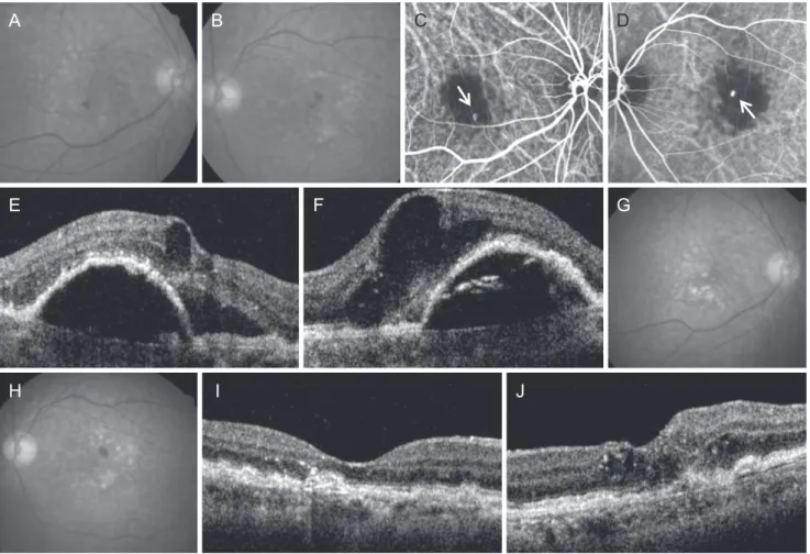
ed with ranibizumab alone. Images of a representative case
of bilateral RAP treated with an anti-VEGF agent are shown
in Fig. 1A-1J. The mean BCVA values of the 38 eyes at
baseline and 3 and 12 months posttreatment were 0.63 ±
0.26, 0.49 ± 0.29, and 0.71 ± 0.34, respectively. The BCVA at
3 months posttreatment was significantly better than the
BCVA at baseline (p = 0.001). However, there was a signifi-
cant decrease in mean BCVA between 3 and 12 months post-
treatment (p < 0.001). Therefore, the BCVA at 12 months
posttreatment was not significantly different from that at
baseline (p = 0.192).
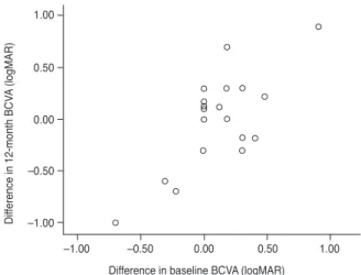
A significant association was found in mean 12-month change in BCVA between the eyes of a pair (p < 0.001, r = 0.739) (Fig. 2). Thus, the eye that exhibited better visual acuity than the paired eye at baseline was likely to continue to exhibit better visual acuity than the other eye at 12 months posttreatment. The CFT measurements were analyzed in 34 eyes of 17 patients (87.2%) with available OCT results at baseline and at 3 and 12 months posttreatment. The CFT values at baseline and 3 and 12 months posttreatment were 372.5 ± 111.4, 225.4 ± 67.6, and 266.7 ± 136.4 µm, respectively.
The mean CFT values at 3 and 12 months posttreatment were significantly lower than those at baseline (p < 0.001, both). There was no significant difference between the mean CFT values at 3 and 12 months posttreatment (p = 0.150).
Subgroup analysis based on baseline BCVA
Of the 19 pairs of eyes included in the study, 13 exhibited different baseline BCVA values in paired eyes (Fig. 3A). In the better visual acuity group (n = 13), the BCVA values at baseline and 3 and 12 months posttreatment were 0.48 ± 0.19, 0.35 ± 0.19, and 0.58 ± 0.29, respectively, and the BCVA at baseline was not significantly different from that at 12 months posttreatment (p = 0.174). These eyes were treated with an average of 4.5 ± 0.8 anti-VEGF injections during the 12-month period. In the worse visual acuity group (n = 13), the BCVA values at baseline and 3 and 12 months posttreatment were 0.83 ± 0.20, 0.64 ± 0.30, and 0.90 ± 0.31, respectively, and the BCVA at baseline was not significantly different from that at 12 months posttreatment (p = 0.460). There was no significant difference in 12-month change in BCVA (p = 0.786) or number of anti-VEGF in- jections (p = 0.111) between the eyes with better and worse visual acuity.
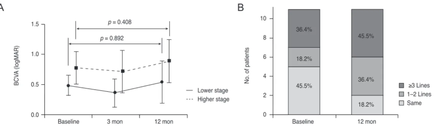
Of the six patients who exhibited equivalent baseline BCVA in both eyes, five exhibited the same stage of RAP in both eyes. Equivalent BCVA values in paired eyes were noted in two patients (33.3%) at 12 months posttreatment.
The remaining four patients exhibited 1 to 2 lines or ≥3 lines of difference in BCVA (two patients [33.3%] each) at 12 months posttreatment (Fig. 3B).
Subgroup analysis based on RAP stage at diagnosis Of the total 19 patients, eight exhibited different stages of RAP in paired eyes at baseline (Fig. 4A). In the lower stage group (n = 8), the BCVA values at baseline and 3 and 12 months posttreatment were 0.48 ± 0.17, 0.36 ± 0.23, and 0.54 ± 0.35, respectively, and the BCVA at baseline was not significantly different from that at 12 months posttreat- ment (p = 0.892). In the higher stage group (n = 8), the BCVA values at baseline and 3 and 12 months posttreat- ment were 0.78 ± 0.27, 0.72 ± 0.35, and 0.89 ± 0.36, respec- tively, and the BCVA at baseline was not significantly dif- ferent from that at 12 months posttreatment (p = 0.408).
There were no significant differences in the 12-month change in BCVA (p = 0.505) and the number of anti-VEGF injections (p = 0.279) between the eyes of the lower and the higher stage groups.
Of the 11 patients that exhibited the same RAP stages in paired eyes at baseline, five (45.5%) exhibited equivalent Table 1. Baseline characteristics of the patients with bilateral
RAP included in the study
Characteristics Value
Age (yr) 75.8 ± 7.7
Sex
Male 4 (21.1)
Female 15 (78.9)
Diabetes mellitus 4 (21.1)
Hypertension 11 (57.9)
Fundus findings (eye)
Soft drusen 34 (89.5)
Reticular pseudodrusen 32 (84.2)
Dot pseudodrusen only
*8 (25.0)
Ribbon pseudodrusen with or without dot
pseudodrusen
*24 (75.0)
RAP stage (eye)
1 17 (44.7)
2 21 (55.3)
Difference in RAP stage between both eyes of a pair
Same stage 11 (61.1)
Different stage 8 (42.1)
Difference in BCVA between both eyes of a pair
Same BCVA 6 (31.6)
Difference <3 lines 5 (26.3)
Difference ≥3 lines 8 (42.1)
Values are presented as the mean ± standard deviation or number (%).
RAP = retinal angiomatous proliferation; BCVA = best-corrected visual acuity.
*