대한외과학회지:제 73 권 제 1 호
□ 원 저 □
Vol. 73, No. 1, July, 2007
27 책임저자:이상호, 부산시 서구 암남동 34번지
602-702, 고신대학교병원 외과 Tel: 051-990-6462, Fax: 051-246-6093 E-mail: [email protected]
접수일:2006년 10월 16일, 게재승인일:2007년 5월 4일
위암 환자의 Lewis 혈액형과 혈청 내 CEA 및 임상적 요소와의 관계
고신대학교 의과대학 외과학교실 류 동 원ㆍ이 상 호
Correlation of the Lewis Blood Group and the CEA Serum Level in Stomach Cancer Patients
Dong Won Ryu, M.D. and Sang Ho Lee, M.D.
Purpose: The purpose of our study was to compare the se-
rum levels of CEA, CA19-9 and H.pylori antibody in patients with the Lewis (a+) blood group with that of the patients with the Lewis (a) blood group. We also compared the outcome of the stomach cancer patients with the Lewis (b+) blood group with that of the stomach cancer patients with the Lewis (b) blood group.
Methods: All of the 94 patients who underwent gastrectomy
for stomach cancer at our hospital were retrospectively re- viewed. The outcomes of the CEA, CA19-9 and H.pylori. an- tibody serum levels, the TNM stage, the ABO blood group and the Rh blood group were compared between the patients with the Lewis blood groups (a+) and (a) and the patients with the Lewis blood groups (b+) and (b), respectively.
Results: The mean serum level of CEA between the patients
with the Lewis blood group (a+) and (a) showed a statistical correlation (mean value: 5.51 ng/ml vs 3.25 ng/ml, re- spectively, P=0.016). But the mean serum level of CA19-9 and H.pylori. antibodies between the patients with the Lewis blood group (a+) and (a) did not show a significant dif- ference. The mean serum levels of CEA, CA19-9 and
H.pylori antibodies between the patients with the Lewis bloodgroup (b+) and (b) did not show a significant difference.
Conclusion: It was revealed that an elevated CEA level was
related with the Lewis blood group (a), but this was not re- lated with the Lewis blood group (b) in stomach cancer patients.
(J Korean Surg Soc 2007;73:27-30)Key Words:
Lewis blood group, Gastric cancer, CEA 중심 단어: 루이스 혈액형, 위암, CEA
Department of Surgery, Kosin University Colleage of Medi- cine, Busan, Korea
서 론
인체 조직에서 루이스 혈액형군(Lewis blood group)과 관 련된 항원, 암배아 항원(carcinoembryogenic antigen, CEA) 등 이 가지는 역할은 세포의 이동, 세포의 분화, 정상 점막조직 의 보호, 호중구의 이동, 세균의 결합 등이 있으며 암과 관 련해서는 암의 분화, 암의 전이와 관련된 것으로 알려져 있 다.(1,2) 특히 점막조직의 보호, 호중구 세포의 결합과 암의 전이와 관련된 이 루이스 혈액형군 관련 항원과 암배아 항 원이 암 세포막에서 동시에 확인하였다는 보고가 있다.(3) 그러므로 항원들이 호중구를 염증조직의 내피에 결합시키 는 작용기전은 암의 전이와 병의 예후에 관련되어 있을 것 으로 생각된다. 그리고 H.pylori에 감염된 환자의 소수에서 만 위와 관련된 병변들이 나타난다는 것은 병변의 발현에 다른 인자가 관여한다는 것을 알 수가 있다. 최근의 연구에 의하면 궤양이 있는 조직과 궤양주위 정상조직과의 ABH and Lewis 혈액형의 표현형 차이를 보면 a, h, b의 표현형이 없어지고 Lewis a의 활동성이 증가되어져 있는 사실도 밝혀 졌다.
저자들은 위암 환자에서 Lewis 혈액형과 혈청 CEA 수치 및 이들과 관련된 CA19-9, H.pylori의 혈청수치의 CEA의 상 호 관련성을 관찰하여서 환자의 예후와 암의 전이와 관련 이 있는지에 대해서 알아보고자 한다.
방 법
1999년 1월 1일부터 2000년 12월 31일까지 고신대학교
외과학교실에서 위암으로 절제술을 받은 환자 중에서
Lewis 혈액형과 CEA와 관련된 항원에 대한 조사가 되어 있
는 94명의 환자를 대상으로 하였다. H.pylori에 대한 혈청
IgG 검사는 Cobas Core EIA kit (Roche, Basel, Switzerland)를
이용하였고 혈청검사 양성은 항체가가 7 U/ml 이상으로, 음
성은 항체가가 7 U/ml 미만으로 하였다. Lewis 혈액형은
DiaMed- ID Micro Typing System으로 확인하였다. 환자의
의무기록지를 통하여 Lewis 혈액형 각각의 CEA, CA19-9,
H.pylori 항체, TNM 병기, ABO 혈액형, Rh 혈액형을 조사하
28 대한외과학회지:제 73 권 제 1 호 2007
Table 1. Clinicopathologic characteristics
No. of patients (n=94)
Sex Male 64
Female 30
ABO type A+ 33
B+ 30
O+ 20
AB+ 11
Lewis blood group a+b+ 3
a+b 10
ab+ 60
ab 21
Table 2. The relationship between Lewis (a+), Lewis (a) and serum level of CEA, CA19-9, H.pylori
Lewis (a+) Lewis (a)
P-value blood group blood group
CEA (ng/ml) 5.51 3.25 0.016
CA19-9 (U/ml) 36.33 35.02 0.949
H.pylori (U/ml) 74.54 58.69 0.435
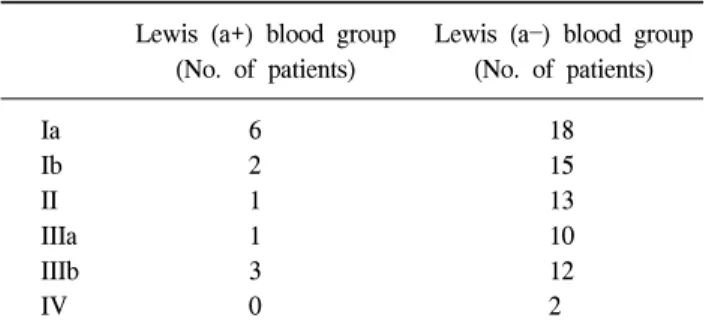
Table 3. The distribution of TNM stages according to Lewis (a) antigen status
Lewis (a+) blood group Lewis (a) blood group (No. of patients) (No. of patients)
Ia 6 18
Ib 2 15
II 1 13
IIIa 1 10
IIIb 3 12
IV 0 2
Table 4. The relationship between Lewis (b+), Lewis (b) and serum level of CEA, CA19-9, H.pylori
Lewis (b+) Lewis (b)
P-value blood group blood group
CEA (ng/ml) 3.42 3.94 0.477
CA19-9 (U/ml) 67.13 48.13 0.241
H.pylori (U/ml) 33.17 39.43 0.685
여 상관관계를 후향적으로 조사하였다. 통계분석은 SPSS window 10.0을 이용하였으며 임상병리학적 인자들에 대한 비교분석은 chi square test를 이용하였다. P-value가 0.05 미 만인 경우에 통계학적으로 유의하다고 판정하였다.
결 과 1) 대상자의 특성
남자가 64예 여자가 30예로 남녀비는 약 2:1이었고 평균 연령은 55세였다. 환자의 ABO 혈액형은 A형이 33예, B형이 30예, O형이 20예, AB형이 11예였으며 Lewis 혈액형은 (a+b+) 3예, (a+b) 10예, (ab+) 60예, (ab) 21예였다(Table 1).
2) Lewis (a+) 혈액형군과 Lewis (a) 혈액형군 사이의
CEA, CA19-9, H.pylori 수치 비교Lewis (a+) 혈액형군은 Lewis (a) 혈액형군에 비해서 혈청 CEA의 값이 높게 측정되었으며 이는 통계학적으로 유의하 게 높았다(평균값 5.51 ng/ml vs 3.25 ng/ml, P=0.016). 반면 H.pylori 항체 혈청 수치(평균값 74.52 U/ml vs 58.69 U/ml, P=0.435) 및 CA19-9 (평균값 36.33 U/ml vs 35.02 U/ml, P=0.949) 혈청 수치에서는 Lewis (a+) 혈액형군과 Lewis (a) 혈액형군 간에 유의한 차이가 없었다. Lewis (a+) 혈액형군 과 Lewis (a) 혈액형군 사이의 남녀 성별분포와 ABO 혈액 형, Rh 혈액형의 분포에서도 통계학적 의미 있는 차이는 없
었다(Table 2).
3) Lewis (a+) 혈액형군과 Lewis (a) 혈액형군과의
TNM 병기 분류Lewis (a+) 혈액형군에서는 병기 Ia가 6예로 가장 많이 분 포되었고 Lewis (a) 혈액형군에서도 병기 Ia가 18예로 가장 많이 분포되어있었다. 양 군 사이의 TNM 병기 분포의 차이 는 통계학적으로 의미 있는 차이는 없었다(P=0.624)(Table 3).
4) Lewis (b+) 혈액형군과 Lewis (b) 혈액형군 간의
CEA, CA19-9, H.pylori 항체의 혈청 수치 비교Lewis (b+) 혈액형 환자는 Lewis (b) 혈액형 환자에 비해 서 혈청 CEA의 수치가 낮게 측정되었으나 통계학적으로 의미 있는 차이는 없었다(평균값 3.42 ng/ml vs 3.94 ng/ml, P=0.477). 두 혈액형군 사이의 혈청CA19-9 수치(평균값 33.17 U/ml vs 39.43 U/ml, P=0.685), H.pylori에 감염된 환자 의 소수에서만 위와 관련된 병변들이 나타난다는 것은 병 변의 발현에 다른 인자가 관여한다는 것을 알 수가 있다.
최근의 연구에 의하면 궤양이 있는 조직과 궤양주위 정상 조직과의 ABH and Lewis 혈액형의 표현형 차이를 보면 a, h, b의 표현형이 없어지고 Lewis a의 활동성이 증가되어져 있는 사실도 밝혀졌다. 항체수치(평균값 52 U/ml vs 25 U/ml, P=0.241)도 통계학적으로 의미 있는 차이는 없었다.
또한 Lewis (b+) 혈액형군과 Lewis (b) 혈액형군 사이의 남
녀 성별분포와 ABO, Rh 혈액형의 분포에서도 통계학적으
로 의미 있는 차이가 없었다(Table 4).
류동원ㆍ이상호:위암 환자의 Lewis 혈액형과 혈청 내 CEA 및 임상적 요소와의 관계
29
Table 6. The distribution of CEA serum level according to Lewis (a+b+), Lewis (a+b), Lewis (a-b+), Lewis (a
b) blood groups
Mean level Minimum level Maximum level
(ng/ml)
Lewis (a+b+) 3.45 10.28 7.25
Lewis (a+b) 1.22 18.93 5.04
Lewis (ab+) 0.67 13.4 3.23
Lewis (ab) 0.60 10.21 3.30
Table 7. The distribution of CA19-9 serum level according to Lewis (a+b+), Lewis (a+b), Lewis (ab+), Lewis (a
b) blood groups
Mean level Minimum level Maximum level
(U/ml)
Lewis (a+b+) 16.5 30.8 21.36
Lewis (a+b) 8.6 145.6 40
Lewis (ab+) 2.8 240 33
Lewis (ab) 1 411.9 38.89
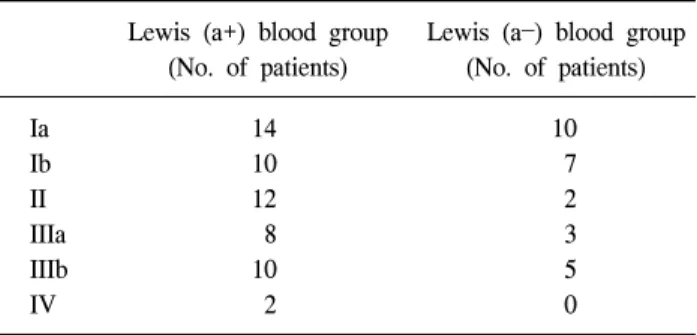
Table 5. The distribution of TNM stages according to Lewis (b) antigen status
Lewis (a+) blood group Lewis (a) blood group (No. of patients) (No. of patients)
Ia 14 10
Ib 10 7
II 12 2
IIIa 8 3
IIIb 10 5
IV 2 0
5) Lewis (b+) 혈액형군과 Lewis (b) 혈액형군 사이의
TNM 병기의 비교Lewis (b+) 혈액형군에서는 병기 Ia가 14예로 가장 많이 분포되었고 Lewis (b) 혈액형군에서도 병기 Ia가 10예로 가 장 많이 분포되어 있었다. 두 군에서의 TNM 병기 분포의 차이는 통계학적으로 의미는 없었다(P=0.451)(Table 5).
6) Lewis (a+b+), Lewis (a+b), Lewis (ab+), Lewis (a
b) 혈액형군 사이의 CEA 혈청 수치 비교
Lewis (a+b+) 혈액형군의 혈청 CEA 평균수치는 7.25 ng/ml였고 Lewis (a+b) 혈액형군의 혈청 CEA 평균수치는 5.04 ng/ml로 두 군 간의 의미 있는 차이는 없었다. 그리고 Lewis (ab+) 혈액형군의 혈청 CEA 평균수치는 3.23 ng/ml 이고 Lewis (ab) 혈액형군의 혈청 CEA 평균수치는 3.30 ng/ml으로 양 군 간의 통계학적 의미 있는 차이는 없었다 (Table 6).
7) Lewis (a+b+), Lewis (a+b), Lewis (ab+), Lewis (a
b) 혈액형군 사이의 CA19-9 혈청 수치 비교
Lewis (a+b+) 혈액형군의 혈청 CA19-9 평균수치는 21.36 U/ml였고, Lewis (a+b-) 혈액형군의 혈청 CA19-9 평균수치 는 40 U/ml로 두 군 간의 의미 있는 차이는 없었다. 그리고 Lewis (ab+) 혈액형군의 혈청 CA19-9 평균수치는 33 U/ml 였고, Lewis (ab) 혈액형군의 혈청 CA19-9 평균수치는 38.89 U/ml로 양 군의 통계학적 의미 있는 차이는 없었다 (Table 7).
고 찰
Lewis 혈액항원은 세포표면을 구성하는 탄수화물복합체 로, 소위 혈액항원으로서 적혈구의 세포표면을 덮고 있다.
즉 적혈구의 표면항원 중의 하나이다.(1) 그리고 이 항원은 인체의 비조혈기관에서도 나타난다. 이 항원이 요즈음 들 어서 주목을 받고 있는 이유 중의 한 가지는 이 항원은 발
생과정을 거치면서 특별한 변화가 관찰되고 그러한 변화 중의 한 가지가 악성 변화를 할 수 있다는 사실이다.(2,3) Lewis 혈액형과 CEA와 관련이 있는 항원들은 인체조직 속 에서 흡착과 관련된 기능을 가지고 있다는 것이다. 또한 이 항원은 정상적인 점막의 분화와 방어에 관여하고 호중구의 이동이나 호중구의 박테리아와의 결합에 관여하고 종양세 포의 분화와 전이에 관여를 한다는 것이다. 즉 이러한 점막 방어와 호중구의 결합이나 종양의 전이에 관련된Lewis 혈 액항원이나 CEA에 관련된 항원들은 세포막의 외막에 같이 표현된다고 알려져 있다. 본 연구에서도 Lewis 혈액형과 CEA의 연관성을 알아보고자 Lewis (a+) 혈액형군과 Lewis (a) 혈액형군 두 군 간의 CEA의 혈청농도를 비교한 조사 에서 통계학적으로 유의하게 Lewis (a+) 혈액형군이 높은 수치를 보였다. 그리고 통계적 의미는 없었지만 Lewis (b+) 혈액형군도 Lewis (b) 혈액형군에 비해 CEA의 혈청수치가 높게 측정되었다.
Lewis system은 원래 체액이나 분비액 즉 타액, 유액, 요, 정액, 양수 및 태변 등에 존재하는 수용성의 항원물질이 이 차적으로 적혈구 표면에 흡착되어 특징적인 항원성을 나타 내는 혈액군이기 때문에 단순히 혈액형(blood group)이라기 보다는 Lewis system이라고도 한다.(4,5)
Lewis 혈액항원은 구조적으로는 주요 혈액형 조직적합항
원인 ABO와 관련이 있다. 적혈구에서 보이는 Lewis 혈액항
원의 표현은 Lewis gene, H gene, secretor genes과의 상호작
용에 의해서 표현된다. 그러나 본 연구에서는 Lewis 혈액형
30 대한외과학회지:제 73 권 제 1 호 2007
과 ABO 혈액형 사이의 환자 분포에서는 통계학적으로 의 미 있는 연관성은 없는 것으로 나타났다.(6,7)
Lewis 항원은 혈중 내의 당지질의 구성성분에 의해서 수 동적으로 결정되고 아직은 어디에서 합성되는지는 알려지 지 않았다. Lewis 혈액항원은 침이나 위액의 분비물에서도 나타난다.(8) Lewis a 항원의 역할은 Lewis x 항원과 그 역할 이 유사한데 이 Lewis x 항원의 역할은 배아의 발생 시에 세포인식을 조절하는 것으로 알려져 있다.(9) 특히 소뇌의 발달과정에서 Lewis x 항원은 과립세포의 이동에 관여하는 것으로 되어있다.(10,11) 그래서 쥐를 대상으로 한 실험에서 Lewis x 항원에 대한 항체를 주입하게 되면 쥐의 소뇌 발달 을 변형시키는 것으로 알려져 있다.(13) 이러한 사실을 통 해서 유추해보면 Lewis 항원은 태아의 발생과정에서 기관 의 형성을 매개하는 세포 간의 유착이나 과립구 세포의 유 착과 이동, 종양세포의 침입과 관련된 것으로 추측할 수 있 다. 좀 더 최근의 보고에 의하면 Lewis a와 Lewis x 항원 사이에는 구조적이고 기능적인 유사성이 있는 것으로 확인 된다.(3,13) 그리고 과립구 세포와 혈소판이 혈관의 내피세 포에 유착되어서 내피세포를 활성화시키고 이로 인해서 종 양세포의 유착과 혈관 외 침투에도 이들 Lewis 항원들이 관 여하는 것으로 되어있다.(3,13) 본 연구에서는 병기별 Lewis 혈액형의 분포의 차이에서 통계학적 유의성은 없었다.
위 점막에 있는 H.pylori가 만성 활동성 위염, 만성 위축성 위염과 관련되어 있다는 것은 알려져 있는 사실이다.(4,13) 그런데 H.pylori에 감염된 환자의 소수에서만 위와 관련된 병변들이 나타난다는 것은 병변의 발현에 다른 인자가 관 여한다는 것을 알 수가 있다. 최근의 연구에 의하면 궤양이 있는 조직과 궤양주위 정상조직과의 ABH and Lewis 혈액 형의 표현형 차이를 보면 a, h, b의 표현형이 없어지고 Lewis a의 활동성이 증가되어져 있는 사실도 밝혀졌다.(12) 위암 환자를 대상으로 한 본 연구에서도 이에 착안하여 H.pylori에 대한 항체 혈청수치를 조사해 보았는데 Lewis (a) 혈액형군에 비해서 Lewis (a+) 혈액형군에서 높게 측정이 되었으나 통계학적 의미는 없었다.
결 론
위암 환자에서 Lewis (a+) 혈액형군의 혈청 CEA 수치가 Lewis (a) 혈액형군에서 보다 높게 나타났다. 이러한 결과 는 Lewis 혈액항원이 위 점막 상피세포의 암성 변화에 관여 할 수 있다는 것을 보여주는 한 모습이다. 그리고 통계학적 으로 의미는 없었으나 Lewis (a+) 혈액형군 환자에서 혈청 H.pylori 항체수치가 높게 측정이 된 것은 Lewis 혈액항원이 만성 위축성 위염과 관련될 수 있다는 것을 보여줄 수 있다.
향후 더 많은 환자를 대상으로 H.pylori에 감염된 환자에서 궤양이나 종양성 변화가 발생한 환자군과 그렇지 않은 환 자군의 Lewis 혈액형 및 CEA를 혈액 및 조직에서 조사하는 것이 환자의 치료에 필요할 것으로 생각한다.
REFERENCES
1) Sanders DS, Kerr MA. Lewis blood group and CEA related antigens; coexpressed cell-cell adhesion molecules with roles in the biological progression and dissemination of tumours.
Mol Patol 1999;52:174-8.
2) Marshall BJ. Helicobacter pylori. Am J Gastroenterol 1994;
89(8 Suppl):S116-28.
3) Atkinson BF, Ernst CS, Herlyn M, Steplewski Z, Sears HF, Koprowski H. Gastrointestinal cancer-associated antigen in im- munoperoxidase assay. Cancer Res 1982;42:4820-3.
4) Coon JS, Weinstein RS. Blood group-related antigens as mark- ers of malignant potential and heterogeneity in human carcinomas. Hum Pathol 1986;17:1089-106.
5) Gerhard M, Lehn N, Neumayer N, Boren T, Rad R, Schepp W, et al. Clinical relevance of the Helicobacter pylori. gene for blood-group antigen-binding adhesion. Proc Natl Acad Sci USA 1999;96:12778-83.
6) McCarthy NC, Simpson JR, Coghill G, Kerr MA. Expression in normal adult, fetal and neoplastic tissues of a carbohyrdate differentiation antigen recognised by antigrnanulocyte mouse monoclonal antibodies. J Clin Pathol 1985;38:521-9.
7) Arends JW, Verstynen C, Bosman FT, Hilgers J, Steplewski Z. Distribution of monoclonal antibody-defined monosialo- ganglioside in normal and cancerous human tissues: an im- munoperoxidase study. Hybridoma 1983;2:219-29.
8) Sun XF, Zhang H. Expression of tumor-related antigens Lewis (a), Lewis (b), X,Y, Span-1 and CEA in relation to differ- entiation and prognosis in rectal adenocarcinomas. APMIS 1996;104:784-8.
9) Burtin P, von Kleist S, Sabine MC, King M. Immunohisto- logical localization of carcinoembryonic antigen and non- specific cross-reacting antigen in gastrointestinal normal and tumoral tissues. Cancer Res 1973;33:3299-305.
10) Dunn BE, Cohen H, Blaser MJ. Helicobacter pylori. Clin Microbiol Rev 1997;10:720-41.
11) Itzkowitz SH, Kim YS. New carbohydrate tumor markers.
Gastroenterology 1986;90:491-4.
12) Henry S, Oriol R, Samuelsson B. Lewis histo-blood group sys- tem and associated secretory phenotypes. Vox Sang 1995;
69:166-82.
13) Marcus DM, Cass LE. Glycosphingolipids with Lewis blood group activity uptake by human erythrocytes. Science 1969;
164:553-4.