ARTICLE
Int J Thyroidol 2017 May 10(1): 5-13 https://doi.org/10.11106/ijt.2017.10.1.5Received October 4, 2016 / Revised November 25, 2016 / Accepted February 24, 2017
Correspondence: Byeong-Cheol Ahn, MD, PhD, Department of Nuclear Medicine, Kyungpook National University Medical Center and School of Medicine, 130 Dongdeok-ro, Jung-gu, Daegu 41944, Korea
Tel: 82-53-420-5583, Fax: 82-53-422-0864, E-mail: [email protected]
Copyright ⓒ 2017, the Korean Thyroid Association. All rights reserved.
This is an open-access article distributed under the terms of the Creative Commons Attribution Non-Commercial License (http://creative- commons.org/licenses/by-nc/4.0/), which permits unrestricted non-commercial use, distribution, and reproduction in any medium, provided the original work is properly cited.
Consideration of Serum Thyrotropin When Interpreting Serum Thyroglobulin Level in Patients with Differentiated Thyroid Cancer
Seung Hyun Son, Chang-Hee Lee, Ji-hoon Jung, Choon-Young Kim, Ju Hye Jeong, Shin Young Jeong, Sang-Woo Lee, Jaetae Lee
and Byeong-Cheol Ahn
Department of Nuclear Medicine, Kyungpook National University Medical Center and School of Medicine, Daegu, Korea
Background and Objectives: The level of thyroid-stimulating hormone (TSH)-stimulated thyroglobulin (Tg) after thyroid hormone withdrawal (THW) is the most sensitive marker for detecting recurrence of differentiated thyroid cancer (DTC). In DTC, Tg production is regulated by TSH; however, TSH values after THW are never identical, even in the same patient. The objective of this study was to evaluate the influence of TSH on Tg levels after THW. Materials and Methods: TSH and Tg concentrations were measured twice at 2 and 3 weeks after THW in 309 patients with DTC. TSH and Tg levels at these time points were compared. The percent change in TSH (ΔTSH) and change in Tg level (%ΔTg) from 2 to 3 weeks after THW were calculated, and Pearson's correlation coefficients were calculated to determine whether ΔTSH could affect %ΔTg. Tg cutoff value for diagnostic imaging was 2 ng/mL. Results: The TSH and Tg values at 3 weeks were significantly higher than those at 2 weeks after THW. Tg values increased significantly to >2 ng/mL after 1 week in 38.5% of the patients with Tg values of 0.2-2 ng/mL at 2 weeks after THW. In patients with Tg values ≥2 ng/mL at 2 weeks after THW, Tg values increased significantly after an additional week of THW. ΔTSH correlated significantly with %ΔTg. Conclusion:
TSH values differed according to time after THW, and Tg values differed significantly according to TSH values.
Therefore, TSH values should be considered carefully when interpreting the meaning of Tg levels in patients with DTC.
Key Words: Thyroid-stimulating hormone, Thyroid function test, Thyroglobulin, Thyroid cancer
Introduction
Differentiated thyroid carcinoma (DTC), the most common type of endocrine malignancy, has an ex- cellent prognosis and 10-year survival rate higher than 90%.1,2) This outcome has been ensured by in- creased diagnostic scrutiny. However, the DTC re- currence rate is 10-30%, and the potential for re- appearance after several years3,4) indicates the need
for prolonged follow-up.
According to the American Thyroid Association guideline,5) a disease-free status comprises all of the following: no clinical evidence of tumor, no imaging evidence of tumor (i.e., no uptake outside the thyroid bed on the initial post-treatment whole-body scan or, if uptake outside the thyroid bed is present, no imag- ing evidence of tumor on a recent diagnostic scan and neck ultrasound), and undetectable serum thyro- globulin (Tg) values during thyroid-stimulating hor-
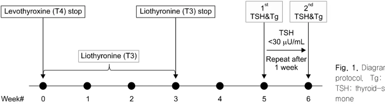
Fig. 1. Diagram of the study protocol. Tg: thyroglobulin, TSH: thyroid-stimulating hor- mone
mone (TSH) suppression and stimulation in the ab- sence of interfering antibodies.
The TSH-stimulated Tg level after successful thyroid ablation is known as a sensitive and reliable marker for surveilling tumor persistence and recurrence,6,7) particularly in patients with low or undetectable Tg values in the absence of TSH stimulation. Frequently, stimulated Tg cutoff values of >1-2.5 ng/mL are used to detect or predict persistent disease.8-12) Al- though some recent studies have reported that Tg stimulation with recombinant human TSH may be more beneficial in patients who have undetectable (unsti- mulated) Tg values, thyroid hormone withdrawal (THW) is more frequently used to measure stimulated Tg.
The adequate TSH value needed to achieve suffi- cient Tg stimulation after THW has not been de- termined, and the commonly used TSH cutoff has been derived from the value considered necessary for radioactive iodine (RAI) imaging. Edmonds et al.13) re- ported a study of seven patients in whom a TSH value
>30 μU/mL was necessary for adequate uptake on an RAI whole-body scan. Other groups have reported that a TSH value of >25-30 μU/mL at 2-3 weeks after THW is sufficient.13-17) Valle et al.18) reported that TSH values of >80-100 μU/mL would serve as a more appropriate cutoff, compared to >30 μU/mL.
Although many institutions apply a TSH cutoff of >30 μU/mL, it remains unclear when TSH or Tg values might plateau and whether Tg values would continue to rise once the TSH cutoff has been achieved. In ad- dition, the same patient might exhibit different TSH val- ues after each THW, even when an identical THW protocol has been applied consistently. We thought that if the TSH values differed between two serial fol- low-up tests, a comparison of Tg values without con-
sidering TSH might not be clinically relevant, given the possibility of a change in Tg levels due to different levels of TSH stimulation.
The aim of this study was to evaluate the influence of TSH levels on Tg and to determine whether we could predict the exact trend in Tg change between two serial follow-up tests in each patient.
Materials and Methods
Patients
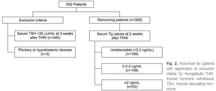
Fig. 1 illustrates the THW protocol used in this study, including a change of levothyroxine to liothyronine for 3 weeks followed by a period of no thyroid hormone treatment for the next 2 weeks. From October 2008 through September 2013, we consecutively enrolled 4967 patients with DTC who underwent THW, and selected 652 (13.1%) with limited TSH elevation (<30 μU/mL) at 2 weeks after THW and repeated TSH measurement at 3 weeks after THW. Only patients with a TSH value >30 μU/mL at 3 weeks after THW were then selected to effectively demonstrate the in- fluence of elevated TSH levels on Tg levels. Subjects with pituitary or hypothalamic disease were also ex- cluded (Fig. 2). Finally, 309 patients were evaluated in this retrospective study, which was approved by the Institutional Review Board at our institution (Kyungpook National University Medical Center and School of Medicine, Daegu, Korea). The requirement for written informed consent by participants for the use of their clinical records in this study was waived because pa- tient information was de-identified prior to analysis.
Fig. 2. Flowchart for patients with application of exclusion criteria. Tg: thyroglobulin, THW:
thyroid hormone withdrawal, TSH: thyroid-stimulating hor- mone
Measurements of Tg and TSH
Radioimmunoassay commercial kits were used to measure serum Tg (Thyroglobulin IRMA; CIS Bio International, Gif sur Yvette, France). Seven calibrators, two controls, and patients’ serum samples were mixed with buffer solution and incubated for 3 hours with ag- itation (400 rpm) at room temperature (18-25oC). The tubes were washed twice with washing solution, after which iodine-125–labeled anti-thyroglobulin was dis- pensed into each tube. The tubes were then in- cubated overnight (16-20 hours) at room temperature (18-25oC) without agitation. The tubes were again washed twice, and the remaining radioactivity bound to the tube was measured with a gamma scintillation counter for 1 minute. This Tg assay had a functional sensitivity of 0.7 ng/mL and analytical sensitivity of 0.2 ng/mL. The limit of detection for Tg in this study was
≥0.2 ng/mL.
Commercial immunoradiometric assay kits were used for TSH measurement (TSH IRMA, BㆍRㆍAㆍHㆍ MㆍS GmbH, Hennigsdorf, Germany). Seven calibra- tors, two controls, and patients’ serum samples were incubated with an excess of two anti-thyrotropin anti- bodies (mouse monoclonal) that recognized different binding sites on the antigen (TSH). One antibody was labeled with iodine-125, and the other was immobi- lized on the inner surface of the tube (coated tube system). The tubes were incubated in an orbital shak-
er (170-250 rpm) for 1 hour at room temperature (18-25oC), after which the remaining excess io- dine-125–labeled anti-thyrotropin antibody was di- luted and completely removed. The tubes were wash- ed twice, and the radioactivity in each tube was measured for 1 minute in a gamma counter. The TSH assay had a functional sensitivity of 0.07 μU/mL.
Statistical Analysis
For analysis, patients were divided into three groups according to their Tg values at 2 weeks after THW (Fig. 2): <0.2 ng/mL (108 patients, 35%), ≥0.2 to
<2 ng/mL (148 patients, 48%), and ≥2 ng/mL (53 patients, 17%). The paired-samples t-test was used to compare differences in TSH and Tg levels from 2 to 3 weeks after THW in each patient. In patients with Tg values ≥2 ng/mL, the absolute change in TSH values (ΔTSH=TSH at 3 weeks after THW−TSH at 2 weeks after THW) and percent change in Tg values [%ΔTg=(Tg at 3 weeks THW−Tg at 2 weeks THW)/
Tg at 2 weeks THW×100] from 2 to 3 weeks after THW were calculated, and Pearson’s correlation co- efficients were calculated to determine the relationship between ΔTSH and %ΔTg. A Tg cutoff value of 2 ng/mL was used to represent clinical significance for diagnostic imaging or further evaluations. MedCalc Statistical Software version 15.6.1 (MedCalc Software bvba, Ostend, Belgium) was used for statistical analysis.
Continuous variables are presented as means±
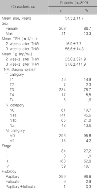
Table 1. Characteristics of the 309 study patients
Characteristics Patients (n=309)
n %
Mean age, years 54.5±11.7
Sex Female Male
268 41
86.7 13.3 Mean TSH (μU/mL)
2 weeks after THW 3 weeks after THW
16.9±7.7 56.6±14.3 Mean Tg (ng/mL)
2 weeks after THW 3 weeks after THW
25.8±321.9 37.8±411.9 TNM staging system
T category T1 T2 T3 T4 Tx
46 7 234 17 5
14.9 2.3 75.7 5.5 1.6 N category
N0 N1a N1b Nx
61 141 65 42
19.7 45.6 21.0 13.6 M category
M0 M1
296 13
95.8 4.2 Stage
I II III IV
84 3 163 59
27.2 1.0 52.8 19.1 Histology
Papillary Follicular
Papillary+follicular
299 9 1
96.8 2.9 0.3 Tg: thyroglobulin, THW: thyroid hormone withdrawal, TSH:
thyroid-stimulating hormone
Fig. 3. Changes in serum thyroglobulin (Tg) values from 2 to 3 weeks after thyroid hormone withdrawal (THW) in all 309 patients.
standard deviations (SDs). A p value <0.05 was con- sidered to indicate a statistically significant difference.
Results
Patient Characteristics
The characteristics of all 309 patients are presented in Table 1. The study cohort included 41 men and 268 women with a mean age of 54.5±11.7 years.
All patients underwent total or near-total thyroi- dectomy, which was completed by central neck dis-
section or modified radical neck dissection in 86% of cases, resulting in the apparent complete resection of the neoplastic tissue. In addition, 93% of patients un- derwent THW for ablation of the thyroid remnant or treatment of persistent thyroid cancer, and 7% under- went THW for thyroid cancer surveillance.
Changes in TSH and Tg Values between 2 and 3 Weeks after THW
Fig. 3 and Supplementary Fig. 1 show the changes in serum Tg values in all 309 patients. In 227 patients (73.5%), the Tg values increased after an additional week of THW. The mean values of TSH and Tg at 2 weeks after THW were 16.9±7.7 μU/mL and 25.8±321.9 ng/mL, respectively, and the corre- sponding values at 3 weeks after THW were 56.6±
14.3 μU/mL and 37.8±411.7 ng/mL, respectively.
Tg values increased significantly after the additional week of THW (p=0.0233).
In a group of 108 patients with Tg values <0.2 ng/mL at 2 weeks after THW, the Tg values in ap- proximately half (49.1%) of the patients remained below 0.2 ng/mL after the additional week of THW, whereas those in the other half (50.9%) increased but remained
<2 ng/mL. Therefore, the Tg values at 3 weeks after THW had no clinical significance in patients with Tg values <0.2 ng/mL at 2 weeks after THW.
In a group of 148 patients with Tg values between 0.2 and 2 ng/mL at 2 weeks after THW, the Tg values
Table 3. Changes in Tg values in accordance with increased TSH values in 53 patients with Tg values of >2 ng/mL at 2 weeks THW
Tg at 3 weeks after THW
Variables
Undetectable n=1 (1.9%) ≥2 ng/mL n=52 (98.1%)
2 weeks after THW
3 weeks
after THW p value 2 weeks
after THW
3 weeks
after THW p value
TSH (μU/mL) 16.0 42.2 - 18.3±7.4 55.0±12.7 <0.0001
Tg (ng/mL) 2.5 1.8 - 151.1±778.9 216.7±992.1 0.0356
Tg: thyroglobulin, THW: thyroid hormone withdrawal, TSH: thyroid-stimulating hormone
Table 2. Changes in Tg values according to increased TSH values after dichotomization by Tg values (cutoff=1 ng/mL) at 2 weeks after THW in 148 patients with Tg values of 0.2-2 ng/mL at 2 weeks THW
Tg at 2 weeks after THW
Variables
<1 ng/mL n=122 (82.4%) ≥1 ng/mL n=26 (17.6%)
2 weeks after THW
3 weeks
after THW p value 2 weeks
after THW
3 weeks
after THW p value
TSH (μU/mL) 16.6±7.9 54.8±13.4 <0.0001 17.9±7.1 58.8±15.6 <0.0001
Tg (ng/mL) 0.4±0.2 1.9±2.6 <0.0001 1.3±0.3 4.8±3.9 0.0001
Tg: thyroglobulin, THW: thyroid hormone withdrawal, TSH: thyroid-stimulating hormone
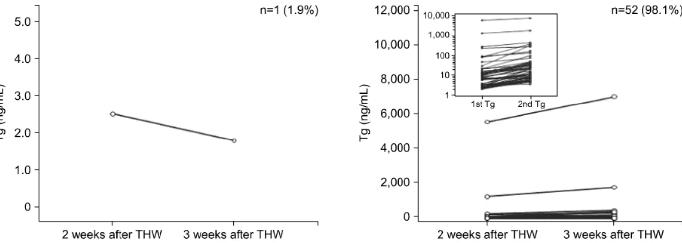
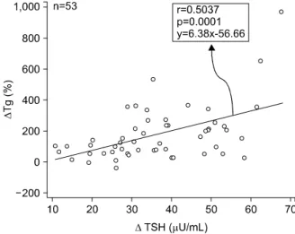
Fig. 4. Pattern of changes in Tg values in accordance with the increase in the thyroid-stimulating hormone values in 53 patients with Tg values of >2 ng/mL at 2 weeks after thyroid hormone withdrawal (THW).
in 121 (81.8%) patients increased after an additional week of THW, along with increases in TSH values (TSH: 19.1±8.0 to 55.1±13.8 μU/mL, p<0.0001;
Tg: 0.5±0.3 to 2.9±3.2 ng/mL, p=0.0001). In these 121 patients, the Tg values of 57 (47.1%) patients in- creased to >2 ng/mL after an additional week of THW, accompanied by increases in TSH values (TSH: 15.0±
7.6 to 56.8±14.5; Tg: 0.9±0.4 to 5.1±3.5, p<
0.0001). We further dichotomized the 148 patients by
their Tg values at 2 weeks after THW—122 patients with Tg values <1 ng/mL and 26 patients with Tg values ≥1 ng/mL—as a sub-analysis to determine the effect of Tg at 2 weeks after THW. Approximately 30% of 122 patients in the former group and approx- imately 77% of 26 patients in the latter eventually ach- ieved a Tg value ≥2 ng/mL after the additional week.
These results indicated that the patients with initially high Tg values achieved a relatively higher rate of in-
Fig. 5. Relationship between thyroglobulin (Tg) and thyroid- stimulating hormone (TSH) in 53 patients with a Tg value of
>2 ng/mL at 2 weeks after thyroid hormone withdrawal.
crease in Tg after the additional week of THW (Table 2).
In a final group of 53 patients with Tg values ≥2 ng/mL, the Tg values of all but one patient remained in the same Tg category (≥2 ng/mL) after an addi- tional week of THW (Fig. 4), and all but one of the 52 patients exhibited increased Tg values after the addi- tional week (Table 3).
To evaluate the correlation between the changes in TSH and Tg values, we subjected 190 patients with detectable Tg values in both the first and second tests to a linear regression analysis and Pearson’s correla- tion analysis. The %ΔTg was found to correlate pos- itively with the ΔTSH (p=0.0001), with a correlation coefficient of 0.2881. We also performed the same analysis in the 53 patients with Tg values ≥2 ng/mL at 2 weeks after THW, a value that is suggestive of tum or recurrence or persistent tum or burden. The
% ΔTg was also found to correlate positively with the ΔTSH (p=0.0001), with a correlation coefficient of 0.5037 (Fig. 5).
We performed a sub-analysis of 22 patients who underwent THW for thyroid cancer surveillance. The Tg values were undetectable in 12 (54.5%) of these 22 patients, and <2 ng/mL in the other 10 (45.5%) patients. Among the former 12 patients, three exhibited increased Tg values that remained <2 ng/mL after an additional week of THW. All but one of the latter 10 patients exhibited increased Tg values after the
additional week of THW, and five (55.6%) of the nine patients’ Tg values increased to >2 ng/mL after the additional week.
Discussion
Generally, serum Tg values are used to predict successful thyroid remnant ablation,19) assess re- sponses to RAI therapy,20) detect recurrent tumors,6,21) and predict disease recurrence22,23) and prognosis.24,25) A few studies have demonstrated that Tg values could increase in accordance with increases in TSH values.18)
The current large-scale study aimed to determine the importance of considering the serum TSH value with regard to Tg stimulation by THW. Our results demonstrated increased Tg values in a majority (73.5%) of patients after an additional week of THW.
Furthermore, 38.5% of the patients with Tg values be- tween 0.2 and 2 ng/mL at 2 weeks after THW ex- hibited increases to >2 ng/mL, suggesting a need for diagnostic imaging or other evaluations. These results are consistent with those of a previous report.18) The current study demonstrated that stimulated Tg levels in response to THW must be interpreted in the context of the corresponding TSH level to avoid drawing false conclusions about the disease status.
Almost all patients with Tg values >2 ng/mL at 2 weeks after THW demonstrated a significant increase in Tg along with an increase in TSH values after an additional week of THW. As Tg synthesis and secre- tion is an active process requiring stimulation by TSH, TSH values should be carefully considered by clini- cians when determining whether Tg levels suggest the need for further diagnostic evaluations. Furthermore, our results also revealed that Tg values did not in- crease to >2 ng/mL at 3 weeks after THW in patients with undetectable Tg values at 2 weeks after THW.
This result signifies that further clinical evaluation may not be needed when Tg values are undetectable at 2 weeks after THW without reference to TSH values.
A previous study18) tried to predict final Tg values using serial evaluations of each patient's Tg values at different time points. However, accurate prediction was
not possible because the rate of increase was quite diverse for each patient. The residual tumor volumes always differed among patients; therefore, the rate of Tg increase in accordance with the increase of TSH value would always differ in each patient. We defined ΔTSH as the absolute change in TSH values and % ΔTg as the percent change in Tg values. Accordingly, using %ΔTg, we could overcome the difference of the rate of increase in each individual patient. Finally, %Δ Tg was found to correlated significantly with ΔTSH.
This result indicates that Tg values should be in- terpreted in light of the TSH value in each individual patient at each individual testing time point.
Even among the 22 patients who underwent THW for thyroid cancer surveillance, nine of 10 patients with Tg values <2 ng/mL exhibited increased Tg values after the additional week of THW, and more than half of these nine patients’ Tg values increased to >2 ng/mL. This result indicates that TSH levels should carefully be considered even among patients in the surveillance group, who were thought to be dis- ease-free.
Meanwhile, the current study was limited by a THW duration of only 3 weeks. It remains unknown how long the TSH and Tg would continue to increase.
Although they used a different THW protocol, Valle et al.18) reported that 4 weeks of THW was not sufficient to reach a final Tg value. The mean TSH values of the current study were lower than the range of 80-100 μU/mL suggested by their study. Nevertheless, in actual clinical situations, 3-4 weeks is a common interval for levothyroxine withdrawal. If levothyroxine is withdrawn for ≥4 weeks, liothyronine may be sub- stituted; in such circumstances, 2-3 weeks is a com- mon time period for liothyronine withdrawal. The cur- rent study did not assess the clinical significance of an increase in Tg values to >2 ng/mL at 3 weeks after THW. In future studies, we will investigate the clinical significance of a Tg increase after an additional week of THW.
In conclusion, the Tg value increases significantly in accordance with the increase in TSH values in pa- tients with initially detectable Tg values. Careful con- sideration should be given to the TSH value when in-
terpreting the meaning of changes in Tg levels in pa- tients with DTC, as TSH values can always vary, even within the same patient and when the values exceed the cutoff (30 μU/mL), and can affect every meas- urement of stimulated Tg.
Acknowledgments
This research was supported by a grant of the Korea Health Technology R&D Project through the Korea Health Industry Development Institute (KHIDI), funded by the Ministry of Health & Welfare, Republic of Korea (grant number: HI15C0001).
Conflicts of Interest
None of the authors have any potential conflicts of interest associated with this research.
References
1) Budak A, Gulhan I, Aldemir OS, Ileri A, Tekin E, Ozeren M. Lack of influence of pregnancy on the prognosis of survivors of thyroid cancer. Asian Pac J Cancer Prev 2013;14(11):6941-3.
2) Lin Y, Li T, Liang J, Li X, Qiu L, Wang S, et al. Predictive value of preablation stimulated thyroglobulin and thyroglobulin/
thyroid-stimulating hormone ratio in differentiated thyroid cancer. Clin Nucl Med 2011;36(12):1102-5.
3) Mazzaferri EL. An overview of the management of papillary and follicular thyroid carcinoma. Thyroid 1999;9(5):421-7.
4) Mazzaferri EL, Kloos RT. Clinical review 128: Current approaches to primary therapy for papillary and follicular thyroid cancer. J Clin Endocrinol Metab 2001;86(4):1447-63.
5) American Thyroid Association (ATA) Guidelines Taskforce on Thyroid Nodules and Differentiated Thyroid Cancer, Cooper DS, Doherty GM, Haugen BR, Kloos RT, Lee SL, et al. Revised American Thyroid Association management guidelines for patients with thyroid nodules and differentiated thyroid cancer. Thyroid 2009;19(11):1167-214.
6) Mazzaferri EL, Robbins RJ, Spencer CA, Braverman LE, Pacini F, Wartofsky L, et al. A consensus report of the role of serum thyroglobulin as a monitoring method for low-risk patients with papillary thyroid carcinoma. J Clin Endocrinol Metab 2003;88(4):1433-41.
7) Schlumberger MJ. Papillary and follicular thyroid carcinoma.
N Engl J Med 1998;338(5):297-306.
8) Brassard M, Borget I, Edet-Sanson A, Giraudet AL, Mundler O, Toubeau M, et al. Long-term follow-up of patients with papillary and follicular thyroid cancer: a prospective study on 715 patients. J Clin Endocrinol Metab 2011;96(5):1352-9.
9) Haugen BR, Pacini F, Reiners C, Schlumberger M, Ladenson
PW, Sherman SI, et al. A comparison of recombinant human thyrotropin and thyroid hormone withdrawal for the detection of thyroid remnant or cancer. J Clin Endocrinol Metab 1999;84(11):3877-85.
10) Kloos RT. Thyroid cancer recurrence in patients clinically free of disease with undetectable or very low serum thyroglobulin values. J Clin Endocrinol Metab 2010;95(12):5241-8.
11) Kloos RT, Mazzaferri EL. A single recombinant human thyrotropin-stimulated serum thyroglobulin measurement predicts differentiated thyroid carcinoma metastases three to five years later. J Clin Endocrinol Metab 2005;90(9):5047-57.
12) Mazzaferri EL, Kloos RT. Is diagnostic iodine-131 scanning with recombinant human TSH useful in the follow-up of differentiated thyroid cancer after thyroid ablation? J Clin Endocrinol Metab 2002;87(4):1490-8.
13) Edmonds CJ, Hayes S, Kermode JC, Thompson BD.
Measurement of serum TSH and thyroid hormones in the management of treatment of thyroid carcinoma with radioiodine.
Br J Radiol 1977;50(599):799-807.
14) Goldman JM, Line BR, Aamodt RL, Robbins J. Influence of triiodothyronine withdrawal time on 131I uptake postthyroi- dectomy for thyroid cancer. J Clin Endocrinol Metab 1980;50(4):
734-9.
15) Hershman JM, Edwards CL. Serum thyrotropin (TSH) levels after thyroid ablation compared with TSH levels after exogenous bovine TSH: implications for 131-I treatment of thyroid carcinoma. J Clin Endocrinol Metab 1972;34(5):814-8.
16) Hilts SV, Hellman D, Anderson J, Woolfenden J, Van Antwerp J, Patton D. Serial TSH determination after T3 withdrawal or thyroidectomy in the therapy of thyroid carcinoma. J Nucl Med 1979;20(9):928-32.
17) Tamai H, Suemastu H, Kurokawa N, Esaki M, Ikemi T, Matsuzuka F, et al. Alterations in circulating thyroid hormones and thyrotropin after complete thyroidectomy. J Clin Endocrinol Metab 1979;48(1):54-8.
18) Valle LA, Gorodeski Baskin RL, Porter K, Sipos JA, Khawaja R, Ringel MD, et al. In thyroidectomized patients with thyroid
cancer, a serum thyrotropin of 30 muU/mL after thyroxine withdrawal is not always adequate for detecting an elevated stimulated serum thyroglobulin. Thyroid 2013;23(2):185-93.
19) Kendler DB, Vaisman F, Corbo R, Martins R, Vaisman M.
Preablation stimulated thyroglobulin is a good predictor of successful ablation in patients with differentiated thyroid cancer.
Clin Nucl Med 2012;37(6):545-9.
20) Tamilia M, Al-Kahtani N, Rochon L, Hier MP, Payne RJ, Holcroft CA, et al. Serum thyroglobulin predicts thyroid remnant ablation failure with 30 mCi iodine-131 treatment in patients with papillary thyroid carcinoma. Nucl Med Commun 2011;32(3):212-20.
21) Eustatia-Rutten CF, Smit JW, Romijn JA, van der Kleij- Corssmit EP, Pereira AM, Stokkel MP, et al. Diagnostic value of serum thyroglobulin measurements in the follow-up of differentiated thyroid carcinoma, a structured meta-analysis. Clin Endocrinol (Oxf) 2004;61(1):61-74.
22) Pelttari H, Valimaki MJ, Loyttyniemi E, Schalin-Jantti C.
Post-ablative serum thyroglobulin is an independent predictor of recurrence in low-risk differentiated thyroid carcinoma: a 16-year follow-up study. Eur J Endocrinol 2010;163(5):757-63.
23) Toubeau M, Touzery C, Arveux P, Chaplain G, Vaillant G, Berriolo A, et al. Predictive value for disease progression of serum thyroglobulin levels measured in the postoperative period and after (131)I ablation therapy in patients with differentiated thyroid cancer. J Nucl Med 2004;45(6):988-94.
24) Lin JD, Huang MJ, Hsu BR, Chao TC, Hsueh C, Liu FH, et al. Significance of postoperative serum thyroglobulin levels in patients with papillary and follicular thyroid carcinomas. J Surg Oncol 2002;80(1):45-51.
25) Webb RC, Howard RS, Stojadinovic A, Gaitonde DY, Wallace MK, Ahmed J, et al. The utility of serum thyroglobulin measurement at the time of remnant ablation for predicting disease-free status in patients with differentiated thyroid cancer:
a meta-analysis involving 3947 patients. J Clin Endocrinol Metab 2012;97(8):2754-63.