Veterinary Science
http://dx.doi.org/10.4142/jvs.2013.14.3.307
Received: 19 Oct. 2012, Revised: 1 Dec. 2012, Accepted: 15 Dec. 2012
Original Article
*Corresponding author: Tel: +82-31-467-1715; Fax: +82-31-467-1883; E-mail: [email protected]
ⓒ 2013 The Korean Society of Veterinary Science.
This is an Open Access article distributed under the terms of the Creative Commons Attribution Non-Commercial License (http://creativecommons.org/licenses/by-nc/3.0) which permits unrestricted non-commercial use, distribution, and reproduction in any medium, provided the original work is properly cited.
The signal sequence of type II porcine reproductive and respiratory syndrome virus glycoprotein 3 is sufficient for endoplasmic reticulum retention
Do-Geun Kim
1, Chang-Seon Song
1, In-Soo Choi
1, Seung-Yong Park
1, Joong-Bok Lee
1, Sang-Soo Lee
2,*
1
Department of Infectious Disease, College of Veterinary Medicine, Konkuk University, Seoul 143-701, Korea
2
Animal and Plant Quarantine Agency, Anyang 430-757, Korea
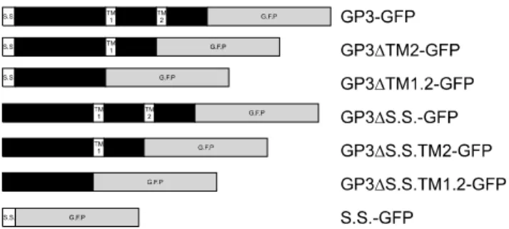
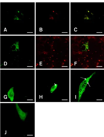
The glycoprotein 3 (GP3) of type II porcine reproductive and respiratory syndrome virus has the characteristic domains of a membrane protein. However, this protein has been reported to be retained in the endoplasmic reticulum (ER) rather than transported to the plasma membrane of the cell. In this study, we performed confocal laser scanning microscopy analysis of variants of GP3 and foundthat the signal sequence of the GP3 led to confinement of GP3 in the ER, while the functional ortransmembrane domain did not affect its localization.
Based on these results, we concludedthat the signal sequence of GP3 contains the ER retention signal, which might play an important role in assembly of viral proteins.
Keywords: endoplasmic reticulum, glycoprotein 3, porcine reproductive and respiratory syndrome virus, retention signal, signal sequence
Introduction
Porcine reproductive and respiratory syndrome (PRRS) has been a worldwide threat to the pig industry since it was first reported in the US [19] and Europe [36]. The etiological agent, PRRS virus (PRRSV), was isolated in Europe in 1991 and in the US in 1992 [12,36]. Weak and delayed protective immune response against PRRSV and lack of proper vaccine are the main reasons for worldwide spread of this virus [24,30,35]. PRRSV belongs to the family Arteriviridae, along with equine arteritis virus (EAV), lactate dehydrogenaseelevating virus (LDV), and simian hemorrhagic fever virus (SHFV) [6]. PRRSV can be divided into European (type 1) and American (type 2) strains based on their sequence homology [25]. PRRSV is
a single positive-stranded RNA virus composed of ten different open reading frames (ORFs). ORF 1a and 1b are the largest ORFs, encompassing approximately 80% of the whole genome. Protein products translated from these sequences are cleaved into several non structural proteins (nsps), some known to be required for the replication of viruses and others with unknown functions [33]. ORF 2a, 3, 4, and 5 are glycoproteins (GPs), while ORF2 b is an envelope (E) protein and ORF 6 and 7 are matrix (M) and nucleocapsid (N) proteins, respectively [14,22,23,26,27].
GPs of PRRSVare known to be critical to infection due to their ability to interact with receptors on the cell surface, including sialoadhesin and CD 163 [6,35,37], and the formation of GP complexes [10,38]. Additionally, GPs are considered to be important antigens inimmune response against PRRSV [13,14,17,23,37].
Although minor GPs (GP2a, 3, 4) are thought to play important roles in infection and formation of the glycoprotein complex of PRRSV, their structures and functions have not been extensively investigated relative to those of GP5, which is classified as a major glycoprotein. The absence of any minor GPs or envelope protein suppresses the protein complex formation or retention of proteins in the ER, which further hinders modification at the Golgi complex for unknown reasons [38]. Furthermore, in a recent study, minor glycoproteins GP2-GP4 were shown to be retained in the ER along with E proteins [39]. GP5 moves to the Golgi for further modification and the signal sequence is cleaved [2], which differs from other GPs.
GP3 is a minor GP that consists of 254 a.a. with a
molecular weight of approximately 42 kDa and contains 7
N-glycosylation sites [14,16,22,27]. In previous studies,
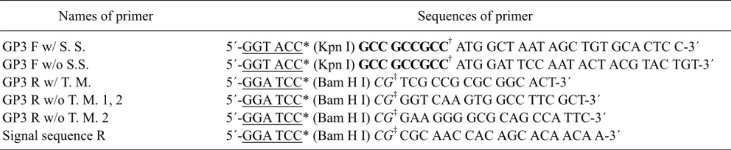
Table 1. Sequences and pairs of primers used for PCR in this study
Names of primer Sequences of primer
GP3 F w/ S. S.
GP3 F w/o S.S.
GP3 R w/ T. M.
GP3 R w/o T. M. 1, 2 GP3 R w/o T. M. 2 Signal sequence R
5´-GGT ACC* (Kpn I) GCC GCCGCC
†ATG GCT AAT AGC TGT GCA CTC C-3´
5´-GGT ACC* (Kpn I) GCC GCCGCC
†ATG GAT TCC AAT ACT ACG TAC TGT-3´
5´-GGA TCC* (Bam H I) CG
‡TCG CCG CGC GGC ACT-3´
5´-GGA TCC* (Bam H I) CG
‡GGT CAA GTG GCC TTC GCT-3´
5´-GGA TCC* (Bam H I) CG
‡GAA GGG GCG CAG CCA TTC-3´
5´-GGA TCC* (Bam H I) CG
‡CGC AAC CAC AGC ACA ACA A-3´
*Underlined sequences are the flanking sites used for construction of the EGFP fusion plasmid. †Bold typed sequences are the integrated Kozak sequence. ‡Italicized sequences are added to prevent frame shift of the fusion protein.