Journal of Bacteriology and Virology 2006. Vol. 36, No. 4 p.293 – 303
큰 플라크를 형성하는 돼지생식기호흡기증후군바이러스의 전체 게놈 RNA의 염기서열 분석
충북대학교 의과대학 미생물학교실, 의학연구소 이 영 민
*Complete Nucleotide Sequence of Genomic RNA of a Large-Plaque Forming Porcine Reproductive and Respiratory Syndrome Virus PL97-1/LP1
Young-Min Lee*
Department of Microbiology College of Medicine and Medical Research Institute Chungbuk National University 12 Gaeshin-Dong, Heungduk-Ku, Cheongju, Chungbuk 361-763, Korea
Received : November 24, 2006 Accepted : December 13, 2006
Porcine reproductive and respiratory syndrome virus (PRRSV) is one of the most important animal pathogens in swine industry worldwide. In this study, we isolated the large-plaque forming variant virus designated PL97-1/LP1 from the parental strain PL97-1, the first Korean strain of PRRSV, isolated from the serum of an infected pig in 1997. We found that the 15411-nucleotide genome of PL97-1/LP1 consisted of a 189-nucleotide 5' untranslated region (UTR), a 15071-nucleotide protein-coding region, and a 151-nucleotide 3'UTR, followed by a poly (A) tail of about 50~60 nucleotides in size. The 5'-end of PL97-1/LP1 began with ATGACGTAT. Comparison of the PL97-1/LP1 genome with the 11 fully sequenced PRRSV genomes currently available revealed sequence similarity from 99.6~99.7% (the North American VR-2332 and two VR-2332-derived vaccine strains MLV RespPRRS/Repro and RespPRRS MLV) to 62.0% (the Dutch Lelystad strain). Phylogenetc analysis revealed that PL97-1/LP1 is most closely related to the North American genotype VR-2332, two VR-2332-derived vaccine strains, and Chinese BJ-4. It is distantly related to the European genotype Lelystad. The entire nucleotide sequence of PL97-1/LP1 was identical to that of the parental virus PL97-1 except for three silent nucleotide substitutions, one in ORF1a (U4230C), one in ORF1b (C10977U), and one in ORF5 (U13976A). This nucleotide sequence has been submitted to the GenBank database under the accession number AY612613.
Key Words: Arteriviruses, Porcine reproductive and respiratory syndrome virus, Full-length genome, Large-plaque
forming virus
서 론
돼지생식기호흡기증후군 (porcine reproductive and respira-
tory syndrome; PRRS)은 약 20년 전에 돼지 축산업 분야에서 'mystery disease'로 처음 밝혀진 이후, 현재 이 분야에서 있어 서 경제적으로 가장 중요한 질병 중의 하나이다 (14,27,36).
이 질병을 일으키는 병원체는 돼지생식기호흡기증후군바이 러스 (PRRSV)로 명명되었다. 현재 유럽에서 최초로 분리된 Lelystad 바이러스 (36)와 북미에서 분리된 VR-2332 바이 러스 (3,6)가 각각 유럽주 (European strain)와 북미주 (North American strain)의 prototype으로 분류되어 있으며, 이들 사 이에는 염기서열 상동성이 약 55~70% 정도로 매우 낮다 (8,
*교신저자: 이영민. 361-763 충북 청주시 흥덕구 개신동 12, 충북대학교 의과대학 미생물학교실, 의학연구소
Phone: 043-261-2863, Fax: 043-272-1603, e-mail: [email protected]
**본 연구는 2005년도 충북대학교 학술연구지원사업의 연구비지원 에 의하여 이루어진 것임.
293
15,17~19,21,22,24). PRRSV는 현재 equine arteritis virus (EAV), lactate dehydrogenase-elevating virus (LDV), simian hemorrhagic fever virus (SHFV)와 더불어 Nidovirales 목 (order), Arteri- viridae 과 (family)에 속한다 (5,31).
PRRSV는 인지질을 가진 매우 작은 엔벨롭바이러스 (enveloped virus)이며, 정다면체 형태의 뉴클레오캡시드 안에 약 15 kb 정도의 양성극성의 단일가닥 RNA 게놈을 가지고 있다 (6). 바이러스의 자가복제에 필요한 효소들로 구성된 비 구조단백질인 replicase를 인코딩하는 유전자들은 게놈 RNA 의 5'-말단으로부터 약 80%를 차지하며, 게놈 RNA로부터 발현되는 서로 중첩되는 부위를 가진 ORF1a와 ORF1b에 의 해서 만들어진다. 이 중에서 ORF1b는 ribosomal frameshift 기작에 의해 발현된다고 보고된 바 있다 (4). 바이러스의 구 조단백질들은 게놈 RNA의 3'-말단으로부터 약 20%를 차지 하며, ORF2a, ORF2b에서부터 ORF7의 7개의 유전자로부터 발현된다. 이들은 감염된 세포 내에서 monocistronic subge- nomic mRNA의 5'-말단으로부터 발현된다 (20,33). PRRSV의 구조단백질 뿐만 아니라 비구조단백질을 포함한 대부분의 단백질에 대한 기능 및 분자생물학적 특성에 관한 연구 결 과는 아직 미미한 상태이다.
PRRSV가 돼지에 감염되면 임상적인 증상으로 심한 생식 감퇴와 호흡기 장애가 나타난다. PRRSV 북미주와 유럽주는 질병 증상에서 분명하게 다른 점을 증명할 수 없다는 사실에 도 불구하고, 그것들은 항원적으로나 유전형적으로 뚜렷한 차이를 가지고 있다 (1,10,19,24,36,37). 또한, 북미주와 유럽 주 내에서도 분리주들 간의 차이를 나타낸다. 예를 들면, 북미주에 속하는 여러 분리주들 사이의 염기서열의 변이는 RNA 중합효소 또는 RNA 재조합의 본질적인 에러에 기인할 수 있으며, 이들의 유전학적 변이는 바이러스의 병원성에 중 요한 차이를 나타낼 수 있다고 보고된 바 있다 (2,11,13,19, 23,35). 자연적인 숙주 또는 세포 내에서 PRRSV의 유전학 적 진화를 연구하기 위해서는 분리 확보된 바이러스의 정확 한 전체 게놈의 염기서열을 분석하는 것이 필요하다. 그러므 로, 본 연구에서는 PRRSV 국내분리주 PL97-1의 consensus sequence (12)와 이로부터 분리된 큰 플라크를 형성하는 PRRSV PL97-1/LP1의 단일 바이러스클론의 전체 게놈 염기 서열을 서로 비교 분석하였다. 분석 결과, PL97-1/LP1의 전 체 염기서열은 모바이러스 PL97-1과 비교하였을 때 3개의 침묵돌연변이 (silent mutation)을 가지고 있는 것을 알 수 있었다. 이 돌연변이들은 바이러스의 비구조단백질 ORF1a (U4230C)와 ORF1b (C10977U), 그리고 바이러스의 구조단백 질 ORF5 (U13976A)에 각각 하나씩 존재하는 것을 발견하 였다. 또한, 본 연구에서는 이렇게 밝혀진 PL97-1/LP1 전체 게놈의 염기서열을 이용하여, 앞으로 PRRSV의 세포내 생활
사 연구를 위해서 이들의 분자생물학적 특성을 심도있게 분 석하였다.
재료 및 방법
1. 세포배양과 바이러스
MARC-145 세포는 5% 우태아혈청 (fetal bovine serum), 불필요 아미노산 (nonessential amino acids), sodium pyruvate, 페니실린/스트렙토마이신이 첨가된 minimal essenial medium (MEM)배지에 배양하였다. 세포배양시 사용한 모든 재료들 은 Gibco BRL Life Tech. USA으로부터 구입하였다. 본 실험 에서 사용된 모바이러스 (parental virus)는 1997년 경기도에 서 만성적으로 호흡기에 병적 증상을 나타내는 감염된 돼지 로부터 분리된 PL97-1 바이러스를 사용하였다 (12). PL97-1 바이러스는 MARC-145 세포에서 3차례 이하로 계대배양된 것을 사용하였으며, MARC-145 세포에서 다양한 크기의 플 라크를 형성하였다 (data not shown). 모든 바이러스는 필요 할 때까지 -80℃에서 보관하였다.
2. 바이러스 클론의 분리
MARC-145 세포 (3×10⁵)을 6-well plate에 접종하였다.
세포 접종 후 12시간이 경과한 다음, PRRSV 모바이러스 PL97-1을 moi = 1에서부터 시작하여 10배로 배양액으로 희 석한 후, 각각의 well에 접종하여 1시간 배양한다. 이렇게 감 염된 세포단층위에 0.5% agarose와 5% 우태아혈청이 들어 있는 MEM배지로 덮어서 37℃, 5% CO2에서 배양하였다. 약 72시간이 경과한 다음, 바이러스의 복제로 인한 플라크를 가 시적으로 확인한 다음, 원하는 플라크를 plaque purification 기법을 적용하여 각각의 플라크를 분리 정제하였다. 이렇게 확보한 큰 플라크를 형성하는 PRRSV를 PL97-1/LP1이라고 명명하였다.
3. 바이러스 RNA의 분리
MARC-145 세포 (5×10⁶)를 P100 조직배양접시에 분주한 후 24시간이 경과한 다음, 분리 정제된 PL97-1/LP1의 seed virus를 접종하였다. 바이러스 접종 후 72시간이 경과한 다 음, 세포배양액에 분비된 바이러스를 수확하기 위해서 얻어 진 배양액을 낮은 속도의 원심 분리를 이용하여 세포의 잔여 물질을 제거하고 바이러스를 포함하는 배양액을 분리하였다.
얻어진 바이러스로부터 바이러스 게놈 RNA의 분리는 약 105 plaque-forming unit의 바이러스를 포함하는 0.5 ml의 배 양액을 lysis buffer (20 mM Tris-HCl, pH 7.5, 5 mM EDTA, 0.5% sodium dodecyl sulfate (SDS), 0.2 mg/ml proteinase K)에 서 45℃에 30분 동안 배양한 후, 페놀/클로로폼을 사용하여
추출하고 에탄올을 이용하여 침전시켰다. 또는 바이러스 게 놈 RNA는 QIAmp viral RNA extraction kit (Qiagen, Dusseldorf, Germany)를 사용하여 간단히 추출할 수 있었다.
4. 역전사반응 및 PCR 증폭
바이러스 게놈 RNA는 Superscript II RNaseH(-) 역전사 효소 (GIBCO/BRL)와 9개의 전체 게놈 염기서열이 밝혀진 PRRSV (Lelystad, NVSL 97-7985, CH-1a, SP, 16244B, PA8, VR-2332, RespPRRS MLV, and BJ-4)의 consensus sequence을 토대로 디자인된 oligonucleotide를 사용한 cDNA 합성의 주 형으로 사용하였다. 역전사반응은 RT buffer (50 mM Tris-HCl, pH 8.3, 75 mM KCl, 10 mM DTT, 3 mM MgCl2, 0.5 mM dNTPs) 에서 42℃에서 1시간 동안 수행하였다. 계속되는 PCR 증 폭은 RT 반응 결과 만들어진 first-strand cDNA 단편으로부 터 double-stranded cDNA product를 얻기 위해서 실행되었다.
PCR 반응은 10 mM Tris, pH 8.3, 1.5 mM MgCl2, 50 mM KCl, 0.01% gelatin, 200 µM dNTPs, 0.4 µM primers, 2.5 unit Pyro- coccus spp.에서 유래된 Pyrobest DNA polymerase (Takara, Japan)를 포함하는 100 µl 반응액에서 이루어졌다. PCR 증폭 은 denaturation (94℃, 30초), annealing (60℃, 30초), extension (72℃, 6분)으로 총 35 사이클을 실행하였으며, 마지막 exten- sion 단계는 72℃에서 10분 동안 진행하였다.
5. 4개의 중첩되는 PRRSV cDNA의 합성
PRRSV 바이러스의 5' 및 3'-말단을 제외한 나머지 전체 게 놈 RNA에 해당하는 cDNA를 합성하기 위해서, 전체 게놈을 4개의 중첩되는 부위로 나누어 cDNA 합성 및 PCR 증폭 (PF1, PF2, PF3, PF4)을 수행하였다. 본 실험에서 사용된 oligonucleotide는 Table 1에 요약하였다 (12). RT-PCR에 사용 된 oligonucleotide는 PF1 cDNA (nt 180-5297)의 경우 PR1RT 과 PR1F/PR1R primer, PF2 cDNA (nt 3708-9108)의 경우 PR2RT과 PR2F/PR2R primer, PF3 cDNA (nt 7688-13051)의 경 우 PR3RT과 PR3F/PR3R primer, 그리고 PF4 cDNA (nt 9610- 15238)의 경우 PR4RT과 PR4F/PR4R primer이다.
6. 바이러스 게놈 RNA의 5'-말단 및 3'-말단의 염기서 열 분석
PRRSV 바이러스 게놈 RNA의 5'-말단의 염기서열을 분 석하기 위해서 기존의 5'RACE 방법을 변형하여 사용하였다 (12). 먼저 first-strand cDNA를 바이러스 게놈 RNA로부터 Superscript II RT와 primer PR50을 사용하여 합성하였다. 합 성된 cDNA로부터 RNaseH를 사용하여 RNA 단편들을 제거 하였으며, 그 결과 남은 first-strand cDNA는 페놀과 클로로폼 을 사용하여 정제하였다. 정제된 cDNA를 OligoT를 사용하
여 연결하였으며, 이렇게 연결된 OligoT는 다음 RT-PCR 반 응 수행시 primer 결합위치를 제공하게 된다. cDNA-OligoT 결합 반응 (ligation reaction)은 40U T4 RNA ligase, 7 µl single- stranded cDNA, 10 pmol OligoT, 20% PEG #6000가 들어있는 40 µl 반응액을 16℃에서 12시간 동안 지속하였다. 결합된 반응액을 주형으로 사용하고 primer PR49와 OligoTR을 사용 하여 PCR 증폭을 수행하였다. 증폭된 약 500 bp cDNA 산물 은 분리 정제한 후 PstI-SacI으로 절단한 후, 같은 제한효소 로 절단된 pRS2 벡터에 삽입하였다. 이렇게 클로닝된 바이 러스의 5'-말단부위의 염기서열을 분석하기 위해서 산물이 삽입된 클론 10개를 염기서열 분석에 사용하였다.
PRRSV 바이러스 게놈 RNA의 3'-말단의 염기서열을 분석 하기 위해서, 합성된 oligonucleotide를 바이러스 게놈 RNA 3'-말단에 결합하는 3'RACE 방법을 사용하였다 (38). Ligation 반응은 10U T4 RNA ligase, 40U RNaseOUT, 10 pmol OligoT, 및 바이러스 게놈 RNA를 포함하는 반응액을 16℃에서 12 시간 동안 수행하였다. 반응 후, OligoT가 결합된 바이러스 RNA를 페놀/클로로폼을 사용하여 정제하였다. 정제된 바이 러스 RNA는 Superscript II 역전사효소와 OligoTR primer를 사용하여 cDNA 합성을 수행하였다. 다음으로 합성된 cDNA 를 주형으로 사용하고 PR41과 OligoTR primer를 사용하여 PCR 증폭을 수행하였다. 이렇게 합성 및 증폭된 PCR 산물 은 약 450 bp 정도였으며, MfeI-PstI 제한효소로 절단한 후 약 380 bp 크기의 산물을 EcoRI-PstI으로 절단된 pRS2 벡터 에 삽입하였다. 삽입된 cDNA의 염기서열을 분석하기 위해 서 10개의 독립된 클론을 염기서열 분석에 사용하였다.
7. 바이러스 RNA 게놈 염기서열의 분석
RT-PCR의 결과 합성된 cDNA를 big dye terminator를 이 용하여 ABI PRISM 370 또는 310 Genetic Analyzer (Applied Biosystems, Cetus, Norwalk, CT)로 게놈의 염기서열을 직접 분석하였다. 분석 결과 얻어진 단편의 염기서열은 중첩되는 부위를 이용하여 DNA Strider 프로그램을 사용하여, PRRSV PL97-1/LP1 전체 게놈 염기서열로 연결하였다.
8. Multiple sequence alignment 및 유전학적 계통도 분석 Multiple sequence alignment와 유전학적 계통도 분석을 위 해서 PRRSV PL97-1/LP1 바이러스를 포함하여 GenBank상 의 전체 게놈의 염기서열이 밝혀진 12개의 다른 PRRSV 국외분리주들을 사용하였다. 이들의 GenBank accession num ber은 다음과 같다. CH-1a (AY 032626), HB-1(sh)/2002 (AY 150312), P129 (AF 494042), NVSL 97-7985 (AF 325691), BJ-4 (AF 331831), RespPRRS MLV (AF 066183), MLV RespPRRS/
Repro (AF 159149), VR-2332 (U 87392), PA8 (AF 176348),
16244B (AF 046869), SP (AF 184212), Lelystad (M 96262), EAV (NC002532). 본 연구논문에서는 분리주들 간의 유전학 적 상관관계의 분석을 위해서 ClustralX 프로그램 (34)을 사 용하여 multiple sequence alignment를 수행하였으며, neighbor joining method (29)을 이용하여 배열된 염기서열의 계통도를 구성하였고, TREEVIEW software (26)를 사용하여 시각화하 였다.
결과 및 고찰
1. PRRSV PL97-1/LP1 게놈 RNA를 나타내는 중첩된 6개의 cDNA 합성
PRRSV PL97-1/LP1 바이러스 게놈 RNA의 5'-말단 및 3'- 말단을 제외한 전체-길이에 해당하는 cDNA를 4개의 중첩 되는 단편으로 나누어 RT-PCR을 수행하였다 (Fig. 1). 본 RT-PCR 반응에 사용된 primer들은 Table 1에 요약하였으며, PCR 반응은 에러율이 낮은 DNA 중합효소를 사용하였다.
증폭된 cDNA 단편들은 5'-말단에서부터 PF1 (nt 180-5297), PF2 (nt 3708-9108), PF3 (nt 7688-13051), 및 PF4 (nt 9610- 15238)으로 표기하였으며, 이들의 길이는 각각 5118 bp, 5401
bp, 5364 bp, 및 5629 bp 이다 (Fig. 1). 이렇게 증폭된 cDNA amplicon은 agarose gel을 이용한 전기영동을 실시하여 이들 의 크기를 확인하였다 (Fig. 2A). 또한 컨트롤로써 역전사반 응 중 역전사효소를 첨가하지 않았을 경우에는 각각의 예상 되는 크기의 상응하는 밴드가 관찰되지 않음으로써, 본 실험 에서 증폭된 cDNA 단편들은 PRRSV에 특이적으로 반응한 primer에 의해서 합성된 것임을 알 수 있었다 (Fig. 2A).
PRRSV PL97-1/LP1 바이러스 게놈 RNA의 5'-말단의 염 기서열은 5'RACE 기법으로 분석하였다. 먼저 PRRSV 게놈 RNA의 5'-말단으로부터 510 nucleotide 떨어진 곳에 상보적 결합을 하는 primer를 사용하여 역전사반응을 수행하였다.
합성된 first-strand cDNA를 인위적으로 합성한 OligoT와 결 합시킨 후, OligoT와 상보적 결합을 할 수 있는 OligoTR과 PRRSV 게놈 RNA의 nt 510-531에 상보적 결합을 하는 PR- 49 primer를 사용하여 PCR 증폭을 수행하였다. PCR 증폭 결과, 약 500 bp 크기의 PCR 산물을 agarose gel 전기영동을 통해서 확인하였다 (Fig. 2B). 예상한 대로 ligation 반응시 OligoT 또는 역전사반응시 역전사효소를 첨가하지 않을 경 우, 약 500 bp 크기의 PCR 산물이 증폭되지 않는다는 것을 알 수 있었다. 따라서, 본 실험에서 증폭된 PCR 산물은 Table 1. Oligonucleotides used in this study
Oligonucleotide Sequencea Postitionb Polarity Purpose
PR1RT 5'-TAGGATGGTGAGGGGGTG 5332~5349 - RT
PR1F 5'-CCCTTTAACCATGTCT 180~195 + PCR
PR1R 5'-CAAAGCAACCAGGTAA 5282~5297 - PCR
PR2RT 5'-GAGCATGTCCTCAAACTT 9168~9185 - RT
PR2F 5'-CCGGATATCGTCGCGG 3708~3723 + PCR
PR2R 5'-CCATATGCTGTGCATA 9093~9108 - PCR
PR3RT 5'-CACATTCCCTATCCCGAA 13074~13091 - RT
PR3F 5'-GTTTAAACTGCTAGCC 7688~7703 + PCR
PR3R 5'-GTGTAGCTGAAGGACA 13036~13051 - PCR
PR4RT 5'-CTAATTGAATAGGTGACT 15342~15359 - RT
PR4F 5'-ATTATGAGGGGAAGAA 9610~9625 + PCR
PR4R 5'-ACGCGGATCAGGCGCA 15223~15238 - PCR
PR41 5'-GGAGAAGCCCCATTTTCC 15038~15055 + PCR
PR49 5'-CGACCCGTACCATTCTTT 476~493 - PCR
PR50 5'-AAAAGTCTTCAGGCTTGG 692~709 - RT
OligoT 5'-CCAGTGTTGTGGCCTGCAGGGCGAATT RNA ligation
OligoTR 5'-GATGAATTCGCCCTGCAGGCCACAACA RT, PCR
a PRRSV-specific sequences are show in boldface type
b Nucleotide position refers to the complete genome sequence of the PRRSV PL97-1 strain
PRRSV 염기서열에 특이적으로 증폭된 것임을 알 수 있었 다. 이렇게 증폭된 cDNA 단편을 클로닝 벡터에 삽입한 후, 염기서열 분석에 이용하였다.
PRRSV 바이러스 게놈 RNA의 3'-말단의 염기서열은 3'RACE을 통해서 분석하는 데 성공하였다. 이를 위해서 정 제된 바이러스 게놈 RNA의 3'-말단에 먼저 OligoT를 결합
시킨 후, 다음 단계의 cDNA 합성과 PCR 증폭시 primer 결 합부위로 이용하였다. 이러한 방법으로 PR41과 OligoTR pri- mer를 사용하여 합성된 cDNA의 PCR 증폭을 수행하였다.
Fig. 2C에 나타낸 것과 같이, PCR 증폭된 산물의 크기는 예상한 것과 같이 약 450 bp 정도였으며, 이것을 클로닝 벡 터에 삽입한 후, 염기서열 분석을 위해서 사용하였다. 증폭 Figure 1. Synthesis of cDNA amplicons encompassing the entire genomic RNA of the PRRSV PL97-1/LP1 strain. The full-length viral genomic RNA is schematically drawn at the top and the four overlapping cDNA amplicons are shown below. PF1, nt 180-5297 PF2, nt 3708-9108 PF3, nt 7688-13051 PF4, nt 9610-15238.
B C
A
Figure 2. Synthesis of cDNA amplicons encompassing the entire genomic RNA of the PRRSV PL97-1 strain. (A) Synthesis by long RT-PCR of four long overlapping cDNA amplicons (PF1, PF2, PF3, and PF4) that cover the full-length viral genomic RNA except for the 5' and 3' termini. Four PRRSV-specific overlapping cDNA amplicons are synthesized in the presence of an RNaseH(-) RT (lanes 1, 3, 5, and 7) but not in its absence (lanes 2, 4, 6, and 8). The products were analyzed on a 1% agarose gel and visualized by staining with ethidium bromide. Lanes 1-2, PF1 amplicons; lanes 3-4, PF2 amplicons; lane 5-6, PF3 amplicons, lanes 7-8, PF4 amplicons. M, 1 kb DNA marker indicated in base pairs. (B) Synthesis of PRRSV-specific amplicons by a modified 5'RACE protocol from the OligoT-ligated PRRSV cDNAs to determine the 5'-terminal sequence. RT reaction used to synthesize the first-strand cDNA corresponding to the 5'-end portion of the genome was conducted in the presence (lanes 1-2) or absence (lane 3) of RNaseH(-) RT. The OligoT was ligated to the 3'-end of first-strand cDNA using T4 RNA ligase in the presence (lanes 1 and 3) or absence (lane 2) of OligoT. The OligoT-ligated first-strand cDNA was then PCR-amplified using a pair of primers. PRRSV-specific cDNA amplicons were analyzed on a 1.5% agarose gel and visualized by staining with ethidium bromide. M, 100 bp DNA ladder indicated in base pairs. (C) Synthesis of PRRSV-specific amplicons from the OligoT-ligated PRRSV genomic RNAs by a modified 3'RACE protocol to determine the 3'-terminal sequence. The OligoT was ligated to the 3'-end of viral genomic RNA using T4 RNA ligase in the presence (lanes 1 and 3) or absence (lane 2) of OligoT. The OligoT-ligated viral RNA was then used for cDNA synthesis in the presence (lanes 1-2) or absence (lane 3) of RNaseH(-) RT. After PCR amplification, PRRSV-specific cDNA amplicons were analyzed on a 1.5% agarose gel and visualized by staining with ethidium bromide.
M, 100 bp DNA ladder indicated in base pairs.
5' 3'
Replicase Proteins
Structural Proteins
180 5297 PF1
3708 PF2 9108
7688 PF3 13051
9610 PF4
15238 1b
4 3
6 7
1a 2a
2b 5
0 2 4 6 8 10 12 14 16
된 cDNA 단편들은 클로닝으로 인한 편견된 결과를 피하고 consensus sequence를 얻기 위해서, PCR 증폭된 cDNA ampli- con을 직접 염기서열 분석에 사용하거나, 또는 바이러스의 5'-말단과 3'-말단의 경우 클로닝된 10개의 독립된 클론을 염기서열 분석에 이용하였다. 염기서열 분석은 양쪽방향으로 진행되었으며, 최소한 같은 염기를 2~3번 반복하여 실험 결 과를 얻었다.
2. PRRSV PL97-1/LP1 RNA 게놈의 구조
바이러스의 전체 게놈 염기서열 분석 결과 다음과 같은 사실을 알 수 있었다. PRRSV PL97-1/LP1 바이러스 게놈은 3'-말단에 존재하는 poly(A)를 제외하고 15,411개의 염기로 구성되어 있다. PRRSV PL97-1/LP1의 염기서열과 게놈 구성 은 다른 Arterivirus와 유사하다는 것을 알 수 있었다. Fig. 3
에 나타낸 것과 같이, PRRSV PL97-1/LP1은 189개의 염기로 구성된 5' 비번역부위 (untranslated region; UTR)과 151개의 염기로 구성된 3'UTR을 가지고 있었다. 서로 중첩되는 부위 를 가진 크기가 큰 ORF1a와 ORF1b는 게놈의 맨 앞쪽에 위치하면서 각각 7,509개와 43,89개의 염기로 이루어져 있었 다. 추가적으로 6개의 서로 겹쳐지는 부위를 가진 ORF들은 ORF1b 아래쪽에 위치한다는 것을 알 수 있었다. 이 ORF들 은 PRRSV 구조단백질들인 minor protein (GP2a, GP3, GP4)들 과 major protein (GP5, M, N)들을 발현한다는 것을 알 수 있 었다. 73개의 아미노산으로 구성된 작은 ORF2b라고 명명된 ORF가 ORF2a 안에서 존재한다는 것을 확인하였다 (Fig. 3).
Arterivirus 과에 속하는 다른 바이러스에서 발견되는 ORF2b 는 먼저 유전학적 상동성을 분석함으로써 예측되었고, EAV infectious cDNA molecular clone을 이용하여 최근에 이 단백 질의 발현이 Arterivirus의 감염 및 자가복제에 필수적인 것 이라는 것이 증명되었다 (32). 특히 이 부위는 Arterivirus 과 에 속하는 다른 바이러스들 사이에서 유전학적 다양성을 나 타냄으로써 각각의 바이러스의 자가복제 및 병원성에 중요 한 역할을 할 것으로 추측된다. 이와 마찬가지로, PRRSV의 북미주와 유럽주에 속하는 바이러스의 분리주들 간에도 매 우 유전학적 다양성을 나타냄으로 중요한 의미를 가지는 것 으로 추측된다. 특히, ORF2a 단백질이 발현되는 subgenomic mRNA2의 발현에 중요한 역할을 하는 ORF2a의 위쪽에 위 치한 leader-body junction은 유럽주에 속하는 Lelystad 바이 러스의 경우 38개의 염기로 구성되어 있으나, PL97-1/LP1의 경우 20개의 염기로 구성되어 있는 것으로 분석 결과 알 수 있었다.
3. PRRSV PL97-1/LP1과 다른 PRRSV 분리주들 간의 유전학적 상관관계
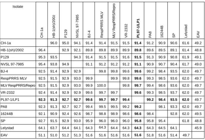
위에서 얻어진 PL97-1/LP1 분리주의 전체 게놈 염기서열을 토대로 지금까지 국외에서 분리된 12개의 서로 다른 PRRSV 국외분리주들과의 유전학적 상관관계를 multiple sequence alignment을 통해서 분석하였다. Fig. 4에 나타낸 것과 같이, PL97-1/LP1은 PRRSV의 북미주의 prototype이며 병원성을 나 타내는 미국 분리주인 VR-2332 (99.6%)와 VR-2332에서 유 래된 modified live attenuated vaccine주인 MLV RespPRRS/
Repro주 (99.7%) 또는 RespPRRS MLV주 (99.6%), 중국분리 주인 BJ-4 (99.6%)와 가장 높은 nucleotide 유사성을 나타내 었다. 이와는 대조적으로, 유럽주의 prototype이며 병원성을 나타내는 Lelystad과는 62%의 유사성만을 나타냄으로써 가 장 낮은 유전학적 상관관계를 나타내었다. 또한 이러한 pair- wise sequence comparison 분석 결과는 phylogenetic tree를 그 려봄으로써 시각적으로 쉽게 구분할 수 있었다. Fig. 5에 나 372
14889-15260 ORF7
525 14375-14899
ORF6
603 13788-14390
ORF5
537 13241-13777
ORF4
765 12696-13460
ORF3
222 12078-12299
ORF2b
771 12073-12843
ORF2a
459 11610-12068
Nsp12
669 10941-11609
Nsp11
1323 9618-10940
Nsp10
2055 7564-9617
Nsp9
11880 ORF1ab
135 7564-7698
Nsp8
777 6787-7563
Nsp7
48 6739-6786
Nsp6
510 6229-6738
Nsp5
612 5617-6228
Nsp4
1338 4279-5616
Nsp3
2943 1336-4278
Nsp2
648 688-1335
Nsp1β
498 189-687
Nsp1α
7509 189 1-189
5'UTR
NT size NT position
Protein ORF
ORF1a
15261-15411 3'UTR
Figure 3. The genome organization and the predicted polype- ptides encoded by the PRRSV PL97-1/LP1 viral genome RNA.
UTR, untranslated region; ORF, open reading frame; Nsp, non- structural protein; NT, nucleotide.
타낸 것과 같이, 본 연구에서 사용한 PL97-1/LP1 바이러스는 VR-2332, VR-2332에서 유래된 2개의 vaccine주 (MLV Resp- PRRS/Repro주와 RespPRRS MLV), 및 BJ-4와 하나의 클러 스터를 형성한다는 것을 알 수 있었다. 본 실험 결과는 PL97-1/LP1의 모바이러스인 PL97-1과의 유전학적 상관관계 분석 결과와 일치한다 (12). 또한, 비록 북미주에 포함되기는 하지만, NVSL 97-7985 및 P129, HB-1(sh)2002, CH-1a와는 서로 다른 클러스터를 형성함으로써, PL97-1/LP1 바이러스 는 이들과는 유전학적으로 측면에서 구별된다는 것을 추측 할 수 있다. 그러나, PL97-1 모바이러스와 비교하였을 때, PL97-1/LP1는 3개의 침묵돌연변이 (silent mutation)을 가지고 있다는 것을 알 수 있었다. 이들 3개의 돌연변이는 바이러스 게놈 전체에 걸쳐서 발견되었으며, 바이러스 비구조단백질 ORF1a (U4230C)와 ORF1b (C10977U), 그리고 바이러스의 구조단백질 ORF5 (U13976A)에 각각 하나씩 존재하는 것을 발견하였다.
4. 5'UTR과 3'UTR의 비교 분석
Nucleotide 상동성 측면에서 PL97-1/LP1의 5'UTR은 북미
주의 prototype인 VR-2332와 유럽주의 prototype인 Lelystad 의 5'UTR과 각각 96.3% 및 44%의 상동성을 나타내었다 (data not shown). Lelystad의 5'UTR은 211개의 염기로 구성되 어 있으며, 190개의 염기로 구성된 VR2332 5'UTR과 189개 의 염기로 구성된 PL97-1/LP1 5'UTR보다 상당히 길다는 것 을 알 수 있었다 (data not shown). 본 실험에서 PRRSV의 5'- 말단의 염기서열을 정확하게 분석하기 위해서 5'RACE 기법 을 사용하였다. 본 실험 결과 PL97-1/LP1은 대부분의 다른 분리주들과 마찬가지로 ATGACGTAT로 시작하는 것을 알 수 있었다 (25).
PL97-1/LP1을 포함하여 대부분의 북미분리주의 3'UTR은 단지 114개의 염기로 구성된 Lelystad주의 3'UTR과는 상당 히 다른 긴 151개의 염기로 구성되어 있다는 것을 알 수 있 었다. PL97-1/LP1의 3'UTR은 VR-2332와 2개의 염기가 다르 게 나타났다. 이들은 바이러스 게놈 3'-말단의 poly(A) 부위 가 시작하는 바로 위쪽에 위치하고 있다. 길이를 제외하고 3'UTR은 5'UTR보다 정렬된 서열 안에서 더 높은 nucleotide 상동성을 보였다. 그러나, 5'UTR과 3'UTR 양쪽의 염기서열 보존 정도는 중요한 시스-작용 유전인자 (cis-acting elements)
Lelystad
SP
16244B
PL97-1/LP1
VR-2332
MLV RespPRRS/Repro
RespPRRS MLV
BJ-4
NVSL 97-7985
P129
HB-1(sh)/2002
49.7 51.4 51.6 51.8 51.6 51.6 51.6 51.6 51.6 51.3 51.2 51.0 51.1 EAV
45.9 64.1
64.5 64.3 64.3 64.3 64.3 64.4
64.3 64.1 64.4 63.7 64.1 Lelystad
48.8 61.8 95.4
95.8 95.8 96.0 96.0 96.0 95.9 93.0 92.9 91.5 92.7 SP
49.5 62.0 92.8 98.4
98.6 98.6 98.9 98.8 98.7 92.6 92.4 90.9 92.1 16244B
49.7 62.0 93.3 98.1 99.2
99.2 99.5 99.5 99.4 92.7 92.7 91.3 92.3 PA8
49.7 62.0 93.5 98.4 99.2 99.4
99.7 99.7 99.6 92.7 92.7 91.3 92.3 PL97-1/LP1
49.7 62.0 93.7 98.5 99.3 99.6 99.7
99.7 99.6 92.9 92.9 91.4 92.4 VR-2332
49.7 62.0 93.6 98.6 99.4 99.7 99.8 100.0
99.9 93.0 92.9 91.5 92.5 MLV RespPRRS/Repro
49.7 62.0 93.6 98.5 99.3 99.6 99.8 99.9 99.9
93.0 92.9 91.5 92.5 RespPRRS MLV
49.7 62.0 93.5 98.4 99.2 99.6 99.6 99.8 99.8 92.9
92.9 91.4 92.5 BJ-4
49.0 61.7 90.4 90.7 90.9 91.1 91.2 91.2 91.2 91.1 94.9
93.8 95.4 NVSL 97-7985
49.1 61.9 90.8 90.9 91.3 91.5 91.6 91.5 91.5 91.4 94.3 93.5
95.3 P129
48.8 61.4 89.1 89.5 89.6 89.8 89.9 89.9 89.8 89.8 92.1 92.9 96.4
HB-1(sh)/2002
49.2 61.6 90.6 90.9 91.2 91.4 91.5 91.5 91.4 91.4 94.1 95.0 96.0 CH-1a
Isolate
EAV
PA8
CH-1a
Figure 4. Pairwise comparisons of full-length genome of PRRSV isolates. Percentage identity of nucleotide and amino acid sequences are shown above and below the diagonal line, respectively.
가 위치할 것으로 추정되는 3'-말단부위에서 가장 높게 나타 났다. PRRSV 바이러스의 3'-말단에 존재하는 poly(A)의 정확 한 개수를 분석하기 위해서, 본 연구에서는 인위적으로 합성 된 PRRSV 염기서열과 무관한 OligoT를 바이러스 RNA 3'- 말단에 결합시킨 후 3'RACE 실험을 수행하였다. 이 방법은 Salles가 고안한 Poly(A) Test (PAT) 방법과는 구별된다 (30).
PAT는 맨 처음 인산화된 oligonucleotide (dT)12-18와 더불어 poly(A)를 포화시킨다. 다음으로 42℃에서 이 oligonucleotide 를 결합시킨다. 이 과정은 서로 보완되는 copy를 가지고 42℃에서 짧은 A/dT 혼성물이 불안정하다는 단점을 가지고 있다. 본 연구에서 수행한 3'RACE 실험 결과, PL97-1/LP1은 모바이러스인 PL97-1과 마찬가지로 약 50~60개의 poly(A) 를 가지고 있다는 것을 알 수 있었다. 따라서, PRRSV의 역 상유전자 시스템의 토대가 되는 infectious cDNA를 합성할 때 poly(A)의 개수를 약 50~60개까지 삽입하는 것이 wild type과 유사한 게놈 RNA를 합성할 수 있는 방법일 것으로 생각된다. 또한, 이러한 poly(A)의 개수가 PRRSV의 자가복
제에 어떠한 영향을 미치는 지는 혹은 자가복제에 어떻게 작용하는 지는 앞으로 수행하여야 할 연구과제 중의 하나라 고 생각한다.
5. ORF1a와 ORF1b 사이에 존재하는 ribosomal frame- shit site 분석
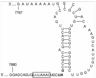
PRRSV ORF1a/1b가 중첩되는 부위에 RNA pseudoknot structure를 형성한다고 예측되며, 이러한 RNA 3차 구조는 PRRSV ORF1b 단백질이 발현하는 매우 중요한 역할을 한다 고 추정된다 (4). ORF1a stop codon의 3개의 염기 바로 위쪽 에 위치하는 7개의 염기 (UUUAAAC)는 서로 다른 PRRSV 분리주들에 상관없이 정확히 보존되어 있었다. 또한, stop codon으로부터 바로 뒷부분에 12개의 Watson-Crick base pairing으로 구성된 stem-loop structure가 형성되었으며, 여기 에는 하나의 A bulge가 가운데 부위에 위치한다는 것을 발 견하였다. 또한, 이와 대조적으로 stem-loop 구조 아래에 형 성되어 있는 염기서열은 다른 PRRSV 분리주에서 몇몇의 돌 연변이를 관찰할 수 있음으로 이 부위는 어느 정도의 돌연 변이는 ribosomal frameshit 기능에 영향을 끼치지 못하는 것 으로 여겨진다. PL97-1/LP1과 VR-2332의 ORF1a/ORF1b 접 합부의 염기서열은 동일하였다 (Fig. 6).
6. 구조 유전자의 비교
ORF2a, ORF2b, 및 ORF3부터 ORF7까지는 바이러스 게 놈의 3'-말단부분에 약 3 kb 정도에 위치하고 있으며, 모든 바이러스의 구조단백질들을 발현한다 (Fig. 1). ORF2b를 제 EAV (NC002532)
Lelystad (Netherlands, 91) NVSL 97-7985 (USA, 97) P129 (USA, 95)
CH-1a (China)
HB-1(sh)/2002 (China, 02)
SP
16244B (USA, 97) PA8 (Canada, 95) PL97-1/LP1 (Korea) VR-2332 (USA) RespPRRS MLV MLV RespPRRS/Repro BJ-4 (China)
0.10.
Figure 5. A phylogenetic tree constructed with the nucleotide sequence of the full-length genome of all 12 available PRRSV isolates. The phylogenetic tree was constructed using the neighbor- joining method in ClustralX method. The scale bar at the bottom of the tree represent the number of nucleotide substitutions per site. The tree was rooted using the nucleotide sequence of EAV, a member of the Arteriviridae family. The strain name is followed by the country and the year of isolation in two digits.
Figure 6. Predicted pseudoknot structure of the ribosomal frame- shift region of PRRSV PL97-1/LP1. The heptanucleotide sequence (UUUAAAC) slippery sequence is in a box. The stop codon UAG of ORF1a is in the boldface type. Numbers indicate nucleotide position.
외하고 각각의 구조단백질은 분리된 subgenomic mRNA로부 터 번역된다 (7). Lelystad에서 확인되는 5'-(U/C/A)AACC-3' 염기서열과는 대조적으로 PRRSV 북미주에서는 mRNA을 발 현을 위해 일치된 intergenic region의 염기서열은 5'-(U/G/A) (U/C/A/G)(A/C)(A/G)(C/U)C-3'이다 (7,24). PL97-1/LP1과 VR- 2332의 북미주들 간에는 이 부위가 완벽하게 잘 보존되어 있었다. PRRSV 북미주들 간에 병원성 비교와 약독화된 균 주들 간의 유전학적 상관관계를 분석하기 위해서 VR-2332, 및 16244B, DK5163-23, DK5163-17, DK3016-12) (16)과 북미 주의 변형된 생백신주 MLV RespPRRS의 구조단백질 유전자 를 이루고 있는 염기와 아미노산 서열을 PL97-1과 비교하여 보았다. Ingelvac PRRSV MLV로써 유럽에서 인증된 Resp- PRRS는 병원성을 가진 북미분리주인 VR-2332에서 유래된 생백신이다 (6). 분석에 사용한 모든 분리주들과 비교하였을 때, 가장 큰 차이점은 PL97-1/LP1은 ORF5 (GP5) 단백질 인 코딩부위에 N-linked glycosylation site로 추정되는 한 개의 NxT/S에 아미노산의 변이가 있음을 알 수 있었다. 네덜란드 분리주 DK5163-17은 GP5의 33번째에 위치하고 있는 아미 노산 잔기 N이 T로 변이된 것을 가지고 있었다. 나머지 네 덜란드 분리주들인 DK5163-17, DK5163-23은 GP5의 34번 째에 위치하고 있는 D가 N으로 치환됨으로써 추정되는 N- glycosylation site가 첨가 되었다. ORF5 (GP5)는 분리주들에 서 일반적으로 다양성을 가진 곳이라고 생각되는 ectodomain 속에 hypervariable region을 가지고 있다 (2,28). 그리고 이 ectodomain은 숙주의 항체들에 의해 selection 되어지는 부위 라고 생각되어 진다 (9).
본 연구에서 PRRSV 한국분리주인 PL97-1로부터 유래된 큰 플라크를 형성하는 PL97-1/LP1의 전체 염기서열을 북미 주인 VR-2332와 비교 분석하였다. PL97-1과 PL97-1/LP1의 전체 게놈 염기서열의 완성은 전 세계에서 분리된 많은 종류 의 PRRSV 국외분리주들과 상세한 유전학적 상관관계 분석 을 가능케 하였다. 본 연구 결과, PL97-1과 이로부터 유래된 PL97-1/LP1은 VR-2332 및 VR-2332에서 유래된 생백신주의 염기 및 아미노산 서열과 가장 유사한 것을 알 수 있었다.
또한, PL97-1과 PL97-1/LP1의 consensus sequence를 비교하 여 보았을 때, 3개의 침묵돌연변이만이 존재함을 알 수 있었 다. 이러한 3개의 침묵돌연변이가 PRRSV 바이러스의 세포 내 자가복제 또는 동물에서의 병원성과 어떠한 관계가 있는 지는 앞으로 역상유전자 시스템을 사용하여 심도있게 분석 하여야 할 과제라고 생각된다.
참 고 문 헌
1) Allende R, Lewis TL, Lu Z, Rock DL, Kutish GF, Ali A,
Doster AR, Osorio FA: North American and European porcine reproductive and respiratory syndrome viruses differ in non-structural protein coding regions. J Gen Virol 80: 307 -315, 1999.
2) Andreyev VG, Wesley RD, Mengeling WL, Vorwald AC, Lager KM: Genetic variation and phylogenetic relationships of 22 porcine reproductive and respiratory syndrome virus (PRRSV) field strains based on sequence analysis of open reading frame 5. Arch Virol 142: 993-1001, 1997.
3) Benfield DA, Nelson E, Collins JE, Harris L, Goyal SM, Robison D, Christianson WT, Morrison RB, Gorcyca D, Chladek D: Characterization of swine infertility and respira- tory syndrome (SIRS) virus (isolate ATCC VR-2332). J Vet Diagn Invest 4: 127-133, 1992.
4) Brierley I, Digard P, Inglis SC: Characterization of an efficient coronavirus ribosomal frameshifting signal: require- ment for an RNA pseudoknot. Cell 57: 537-547, 1989.
5) Cavanagh D: Nidovirales: a new order comprising Corona- viridae and Arteriviridae. Arch Virol 142: 629-633, 1997.
6) Collins JE, Benfield DA, Christianson WT, Harris L, Hen- nings JC, Shaw DP, Goyal SM, McCullough S, Morrison RB, Joo HS, Gorcyca DE, Chladek DW: Isolation of swine infertility and respiratory syndrome virus (isolate ATCC VR- 2332) in North America and experimental reproduction of the disease in gnotobiotic pigs. J Vet Diagn Invest 4: 117-126, 1992.
7) Conzelmann KK, Visser N, Van Woensel P, Thiel HJ:
Molecular characterization of porcine reproductive and respi- ratory syndrome virus, a member of the arterivirus group.
Virology 193: 329-339, 1993.
8) Gagnon CA, Dea S: Differentiation between porcine repro- ductive and respiratory syndrome virus isolates by restriction fragment length polymorphism of their ORFs 6 and 7 genes.
Can J Vet Res 62: 110-116, 1998.
9) Gonin P, Pirzadeh B, Gagnon CA, Dea S: Seroneutraliza- tion of porcine reproductive and respiratory syndrome virus correlates with antibody response to the GP5 major envelope glycoprotein. J Vet Diagn Invest 11: 20-26, 1999.
10) Halbur PG, Paul PS, Frey ML, Landgraf J, Eernisse K, Meng XJ, Lum MA, Andrews JJ, Rathje JA: Comparison of the pathogenicity of two US porcine reproductive and respiratory syndrome virus isolates with that of the Lelystad virus. Vet Pathol 32: 648-660, 1995.
11) Halbur PG, Paul PS, Meng XJ, Lum MA, Andrews JJ,
Rathje JA: Comparative pathogenicity of nine US porcine reproductive and respiratory syndrome virus (PRRSV) isolates in a five-week-old cesarean-derived, colostrum-deprived pig model. J Vet Diagn Invest 8: 11-20, 1996.
12) Kang SY, Yun SI, Park HS, Park CK, Choi HS, Lee YM:
Molecular characterization of PL97-1, the first Korean isolate of the porcine reproductive and respiratory syndrome virus.
Virus Res 104: 165-179, 2004.
13) Kapur V, Elam MR, Pawlovich TM, Murtaugh MP:
Genetic variation in porcine reproductive and respiratory syndrome virus isolates in the midwestern United States. J Gen Virol 77: 1271-1276, 1996.
14) Keffaber KK: Reproductive failure of unknown etiology. Am Assoc Swine Pract Newslett 1: 1-9, 1989.
15) Kwang J, Kim HS, Joo HS: Cloning, expression, and sequ- ence analysis of the ORF4 gene of the porcine reproductive and respiratory syndrome virus MN-1b. J Vet Diagn Invest 6:
293-296, 1994.
16) Madsen KG, Hansen CM, Madsen ES, Strandbygaard B, Botner A, Sorensen KJ: Sequence analysis of porcine repro- ductive and respiratory syndrome virus of the American type collected from Danish swine herds. Arch Virol 143: 1683 -1700, 1998.
17) Mardassi H, Mounir S, Dea S: Identification of major differences in the nucleocapsid protein genes of a Quebec strain and European strains of porcine reproductive and respiratory syndrome virus. J Gen Virol 75: 681-685, 1994.
18) Meng XJ, Paul PS, Halbur PG: Molecular cloning and nucleotide sequencing of the 3-terminal genomic RNA of the porcine reproductive and respiratory syndrome virus. J Gen Virol 75: 1795-1801, 1994.
19) Meng XJ, Paul PS, Halbur PG, Lum MA: Phylogenetic analyses of the putative M (ORF 6) and N (ORF 7) genes of porcine reproductive and respiratory syndrome virus (PRRSV):
implication for the existence of two genotypes of PRRSV in the USA and Europe. Arch Virol 140: 745-755, 1995.
20) Meulenberg JJM, Hulst MM, de Meijer EJ, Moonen PLJM, den Besten A, de Kluyver EP, Wensvoort G, Moormann RJM: Lelystad virus, the causative agent of porcine epidemic abortion and respiratory syndrome (PEARS), is related to LDV and EAV. Virology 192: 62-72, 1993.
21) Morozov I, Meng XJ, Paul PS: Sequence analysis of open reading frames (ORFs) 2-4 of a US isolate of porcine repro- ductive and respiratory syndrome virus. Arch Virol 140: 1313
-1319, 1995.
22) Murtaugh MP, Elam MR, Kakach LT: Comparison of the structural protein coding sequences of the VR-2332 and Lelystad virus strains of the PRRS virus. Arch Viro 140:
1451-1460, 1995.
23) Murtaugh MP, Faaberg KS, Laber J, Elam M, Kapur V:
Genetic variation in the PRRS virus. Adv Exp Med Biol 440:
787-794, 1998.
24) Nelsen CJ, Murtaugh MP, Faaberg KS: Porcine repro- ductive and respiratory syndrome virus comparison: divergent evolution on two continents. J Virol 73: 270-280, 1999.
25) Oleksiewicz MB, Botner A, Nielsen J, Storgaard T: Deter- mination of 5-leader sequences from radically disparate strains of porcine reproductive and respiratory syndrome virus reveals the presence of highly conserved sequence motifs. Arch Virol 144: 981-987, 1999.
26) Page RDM: Treeview: an application to display phylogenetic trees on personal computers. Comput Appl Biosci 12: 357 -358, 1996.
27) Paton DJ, Brown IH, Edwards S, Wensvoort G: 'Blue ear' disease of pigs. Vet Rec 128: 617, 1991.
28) Rowland RR, Steffen M, Ackerman T, Benfield DA: The evolution of porcine reproductive and respiratory syndrome virus: quasispecies and emergence of a virus subpopulation during infection of pigs with VR-2332. Virology 259: 262 -266, 1999.
29) Saitou N, Nei M: The Neighbor-joining method: a new method for reconstructing phylogenetic trees. Mol Biol Evol 4: 406 -425, 1987.
30) Salles FJ, Strickland S: Rapid and sensitive analysis of mRNA polyadenylation states by PCR. PCR Methods Appl 4: 317-321, 1995.
31) Snijder EJ, Meulenberg JJM: The molecular biology of arteriviruses. J Gen Virol 79: 961-979, 1998.
32) Snijder EJ, Meulenberg JJM: Arteriviruses. In: Knipe, D.M., Howley, P.M. (Eds.), Fields Virology, fourth ed., vol. 1. Lippin- cott Williams and Wilkins, PA, pp. 1205-1220, 2001.
33) Snijder EJ, van Tol H, Pedersen KW, Raamsman MJ, de Vries AA: Identification of a novel structural protein of arteri- viruses. J Virol 73: 6335-6345, 1999.
34) Thompson JD, Gibson TJ, Plewniak F, Jeanmougin F, Higgins DG: The ClustalX windows interface: flexible stra- tegies for multiple sequence alignment aided by quality tools.
Nucleic Acids Res 25: 4876-4882, 1997.
35) Ward CD, Stokes MA, Flanegan JB: Direct measurement of the poliovirus RNA polymerase error frequency in vitro. J Virol 62: 558-562, 1988.
36) Wensvoort G, Terpstra C, Pol JM, ter Laak EA, Bloe- mraad M, de Kluyver EP, Kragten C, van Buiten L, den Besten A, Wagenaar F, Broekhuijsen JM, Moonen PLJM, Zetstra T, de Boer EA, Tibben HJ, de Jong MF, vant Veld P, Groenland GJR, van Gennep JA, Voets MT, Verheijden JHM, Braamskamp J: Mystery swine disease in the Nether-
lands: the isolation of Lelystad virus. Vet Q 13: 121 -130, 1991.
37) Wootton SK, Nelson EA, Yoo D: Antigenic structure of the nucleocapsid protein of porcine reproductive and respiratory syndrome virus. Clin Diagn Lab Immunol. 5: 773-779, 1998.
38) Yun SI, Kim SY, Rice CM, Lee YM: Development and application of a reverse genetics system for Japanese ence- phalitis virus. J Virol 77: 6450-6465, 2003.