ISSN 1225-6552, eISSN 2287-7630 http://dx.doi.org/10.7853/kjvs.2014.37.3.143
< Original Article >
Veterinary Service
Available online at http://kjves.org
*Corresponding author: Shien-Young Kang, Tel. +82-43-261-2598, Fax. +82-43-267-2595, E-mail. sykang@cbu.ac.kr
돼지생식기호흡기증후군바이러스 ORF7 유전자 발현 및 단크론항체 생산
이승철
1ㆍ박가혜ㆍ이경원ㆍ류민상ㆍ강신영*
충북대학교 수의과대학, 중앙백신연구소1
Expression of porcine reproductive and respiratory syndrome virus (PRRSV) ORF7 gene and monoclonal antibody production
Seung-Chul Lee
1, Ga-Hye Park, Kyeong-Won Lee, Min-Sang Ryu, Shien-Young Kang*
College of Veterinary Medicine, Chungbuk National University, Cheongju 361-763, Korea
1Choongang Vaccine Laboratories, Daejeon 305-348, Korea
(Received 31 July 2014; revised 28 August 2014; accepted 4 September 2014)
Abstract
Porcine reproductive and respiratory syndrome virus (PRRSV) is the etiological agent of PRRS charac- terized by reproductive losses in sows and respiratory disorders in piglets. The PRRSV is a small envel- oped virus containing a positive-sense, single-stranded RNA genome and divided into two genotype, type 1 (European) and type 2 (North American), respectively, by nucleotide identity. In this study, ORF7 gene of the type 1 and type 2 PRRSV was cloned and expressed in Baculovirus expression system. Also, monoclonal antibodies (MAbs) against ORF7 were produced and characterized. The ex- pressed ORF7 proteins in the recombinant virus were confirmed by indirect fluorescence antibody (IFA) test using His6 and PRRSV-specific antiserum. A total of eight MAbs were produced and characterized.
One (3G12) MAb was type 1 PRRSV ORF7-specific and two (6B10 and 16H8) were type 2 PRRSV ORF7-specific. Other five (1A1, 2A4, 4B4, 12C4 and 13F11) MAbs reacted with both type 1 and type 2 PRRSV. Some PRRSV ORF7-specific MAbs recognized the porcine tissues infected with PRRSV by IFA or immunohistochemistry (IHC) assay. From this experiment, it was confirmed that MAbs produced in this study were PRRSV ORF7-specific and could be used as reliable reagents for type 1/type 2 PRRSV detection.
Key words : Porcine reproductive and respiratory syndrome virus (PRRSV), Open reading frame 7
(ORF7) gene, Expression, Monoclonal antibody
서 론
돼지생식기호흡기증후군바이러스(Porcine reproductive and respiratory syndrome virus: PRRSV)는 Nidovirales 목 (Order) Arteriviridae과(Family) Arterivirus속(Genus)으로 분류되는 바이러스로 임신 말기에 유사산 그리고 이유자 돈 및 육성돈에서 호흡기 문제 등을 유발하여 경제적 파
급효과가 매우 큰 질병의 원인체로 알려져 있다(Cava- nagh, 1997). 최근에는 기존의 원인체 바이러스보다 병원 성이 높은 바이러스가 전 세계적으로 순환되고 있으며, 이들의 유전자는 빠른 속도로 변이되고 재조합되면서 원 인체 바이러스의 유전자가 높은 다양성을 보이는 것으로 보고되었다(Frossard 등, 2013; Zhou 등, 2011).
PRRSV는 유전자의 차이에 따라 2개의 유전자형
(Type 1/European과 type2/North American)으로 구분되
며, 두 가지 유전자형 사이에는 대략 50∼70%의 염
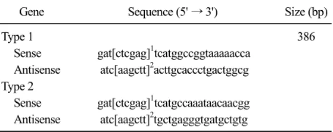
Table 1.Oligonucleotide primers used in this study
Gene Sequence (5' → 3') Size (bp)
Type 1 386
Sense gat[ctcgag]1tcatggccggtaaaaacca Antisense atc[aagctt]2acttgcaccctgactggcg Type 2
Sense gat[ctcgag]1tcatgccaaataacaacgg Antisense atc[aagctt]2tgctgagggtgatgctgtg [ ]1: Xho I site, [ ]2: Hind III site.
기서열 상동성과 50∼80%의 아미노산 상동성을 보이 고 있으며 각각의 유전자형 내에서 염기서열은 평균 12.5∼15% 그리고 최대 21∼30%의 유전적 차이가 있 는 것으로 알려져 있다(Forsberg, 2005; Drigo 등, 2014). PRRSV 유전자는 single-strand, positive-sense RNA로 크기는 15 Kb 정도이고 9개의 overlapping된 open reading frame (ORF: 1a, 1b, 2a, 3, 4, 5, 6 and 7) 을 가지고 있다. ORF1a와 ORF1b는 바이러스 복제와 전사에 필요한 14개의 비구조단백질을 coding한다 (Dokland, 2010). 피막을 가지고 있는 바이러스 입자 에는 3개의 중요한 단백, 즉 glycosylated envelope pro- tein (GP5, 25 kDa), unglycosylated membrane protein (M, 18 kDa), 그리고 nucleocapsid protein (N, 15 kDa) 등의 구조단백질이 있으며 이들 구조단백질은 각각 ORF5, ORF6 그리고 ORF7에 의하여 coding된다 (Jankova와 Celer, 2012). GP5 단백은 숙주세포의 수용 체 인식에 관여하여 M 단백과 함께 PRRSV 중화항체 형성에 관여하는 것으로 알려져 있다(Barfoed 등, 2004). M 단백은 PRRSV의 구조단백질 중에서 유전 학적으로 가장 conserve한 것으로 보고되었으며, 바이 러스의 assembly와 budding에 있어서 중요한 역할을 하는 것으로 알려져 있다(Hu 등, 2012; Jiang 등, 2006). ORF7에 의하여 coding되는 N 단백은 PRRSV 입자에서 가장 많은 부분을 차지하고 바이러스 입자 형성 시 바이러스 RNA와 작용하며 바이러스 복제과 정 중에 감염된 세포의 세포질뿐만 아니라 핵 내에서 도 확인된다(Lee 등, 2006). N 단백은 방어에 관여하 지는 않지만 복제과정 중에 많이 발현되어 PRRSV 진단에 있어서 주된 target 단백질로 알려져 있다.
PRRSV 감염기간 동안 높은 역가로 생산되는 N 단 백을 기본으로 한 ELISA kit가 개발되어 상용화되어 있으며, 이 kit는 감염이 의심되는 돼지의 혈청에서 PRRSV 특이항체의 변화 추이를 확인하기 혈청학적 검사 목적으로 전 세계적으로 널리 사용되고 있다 하 지만 가격이 비싼 것이 단점으로 알려져 있다. 최근에 는 PRRSV 감염을 확인할 수 있을 뿐만 아니라 type 1 과 type 2 PRRSV를 감별할 수 있는 RT-PCR법이 널리 사용되고 있어 단크론항체를 이용한 PRRSV 진단법 은 크게 관심을 끌지 못하고 있다. 하지만 바이러스 특이 단크론항체는 epitope mapping과 같은 바이러스 기초 연구에 널리 활용되고 있다. 특히, 국내에서 PRRSV 기초연구에 유용하게 사용될 수 있는 PRRSV 특이 단크론항체의 확보가 용이하지 않는 실정이다.
본 연구에서는 type1과 type2 PRRSV의 ORF7 유전
자를 Baculovirus expression체계에서 발현하고, ORF7 발현단백질에 특이적인 단크론항체를 생산하여 특성 을 규명하고자 하였다.
재료 및 방법
바이러스 및 세포
Type 1 PRRSV Lelystad와 CE-1 strain은 (주)중앙백 신연구소에서 제공받았으며 type 2 PRRSV PL97-1, CNV 그리고 CA-2 strain은 각각 농림축산검역본부, 충남학교 수의과대학 그리고 (주)중앙백신연구소에서 제공받아 사용하였다. 모든 PRRSV는 5% 소태아혈청 (Fetal bovine serum: FBS)과 gentamycin이 첨가된
-minimum essential medium (-MEM) 배지를 이용하 여 MARC-145 세포에서 증식시켰다. 단크론항체 생 산에 사용한 SP2/O 세포는 10% FBS가 함유된 RPMI 1640 배지를 사용하여 37
oC, 5% CO
2조건 하에서 배 양하였다.
ORF7 유전자 크로닝 및 발현
Type 1과 type 2 PRRSV ORF7 유전자를 크로닝하 고 발현하기 위하여 Lelystad (type 1)와 PL97-1 (type 2) strain을 각각 사용하였으며 RT-PCR법을 이용하여 각각 증폭하였다. ORF7 유전자 증폭에 사용할 primer 는 GenBank의 염기서열을 참고하여 제한효소 Xho I 과 Hind III 절단부위가 포함되도록 Table 1과 같이 제 작하여 다음과 같이 RT-PCR을 수행하였다. 즉, 추출 한 viral RNA를 주형으로 2.5 mM dNTP 3 L, 10x PCR buffer 2.5 L, MMLV RNA reverse transcriptase (Invitrogen, USA) 200U 0.5 L, 각각의 primer 0.25
L, RNA 시료 10 L와 함께 총량 25 L의 RT re-
action mixture를 만들고, 20
oC에서 10분, 42
oC에서 60 분 그리고 95
oC에서 5분간 반응시켜 cDNA를 합성하 였다. 합성된 cDNA template 5 L, 2.5 mM dNTP 2
L, 10x PCR buffer 2.5 L, 50mM MgCl
21 L, pri- mer 10 pM을 각각 1 L씩 첨가하고, Taq DNA poly- merase (Invitrogen) 0.2 L, Dnase free water를 첨가하 여 총 20 L의 PCR mixture를 제조하여 94
oC에서 1 분, 60
oC에서 1분, 72
oC에서 1분 반응하여 35회 증폭 하고 72
oC에서 7분간 post PCR을 실시하였다. 증폭한 반응산물은 1.5% agarose gel에서 전기영동한 후 증폭 유무를 확인하였다.
RT-PCR법으로 증폭된 각각의 ORF7 amplicon은 제 한효소
Xho I과 Hind III로 처리하고 전기영동한 후,QIAquick gel extraction kit (Qiagen GmbH, Germany)를 이용하여 제조사의 방법에 따라 정제하였다. 정제된 각각의 ORF7 유전자는 제조사의 술식에 따라 pBlueBac4.5/V5-His TOPO vector (Invitrogen)에 ligation 하고
E.coil DH5 competent cell에 transformation하여ampicillin (50 g/mL)이 포함된 LB plate에 도말하여 37
oC에서 24시간 동안 배양하였다. 배양된 집락 중 형 질전환으로 형성된 푸른색의 단일 집락을 여러 개 채 취하여 LB broth에 접종하고 37
oC shanking incubator 에서 24시간 진탕 배양하였다. 크로닝 유무를 확인하 기 위해 QIAGEN plasmid mini kit (QIAGEN
Ⓡ)를 사용 하여 제조사의 술식에 따라 plasmid를 추출하고 제한 효소를 처리하여 1% agarose gel에서 전기영동하여 ORF7 삽입여부를 확인하고 각각 pBlueBac-ORF7/Eu 와 pBlueBac-ORF7/Am으로 명명하였다.
Type 1과 type 2 PRRSV ORF7 유전자가 크로닝된 각각의 baculovirus transfer vecor pBlueBac-ORF7/Eu와 pBlueBac-ORF7/Am는 Bac-N-blue transfection kit
Ⓡ(Invitrogen)를 사용하여 Bac-N-blue wild type DNA와 함께 제조사의 술식에 따라 Sf9 세포로 cotransfection 하였다. 재조합된 바이러스는 28
oC에서 7일간 배양 후 plaque picking하여 순수분리하여 각각 rORF7/Eu와 rORF7/Am으로 명명하였다. Plaque법으로 순수분리한 재조합 baculovirus rORF7/Eu와 rORF7/Am은 rabbit an- ti-His
6특이항체(Bethyl laboratories, USA)와 PRRSV antibody test kit(IDEXX PRRS X3, Switzerland)를 사용 하여 PRRSV 감염 양성으로 확인된 돼지혈청을 사용 하여 Yoon 등(2012)의 방법에 따라 indirect fluo- rescence antibody (IFA)법을 수행하여 각각의 ORF7 발 현단백질이 정상적으로 발현되는가를 확인하였다.
ORF7 발현단백질 특이 단크론항체
Type 1과 type 2 PRRSV ORF7 발현단백질에 특이 적인 단크론항체를 생산하기 위해 Lelystad와 CNV strain을 각각 사용하였으며, 농축한 항원은 8주령 BALB/c 마우스의 foot-pad에 4일 간격으로 1차 접종 에는 Freund’s complete adjuvant와 그리고 이후에는 Freund’s incomplete adjuvant와 혼합하여 3회 접종하 여 면역하였다.
세포융합은 Kang 등(1989)과 Yoon 등(2012)의 방법을 사용하여 수행하였다 . 즉, PRRSV로 면역한 마우스의 비 장과 서혜림프절로부터 준비한 세포와 SP2/0 세포를 polyethylene glycol (PEG1500: Roche diagnostics, Ger- many)을 사용하여 융합하였다. 융합된 세포는 20% FBS 와 HAT (50 M hypoxanthine, 0.4 M aminopterin, 16 M thymidine)가 첨가된 RPMI 1640 배지에 부유시켜 96-well plate에 100 L씩 분주하여 37
oC, CO
2incubator에 배양하 였다. 배양 후 3일에 HAT 100 L를 첨가하고 5, 7, 9일째 HT 배지로 교환하여 융합된 세포의 증식성을 확인하였 다. 융합된 세포의 colony가 30% 이상 증식되면 상층액 을 채취하여 Sf9 세포에 rORF7/Eu와 rORF7/Am를 감염 시켜 준비한 plate를 사용하여 IFA법으로 PRRSV ORF7 발현단백질에 특이적으로 반응하는 단크론항체를 선별 하였다 . PRRSV ORF7 발현단백질에 대하여 양성으로 선 별된 hybridoma는 Fuller 등(2001)의 limiting dilution법으 로 크로닝하고 , Ig isotyping kit (Sigma, USA)를 사용하여 단크론항체의 isotype을 확인하였다.
생산된 단크론항체와 PRRSV 사이의 교차반응성은 Yoon 등(2012)의 방법에 따라 IFA법으로 확인하였으 며 type 1 PRRSV로 Lelystad와 CE-1 strain 그리고 type 2 PRRSV로 PL97-1, CNV, CA-2 strain을 사용하 였다. 표준 PRRSV ORF7 발현단백질 특이 단크론항 체로 SDOW17 (Rural technologies, USA)을 구입하여 사용하였다.
단크론항체의 진단적 활용
생산된 PRRSV ORF7 특이 단크론항체의 진단적 활
용성을 확인하기 위하여 RT-PCR법으로 type 1과 type
2 PRRSV 감염이 확인된 돼지로부터 조직(림프절, 폐,
편도)을 채취하여 IFA와 면역조직화학(Immunohisot-
chemistry: IHC) 염색법을 수행하였다. IFA는 감염된
조직을 동결절편하여 PRRSV ORF7 특이 단크론항체
로 처리한 후, FITC conjugated anti-mouse antibody
Fig. 1. Confirmation of expressed ORF7 protein in recombinant viruses. SF9 cells infected with rORF7/Am and rORF7/Eu were re- acted with rabbit anti-His6 and porcine anti-PRRSV serum, re- spectively,and then stained with FITC-conjugated secondary antibody.
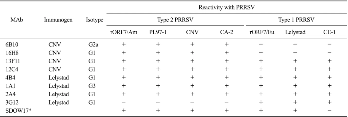
Table 2. Characteristics of porcine reproductive and respiratory syndrome virus (PRRSV) ORF7-specific monoclonal antibodies (MAb)
MAb Immunogen Isotype
Reactivity with PRRSV
Type 2 PRRSV Type 1 PRRSV
rORF7/Am PL97-1 CNV CA-2 rORF7/Eu Lelystad CE-1
6B10 CNV G2a + + + + - - -
16H8 CNV G1 + + + + - - -
13F11 CNV G1 + + + + + + +
12C4 CNV G1 + + + + + + +
4B4 Lelystad G1 + + + + + + +
1A1 Lelystad G3 + + + + + + +
2A4 Lelystad G1 + + + + + + +
3G12 Lelystad G1 - - - - + + +
SDOW17* + + + + + + -
*Reference PRRSV ORF7-specific MAb.
(Kirkegaard & Perry laboratories, USA)를 처리하여 반 응시키고 형광현미경으로 관찰하였다. IHC 염색을 위 하여 감염조직을 10% neutral buffered formalin 용액으 로 48시간 고정 후, 일반적인 조직 처리과정을 거쳐 조 직절편을 준비하였다. 염색 전에 조직절편은 xylene을 사용하여 wax를 제거하였으며 0.5% 과산화수소가 함 유되어 있는 methanol로 30분간 처리하여 조직 내의 peroxidase의 활성을 제거하고 5% normal goat serum으 로 30분간 처리하여 항체의 비특이적 결합을 배제하였 다. 1차 항체로 PRRSV ORF7 특이 단크론항체를 실온 에서 30분간 반응시킨 후, 2차 항체로 biotinylated goat anti-mouse antibody (Vector laboratories, USA)로 30분간 반응시켰다. 같은 방법으로 세척하고 ABC reagent (avidin DH-biotinylated horseradish peroxidase complex, Vector laboratories)로 실온에서 30분간 반응시킨 후, 발 색액(0.6 mg/mL DAB, 0.03% H
2O
2, 0.01M PBS, pH 7.2) 으로 5∼10분간 반응시키고 증류수로 세척하였다.
결 과
ORF7 유전자 크로닝 및 발현
Type 1과 type 2 PRRSV ORF7 유전자를 크로닝하 기 위하여 Lelystad와 PL97-1 strain으로부터 RNA를 추출하여 ORF7 유전자 특이 primer로 RT-PCR을 실 시하여 전기 영동한 결과, 386 bp에 해당하는 유전자 증폭산물을 확인할 수 있었다. 또한, baculovirus trans- fer vector인 pBlueBac-ORF7/Eu와 pBlueBac-ORF7/Am 로부터 DNA를 추출하여 제한효소 Xho I과 Hind III로 처리하고 전기영동한 결과, 삽입된 ORF7 유전자 크 기에 해당하는 386 bp에서 제한효소 소화절편이 확 인되었다. Plaque법으로 순수 분리한 recombinant ba- culovirus에서 ORF7 유전자가 발현되는지 확인하기 위하여 rabbit anti-His
6특이항체와 돼지 anti-PRRSV 혈청을 사용하여 IFA를 실시한 결과, 재조합 baculo- virus rORF7/Eu와 rORF7/Am가 감염된 Sf9 세포에서 만 형광을 나타낸 반면 mock-infected Sf9 세포에서는 어떠한 형광도 나타내지 않았다(Fig. 1).
ORF7 발현단백질 특이 단크론항체
PRRSV로 면역시킨 마우스를 이용한 세포융합을 통
해 총 8개의 ORF7 발현단백질 특이 단크론항체를 생산
하였으며 이들의 특성은 Table 2와 같다. 4개(6B10,
16H8, 13F11, 12C4)의 단크론항체는 면역원으로 type 2
PRRSV CNV strain을 사용한 세포융합에서 생산되었으
며, 나머지 4개(4B4, 1A1, 2A4, 3G12)의 단크론항체는
type 1 PRRSV Lelystad strain을 사용한 세포융합에서 생
Fig. 2. Reactivity patterns of PRRSV ORF7-specific monoclonal an- tibodies by indirect fluorescence antibody (IFA) test. MARC145 cells infected with type 1 (CE-1) and type 2 (CA-2) PRRSV were reacted with PRRSV ORF7-specific monoclonal antibodies (16H8, 12C4, 3G12) and then stained with FITC- conjugated secondary antibody.
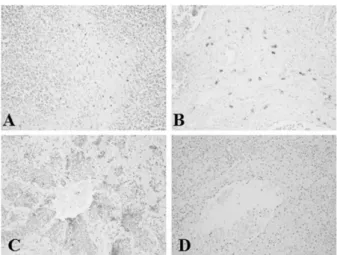
Fig. 4.Immunohistochemical staining of porcine tissues with PRRSV ORF7-specific monoclonal antibody. Lymph node (A), tonsil (B) and lung (C, D) from PRRSV infected pigs were reacted with PRRSV ORF7-specific monoclonal antibody 12C4 (A, B, C) and 3G12 (D), respectively.
Fig. 3.Confirmation of PRRSV infection by indirect fluorescence antibody (IFA) test using PRRSV ORF7-specific monoclonal antibody.
Lymph node (A), tonsil (B) and lung (C, D) from PRRSV infected pigs were reacted with PRRSV ORF7-specific monoclonal antibody 12C4 (A, B, C) and 3G12 (D), respectively.
산되었다. 생산된 단크론항체의 isotype은 IgG1이 6개로 가장 많았으며 IgG2a와 IgG3가 각각 하나로 나타났다.
생산된 단크론항체와 type 1과 type 2에 속하는 PRRSV 사이의 교차반응성을 IFA법으로 확인한 결과, 3개의 Group (I, II, III)으로 구분되었는데 Group II에 속하는 5 개(1A1, 2A4, 4B4, 12C4, 13F11)의 단크론항체는 type 1 과 type 2 PRRSV 모두와 반응하였다. 반면에 Group I에 속하는 2개(6B10, 16H8)의 단크론항체는 type 2 PRRSV 에만 반응하고 , Group III에 속하는 단크론항체 3G12는 type 1 PRRSV에만 반응하는 것으로 나타났다(Table 2, Fig. 2). 이러한 결과로 본 연구에서 PRRSV 각각의 type 에 특이적으로 반응하는 단크론항체와 각각의 type에 공통적으로 반응하는 단크론항체가 모두 생산되었다.
단크론항체의 진단적 활용
PRRSV ORF7 발현단백질에 특이적인 단크론항체 의 진단적 활용성을 확인하기 위하여 PRRSV에 감염 된 돼지의 림프절, 폐 그리고 편도를 이용하여 IFA와 IHC 염색을 수행한 결과, type 2 PRRSV ORF7 발현 단백질에 특이적으로 반응하는 단크론항체인 16H8의 경우, 같은 type의 PRRSV에 감염된 돼지 조직에서 모두 양성반응이 나타났으나 type 1 PRRSV에 감염된 돼지 조직에서는 반응이 나타나지 않았다. 반면에 type 1 PRRSV ORF7 발현단백질 에 특이적으로 반응 하는 단크론항체인 3G12의 경우, type 1과 type 2 PRRSV에 감염된 돼지 조직 모두에서 반응이 나타나 지 않았다. 하지만 type 1과 type 2 PRRSV ORF7 발 현단백질에 공통적으로 반응하는 단크론항체인 12C4
의 경우, type 1과 type 2 PRRSV에 감염된 돼지 조직 모두에서 반응이 강하게 나타났다(Fig. 3, Fig. 4).
고 찰
바이러스에서 방어에 관여하는 유전자를 prokary-
otic/eukaryotic expression system을 이용하여 발현시키
고, 발현된 단백질을 백신개발에 사용하는 연구가
Norovirus, Papillomavirus 그리고 Hepatitis B virus와 같
이
in vitro에서 증식되지 않는 바이러스를 포함한 많은 바이러스에서 활발하게 진행되고 있다(Parra 등, 2012; Park 등, 2008; Michel과 Tiollais, 2010). PRRSV에 서도 백신개발 및 진단법 개발 목적으로 서로 다른 유 전자에 대한 단백질 발현 연구가 보고되었다. PRRSV ORF7 발현단백질은 PRRSV 단백 중에서 가장 면역원 성이 높아 돼지에 백신 또는 야외 감염이 일어났을 때 다른 ORF 발현단백질보다 빠르고 높은 항체 형성능을 보이고, 반복적인 백신 또는 백신 후 야외 감염 시 높 은 수준으로 항체가 형성 된다(Barfoed 등, 2004). 또한 체액성 면역과 세포성 면역을 증가시키고 감염된 동물 에서 viral load를 감소시키는 역할을 한다고 보고되었 다. ORF7 발현단백질의 이러한 기능적 특성은 경구투 여가 가능한 Salmonella 발현 system을 통해 전신면역 과 점막면역을 증가시킴으로서 PRRSV의 면역을 증가 시키는 연구도 진행되고 있다(Han 등, 2011). 또한, PRRSV ORF7 발현단백질은 viral protein에서 20∼40%
정도의 비중을 차지하는 가장 많은 단백질(Barfoed 등, 2004)로 진단적 활용에 많이 사용되고 있다. 본 실험에 서는 Baculovirus expression system을 이용해 type 1과 type 2 PRRSV ORF7 유전자를 발현하고 특성을 규명 하였다. 토끼 anti-His
6특이항체와 돼지 anti-PRRSV 혈 청을 사용하여 확인한 결과, ORF7 발현단백질이 정상 적으로 발현되었음을 확인하였으며, 현재 ORF7 발현 단백질을 순수분리하여 PRRSV 특이항체를 검색할 수 있는 ELISA법을 수행하고 있다. ELISA 방법은 PRRSV의 혈청학적 진단법에서 가장 흔히 사용되고 있는 방법으로 국내에서도 진단적 활용에 많이 사용되 고 있으며, 발현된 N 단백을 이용한 PRRSV ELISA kit 가 개발되었으며 상업적으로 판매되고 있다(Denac 등, 1977; Kreutz와 Mengeling, 1997; Cong 등, 2013).
PRRSV에 특이적으로 반응하는 단크론항체는 PRRSV 연구에 매우 중요하게 사용되고 있다. 중화능 력이 있는 단크론항체는 바이러스 중화와 관련된 항 원결정기를 확인하는데 필수적으로 요구되며 nucleo- capsid(N) 단백에 특이적인 단크론항체는 진단법 개 발에 널리 이용되고 있다. 이러한 이유로 PRRSV ORF4, ORF5 그리고 ORF7에 특이적인 단크론항체가 생산되었고 이들을 이용하여 각각의 단백질에 대한 epitope mapping 연구가 보고되었다(Nelson 등, 1993;
Drew 등, 1995; Pizaden과 Dea, 1997; Rodriguez 등, 1997; Weiland 등, 1999; Van Breedam 등, 2011). 본 실험에서는 type 1과 type 2 PRRSV ORF7에 특이적
으로 반응하는 단크론항체를 생산하고, 생산된 단크 론항체의 type에 따른 반응성을 확인함으로써 단크론 항체의 연구적 활용성과 진단적 응용성을 확인하고 자 하였다. PRRSV PL97-1과 Lelystad strain을 면역원 으로 하여 총 8개의 항체를 생산하였으며 5개의 단크 론항체는 type 1과 type 2 PRRSV에 공통적으로 반응 하는 것으로 확인되었으며 나머지 단크론항체는 각 각의 type PRRSV에 반응하는 것으로 나타나 PRRSV N 단백에는 적어도 2개 이상의 항원결정기가 존재할 것으로 추정할 수 있었다. Nelson 등(1993)은 type 2 PRRSV에 특이적인 단크론항체가 N 단백에 특이적이 고, 일부 단크론항체는 type 2 PRRSV에만 반응하였으 나 일부 단크론항체는 type 1과 type 2 PRRSV N 단백 의 conserved epitope에 반응하는 것으로 보고하였다.
Drew 등(1995)은 type 1 PRRSV에 대한 단크론항체를 생산하여 특성을 규명한 결과, 4개의 단크론항체가 N 단백에 특이적이고 이들 단크론항체는 type 1 PRRSV 에만 반응하는 것으로 보고하였다. Rodriguez 등(1997) 은 단크론항체를 이용한 epitope mapping 연구에서 3 개의 항원결정기가 존재하고 있다고 보고하였다.
Van Breedam 등(2011)은 2개의 N 단백 특이 단크론
항체는 type 1과 type 2 PRRSV 공통적으로 반응하고
하나의 단크론항체는 type 1 PRRSV하고만 반응하여
type 1과 type 2 PRRSV를 감별하는데 활용될 수 있으
며, 조직병리학적으로 처리된 조직에서도 PRRSV를
확인할 수 있어 진단적으로 가치가 있음을 보고하였
다. 본 연구에서 type 2 PRRSV ORF7 발현단백질에
특이적으로 반응하는 단크론항체 16H8의 경우, 같은
type의 PRRSV에 감염된 돼지 조직에서 모두 양성반
응이 나타나고, type 1 PRRSV에 감염된 돼지 조직에
서는 반응이 나타나지 않아 type 1과 type 2 PRRSV를
감별하는데 활용될 수 있을 것으로 사료된다. 하지만,
type 1 PRRSV ORF7 발현단백질에 특이적으로 반응
하는 단크론항체 3G12의 경우, type 1과 type 2
PRRSV에 감염된 돼지 조직 모두에서 반응이 나타나
지 않았는데 이는 3G12의 activity가 낮은 것으로 추
정되며, type 1과 type 2 PRRSV를 감별을 위하여 보
다 반응성이 강한 단크론항체의 확보가 필요할 것으
로 생각된다. 본 실험에서 생산된 단크론항체는 국내
에서 순환되고 있는 바이러스의 분리와 병인학적 연
구 그리고 기본적인 바이러스에 대한 연구를 위한 좋
은 도구로서 활용될 것으로 사료된다.
감사의 글
이 논문은 2012학년도 충북대학교 학술연구지원사 업의 연구비 지원에 의하여 연구되었습니다.
참 고 문 헌
Barfoed AM, Blixenkrone-Moller M, Jensen MH, Botner A, Kamstrup S. 2004. DNA vaccination of pigs with open reading frame 1-7 of PRRS virus. Vaccine 22: 3628- 3641.
Cavanagh D. 1997. Nidovirales: a new order comprising Corona- viridae and Arteriviridae. Arch Virol 142: 629-633.
Cong Y, Huang Z, Sun Y, Ran W, Zhu L, Yang G, Ding X, Yang Z, Huang X, Wang C, Ding Z. 2013. Development and application of a blocking enzyme-linked im- munosorbent assay (ELISA) to differentiate antibodies against live and inactivated porcine reproductive and res- piratory syndrome virus. Virology 444: 310-316.
Denac H, Moser C, Tratschin JD, Hofmann MA. 1997. An in- direct ELISA for the detection of antibodies against por- cine reproductive and respiratory syndrome virus using recombinant nucleocapsid protein as antigen. J Virol Methods 65: 169-181.
Dokland T. 2010. The structural biology of PRRSV. Virus Res 154: 86-97.
Drew TW, Meulenberg JJM, Sands JJ, Paton DJ. 1995. Produc- tion, characterization and reactivity of monoclonal anti- bodies to porcine reproductive and respiratory syndrome virus. J Gen Virol 76: 1361-1369.
Drigo M, Franzo G, Gigli A, Martini M, Mondin A, Gracieux P, Ceglie L, 2014. The impact of porcine reproductive and respiratory syndrome virus genetic heterogeneity on mo- lecular assay performances. J Virol Methods 202: 79-86.
Forsberg R. 2005. Divergence time of porcine reproductive and respiratory syndrome virus subtypes. Mol Virol Evol 22:
2131-2134.
Frossard JP, Fearnley C, Naidu B, Errington J, Westcott DG, Drew TW. 2012. Porcine reproductive and respiratory syndrome virus: antigenic and molecular diversity of British isolates and implications for diagnosis. Vet Microbiol 158: 308-315.
Frossard JP, Hughes GJ, Westcott DG, Naidu B, Williamson S, Woodger NG, Steinbach F, Drew TW. 2013. Porcine re- productive and respiratory syndrome virus: genetic di- versity of recent British isolates. Vet Microbiol 162:
507-518.
Fuller SA, Takahashi M, Hurrel JG. 2001. Cloning of hybridoma cell lines by limiting dilution. Chapt 11:Unit 11.8. In:
Ausubel FM(ed.). Current Protocol in Molecular Bio- logy. Wiley-Interscience.
Han YW, Kim SB, Rahman M, Uyangaa E, Lee BM, Kim JH,
Park KI, Hong JT, Han SB, Eo SK. 2011. Systemic and mucosal immunity induced by attenuated Salmonella en- terica serovar Typhimurium expressing ORF7 of porcine reproductive and respiratory syndrome virus. Comp Immunol Microbiol Infect Dis 34: 335-345.
Hu J, Ni Y, Meng XJ, Zhang C. 2012. Expression and purifica- tion of a chimeric protein consisting of the ectodomains of M and GP5 proteins of porcine reproductive and res- piratory syndrome virus (PRRSV). J Chromatogr B Analyt Technol Biomed Life Sci 911: 43-48.
Jankova J, Celer V. 2012. Expression and serological reactivity of Nsp7 protein of PRRS genotype I virus. Res Vet Sci 93: 1537-1542.
Jiang Y, Xiao S, Fang L, Yu X, Song Y, Niu C, Chen H. 2006.
DNA vaccines co-expressing GP5 and M proteins of porcine reproductive and respiratory syndrome virus (PRRSV) display enhanced immunogenicity. Vaccine 24:
2869-2879.
Kang SY, Saif LJ, Miller KL. 1989. Reactivity of VP4-specific monoclonal antibodies to a serotype 4 porcine rotavirus with distinct serotypes of human (symptomatic and asymptomatic) and animal rotaviruses. J Clin Microbiol 27: 2744-2750.
Kreutz LC, Mengeling WL. 1997. Baculovirus expression and im- munological detection of the major structural pproteins of porcine reproductive and respiratory syndrome virus.
Vet Microbiol 59: 1-13.
Lee C, Hodgins D, Calvert JG, Welch SK, Jolie R, Yoo D. 2006.
Mutations within the nuclear localization signal of the porcine reproductive and respiratory syndrome virus nu- cleocapsid protein attenuate virus replication. Virology 346: 238-250.
Michel ML, Tiollais. 2010. Hepatitis B vaccine: protective effi- cacy and therapeutic potential. Pathol Biol (Paris) 58:
288-295.
Nelson EA, Christopher-Hennings J, Drew T, Wensvoort G, Collins JE, Benfield DA. 1993. Differentiation of U.S.
and European isolates of porcine reproductive and respi- ratory syndrome virus by monoclonal antibodies. J Clin Microbiol 31: 3184-3189.
Park MA, Kim HJ, Kim HJ. 2008. Optimum conditions for pro- duction and purification of human papillomavirus type 16 Li protein from Saccharomyces cerevisiae. Protein Expr Purif 59: 175-181.
Parra GI, Bok K, Taylor R, Haynes JR, Sosnovtsev SV, Richardson CR, Green KY. 2012. Immunogenicity and specificity of norovirus consensus GII.4 virus-like par- ticles in monovalent and bivalent vaccine formulations.
Vaccine 30: 3580-3586.
Pirzadeh B, Dea S. 1997. Monoclonal antibodies to the ORF5 product of porcine reproductive and respiratory syn- drome virus define linear neutralizing determinants. J Gen Virol 78: 1867-1873.
Rodriguez MJ, Sarraseca J, Garcia J, Sanz A, Plana-Duran J, Casal JI. 1997. Epitope mapping of the nucleocapsid
protein of European and North American isolates of re- productive and respiratory syndrome virus. J Gen Virol 78: 2269-2278.
Van Breedam W, Costers S, Vanhee M, Gagnon CA, Rodri- guez-Gomez IM, Geldhof M, Verbeeck M, Van Doorsselaere J, Karniychuk U, Nauwynck HJ. 2011.
Porcine reproductive and respiratory syndrome virus (PRRSV)-specific mAbs: supporting diagnostics and pro- viding new insights into the antigenic properties of the virus. Vet Immunol Immunopathol 141: 246-257.
Venteo A, Rebollo B, Sarraseca J, Rodriguez MJ, Sanz A. 2012.
A novel double recognition enzyme-linked immunosor- bent assay based on the nucleocapsid protein for early detection of European porcine reproductive and respira- tory syndrome virus infection. J Virol Methods 181:
109-113.
Weiland E, Wieczorek-Krohmer M, Kohl D, Conzelmann KK, Weiland F. 1999. Monoclonal antibodies to the GP5 of Porcine reproductive and respiratory syndrome virus are more effective in virus neutralization than monoclonal antibodies to the GP4. Vet Microbiol 66: 171-186.
Yoon YS, Lee SC, Woo SK, Cho KO, Kang SY. 2012. Monoclo- nal antibodies against porcine group C rotavirus VP6.
Korean J Vet Serv 35: 175-182.
Zhou Z, Ni J, Cao Z, Han X, Xia Y, Zi Z, Ning K, Liu Q, Cai L, Qiu P, Deng X, Hu D, Zhang Q, Fan Y, Wu J, Wang L, Zhang M, Yu X, Zhai X, Tian K. 2011. The epidemic status and genetic diversity of 14 highly pathogenic por- cine reproductive and respiratory syndrome virus (HP- PRRSV) isolates from China in 2009. Vet Microbiol 150: 257-269.


