J O U R N A L O F
Veterinary Science
Short Communication
J. Vet. Sci. (2010), 11(2), 169171 DOI: 10.4142/jvs.2010.11.2.169
*Corresponding author
Tel: +82-53-950-7365; Fax: +82-53-955-5522 E-mail: [email protected]
†First two authors contributed equally to this study.
Development and characterization of stable cell lines constitutively expressing the porcine reproductive and respiratory syndrome virus nucleocapsid protein
Mingeun Sagong
1,†, Choi-Kyu Park
2,†, Seong-Hee Kim
2, Sung-Up Moon
3, Seong-Cheol Cho
3, Changhee Lee
1,*
1
Department of Microbiology, College of Natural Sciences, Kyungpook National University, Daegu 702-701, Korea
2
Animal Disease Diagnostic Center, National Veterinary Research and Quarantine Services, Anyang 430-757, Korea
3
Livestock Policy Division, Jeju Special Self-Governing Province, Jeju 690-700, Korea
Despite global efforts to control porcine reproductive and respiratory syndrome virus (PRRSV) infection, the virus continues to cause economic problems in the swine industry worldwide. In this study, we attempted to generate and characterize a panel of stable BHK cell lines that constitutively express the nucleocapsid (N) protein of type 1 or type 2 PRRSV. The established BHK cell lines were found to react well with N-specific antibodies as well as the hyperimmune serum of pigs raised against each genotype of PRRSV. Taken together, the data implicate a potential usefulness for the newly generated stable cell lines as a diagnostic reagent for PRRSV serology.
Keywords: diagnostic reagent, nucleocapsid protein, PRRSV, stable BHK cell line
Porcine reproductive and respiratory syndrome (PRRS) emerged almost simultaneously in North America and Europe.
This disease has since plagued nearly all pig-producing countries with a massive economic impact on the swine industry worldwide [7]. The causative agent, PRRS virus (PRRSV), is a small enveloped, single-stranded, positive- sense RNA virus belonging to the family Arteriviridae in the order Nidovirales [9]. PRRSV is divided into two major genotypes, the European (EU; type 1) genotype and the North American (NA; type 2) genotype. These two genotypes were formerly located on different continents [5] but intermingled existence of the two genotypes has now been identified in many parts of the world, including Korea [4,8].
This situation leads to potentially problematic consequences for PRRSV diagnostics and management, since current
serological tools are usually incapable of identifying whether a pig is infected with a single genotype or co-infected with two genotypes.
The nucleocapsid (N) protein of PRRSV encoded in ORF7 is a small basic multifunctional protein with a molecular weight of 15-kDa [9]. The PRRSV N protein is the most abundant protein in infected cells, constituting approximately 40% of the protein content of the virion, and is known to be highly immunogenic in the natural host [3].
Furthermore, the N protein is encoded by a region of the viral genome that is relatively well conserved between the two genotypes [6]. Thus, these properties enable the N gene to be a proper candidate for the detection of antibodies against PRRSV. In the present study, we aimed to generate a panel of stable BHK cell lines constitutively expressing N proteins from EU and NA genotypes of PRRSV and subsequently to assess their potential use as a diagnostic reagent in immunofluorescence assay (IFA) and immunoperoxidase monolayer assay (IPMA) techniques.
The viral RNA was extracted from each virus stock of Korean type 1 and 2 PRRSV strains, KNU-07 and PL97-1 [1,4], by using the Viral RNA Mini Kit (Qiagen, Germany).
RT-PCR was performed to amplify the full-length ORF7
gene from EU (KNU-07) and NA (PL97-1) genotypes with
the following corresponding primer pairs: KNU07-ORF7-F
(5´-GCCGGGATCCACCATGGCCGGTAGAAAC-3´),
KNU07-ORF7-R (5´-GCCGGGATCCATTTGCATCCTG
ACTGG-3´), PL97-ORF7-F (5´-GCCGGGATCCACCAT
GCCAAATAACAACGGC-3´) and PL97-ORF7-R (5´-GC
CGGGATCCTGCTGAGGGTGATGC-3´), where underlines
indicate the BamHI restriction enzyme sequence. Each PCR
amplicon was initially inserted individually into a
pBudCE4.1 Vector (Invitrogen, USA) that contains six
repetitive histidine codons. Individual His-tagged PRRSV
ORF7 cDNA fragments were then subcloned independently
into a pFB-Neo Retroviral Vector (Stratagene, USA) using
SalI and EcoRI restriction sites.
170 Mingeun Sagong et al.
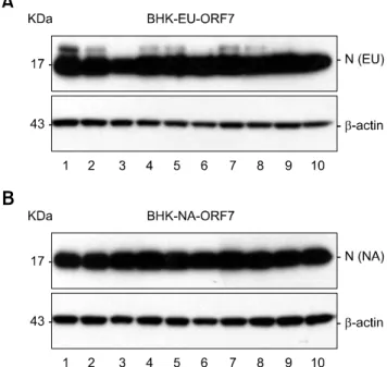
Fig. 1. Constitutive expression of the genotype-specific nucleo- capsid (N) protein in BHK-EU-ORF7 (A) and BHK-NA-ORF7 (B) cells. Expression levels of the N protein in individual stable cell clones were determined by western blot analysis. Each lane represents individual neomycin-resistant BHK cell clones.
Fig. 2. Immunofluorescence assay using the genotype-specific monoclonal antibodies. BHK-EU-ORF7 and BHK-NA-ORF7 cells were incubated individually with each genotype-specific antibody. A and D
α-EU: EU genotype-specific monoclonal antibody (MAb), B and E α-NA: NA genotype-specific MAb, C and F α-EU + NA: genotype-common MAb. ×40.
Fig. 3. Immunoperoxidase monolayer assay. Cells were incubated with the genotype-specific MAbs or PRRSV-specific hyperimmune sera followed by staining using Vectorstain ABC peroxidase and DAB substrate kits. G and J α-LV: anti-LV pig serum, H and K
α-VR-2332: anti-VR-2332 pig serum, I and L Pre-immune: normal pre-immunized pig serum. ×10.
The retrovirus gene transfer system (Stratagene, USA) was applied to generate BHK cell lines constitutively expressing the recombinant ORF7 gene [2]. The selected cell clones (BHK-EU-ORF7 and BHK-NA-ORF7) in the presence of 800 μ g/ml G418 (Invitrogen, USA) were initially subjected to PCR to identify the PRRSV ORF7 gene integration, followed by RT-PCR and nucleotide sequencing to determine N gene expression at the mRNA level. The full-length N genes of about 400 bp were identifiable from individual BHK-EU-ORF7 and BHK-NA-ORF7 cell clones (data not shown). Next, N protein expression at the protein level was tested by western blot analysis [2] and robust levels of
approximately 15-kDa N were detected in all selected BHK cell clones (Figs. 1A and B, upper panels). Each BHK-EU- ORF7 or BHK-NA-ORF7 cell clone that expressed the highest level of N protein was chosen for subsequent studies.
The BHK-EU-ORF7 and BHK-NA-ORF7 cell lines were further examined for the subcellular expression of N gene by IFA [2]. As expected, the specific cytoplasmic and nucleolus staining was clearly evident when the cells were reacted with the anti-N monoclonal antibody (MAb) SDOW17, confirming the constant high level expression of the N protein. In addition, the overall growth kinetics of each N gene-expressing cell line was found to be similar to that of the parental BHK-21 cells, indicating that the N expression has no effect on cell growth (data not shown).
This result further suggests their potential use as a suitable
Development of PRRSV N expressing cells 171