1)
본 논문의 요지는 2004년 제54차 대한소아과학회 추계학술대회에서 포스터 발표하였음.
책임저자 : 이준호, 포천중문의과대학교 분당차병원 소아과
Tel : 031)780-5237, Fax : 031)780-5239, E-mail : [email protected]
Clinical Characteristics of the
Epidemic Mycoplasma pneumoniae Pneumonia Outbreak in 2003∼2004
Hye-Oak Kwon, M.D., Shin-I Park, M.D. and Jun-Ho Lee, M.D.
Department of Pediatrics, Bundang CHA General Hospital, College of Medicine Pochon CHA University, Sungnam, Korea
= Abstract =
Purpose : A wide, epidemic outbreak of M. pneumoniae pneumonia occurred throughout Korea in late 2003. Compared with previous years, the 2003 outbreak resulted in more se- vere cases and in an increased incidence of extrapulmonary symptoms and/or complications.
We compared the clinical characteristics for M. pneumoniae pneumonia of 2003 to those of the past years.
Methods : One hundred six children diagnosed with M. pneumoniae pneumonia by sero- logic tests at Bundang Cha General Hospital between Aug 2003 to April 2004 were enrolled.
Medical records were reviewed retrospectively, for clinical, laboratory and radiological aspect as well as complications. The pleural effusions of 3 patients who underwent thoracentesis were also analyzed.
Results : The duration of fever, cough, rhinorrhea, and sore throat was 8.2±4.7, 22.1±
4.8, 8.4±2.1, 4.3±1.2 days, respectively. The incidence (percentage) and duration of abdom- inal pain, vomiting, diarrhea, headache, skin rash, arthralgia was 5.1±2.5 (21.9%), 3.4±2.1 (17.1%), 4.3±1.8 (16.2%), 3.5±2.1 (14.4%), 5.5±0.7 (5.9%) and 4.6±1.3 days (4.9%), re- spectively. The mean duration of admission and treatment were 7.4±4.3 days and 21.6±11.1 days. Higher values of CRP and ESR on admission were positively correlated with the dura- tion of fever and length of admission. The findings of pleural effusion were similar to those seen in TB pleurisy. Complications, including myocarditis (2 cases), arthritis (3 cases), vascu- litis (5 cases), asthma (3 cases), ARDS (1 case), and DIC (2 cases) were observed in 14.1%
of patients.
Conclusion : We found a number of characteristics of M. pneumoniae pneumonia among cases from late 2003 that were different from those of previous years. This outbreak resulted in more severe cases and in an increased incidence of extrapulmonary symptoms and/or com- plications. A multicenter study is needed to verify the changes in clinical characteristics observed during the 2003 outbreak from previous ones.
. Key Words : Mycoplasma pneumoniae pneumonia, Epidemic outbreak
Introduction
Mycoplasma pneumoniae is a respiratory pathogen that primarily causes bronchitis and pneumonia as well as pharyngitis, croup, and bronchiolitis.
Among school-aged children, M. pneumoniae is the most common causative organism of community- acquired pneumonia, and M. pneumoniae pneumonia is prevalent among even younger children who begin daycare at an earlier age1, 7). While the sporadic spread of M. pneumoniae pneumonia occurs each year, epidemic outbreaks occur every 4∼7 years. In Korea, epidemics of M. neumoniae are known to occur every 3∼5 years and data on the characteris- tics of these epidemics have been widely published.
According to reports6, 7) from metropolitan areas pri- or to 1999, the occurrences of M. pneumoniae pneu- monia occurred at 3-year intervals with durations of 1 year. The majority of studies showed that M.
pneumoniae infections have a benign, uncomplicated clinical course and few complications. During late 2003 and early 2004, a nation-wide outbreak of M.
pneumoniae infection occurred. Compared with the previous outbreaks, this one resulted in more severe cases and in an increased incidence of extrapulmo- nary symptoms and/or complications. Therefore, a retrospective study was undertaken to determine the clinical features of patients presenting to our hospital during the outbreak.
Materials and Methods
1. Subjects
A retrospective analysis of the medical records of 106 patients (Group 2) diagnosed with M. pneumo- niae pneumonia and admitted to Bundang Cha Gen- eral Hospital between Aug 2003 and April 2004 was done. Clinical characteristics were compared with 42 patients (Group 1) diagnosed with M. pneu- moniae pneumonia from Jan 2002 to Dec 2002.
2. Diagnostic criteria
The diagnoses were confirmed by serological tests of mycoplasma antibody titers, which were measured using a Serodia-Myco II gelatin particle agglutinin test kit (Fujirebio Co., Japan), as an indirect hemag- glutination test. The stated diagnostic criteria re- quired that all of the following were present : ① respiratory symptoms, and ② either a single myco- plasma antibody titer rise to 1 : 320 or greater at the time of admission, or paired samples showing at least a 4-fold rise over a 1∼2 week period.
3. Clinical, radiological and laboratory measurements
Medical records were reviewed retrospectively for : ① the duration of fever, respiratory symptoms, admission, and treatment, ② the incidence of extra- pulmonary symptoms, and ③ the occurrence of com- plications.
Laboratory data such as CRP, ESR, leukocyte count, and mycoplasma antibody titers from blood samples taken both at the time of admission and follow up were evaluated. Radiological variables in- cluded the site of pulmonary infiltration shown on chest X-rays and the incidence of pleural effusion.
The pleural effusions of 3 patients who underwent thoracentesis were analyzed. In addition, our analysis included data from chest CT scans performed on 4 patients who had continuous rales on auscultation, over a 3 month period.
4. Statistical analysis
Data were expressed as mean±standard deviation.
Statistical analysis was performed using SPSS soft- ware ver. 10.0 and Student's t-test. A P-value <0.05 was considered significant.
Results
1. Clinical manifestations
During this period, 78% of patients were children aged between 1 and 5 years, with the peak inci- dence at 4 to 5 years old. The mean age of en- rolled patients was 50±10 months, and the male to female ratio was 1.3 : 1. The outbreak was concen- trated in the late fall and winter. These findings did not differ from those of previous studies. The pa- tients were febrile in 83.5% of cases at the time of admission, and the mean duration of fever was 8.2
±2.7 days. Respiratory symptoms involved cough (96.2%), rhinorrhea (48.6%), sore throat (39.4%), and respiratory difficulty (12.3%) with durations of 22.1±4.8 days, 8.4±2.1 days, 4.3±1.8 days, 3.8±
3.4 days, respectively (Table 2). The incidence of respiratory difficulty seemed to be higher and the duration of cough appeared to be longer than the previous year. Though macrolide was continuously prescribed in patients with a protracted cough of over 2 week's duration until the cough subsided, the extension of treatment duration did not seem to shorten the duration of cough. In 73.5% of patients,
extrapulmonary symptoms were observed, including abdominal pain (21.9%), vomiting (17.1%), diarrhea (16.2%) and the mean duration was 5.1±2.5 days, 3.4±2.1 days, 4.3±1.8 days, respectively (Table 2).
The appearance of skin eruption was variable and involved macules, maculopapular rash, urticaria, and petechiae. The mean duration of hospitalization was 7.4±4.3 days, and the mean duration of treatment was 21.6±11.1 days. All patients were treated with either roxithromycin (n=92) or clarithromycin (n=14).
17% of patients were initially treated with beta-lac- tam penicillin and/or third cephalosporin, and the prescription of macrolide was added after the diag- nosis of M. pneumoniae pneumonia. Those without initial treatment of beta-lactam were started with macrolide. On auscultation, crackles (87.8%) and wheezing (21.5%) were audible, and decreased breath sounds were observed in 20.3% of the pa- tients. We compared these data with those from the preceding year. The clinical and laboratory features are broadly similar. However, we found both the se- verity and duration of the symptoms to be much greater than those of preceding year (Table 1).
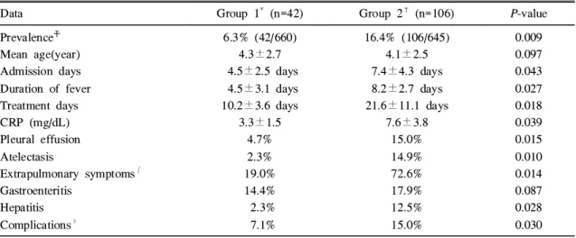
Table 1. Comparison of Clinical Features in Group 1 vs Group 2
Data Group 1* (n=42) Group 2†(n=106) P-value
Prevalence☨ Mean age(year) Admission days Duration of fever Treatment days CRP (mg/dL) Pleural effusion Atelectasis
Extrapulmonary symptoms∫ Gastroenteritis
Hepatitis Complications∥
6.3% (42/660) 4.3±2.7 4.5±2.5 days 4.5±3.1 days 10.2±3.6 days
3.3±1.5 4.7%
2.3%
19.0%
14.4%
2.3%
7.1%
16.4% (106/645) 4.1±2.5 7.4±4.3 days 8.2±2.7 days 21.6±11.1 days
7.6±3.8 15.0%
14.9%
72.6%
17.9%
12.5%
15.0%
0.009 0.097 0.043 0.027 0.018 0.039 0.015 0.010 0.014 0.087 0.028 0.030
*Patients admitted with M. pneumoniae pneumonia in 2002, †Patients enrolled in our study with M. pneu- moniae pneumonia since late 2003, ☨Prevalence of M. pneumoniae pneumonia, ∫, ∥Prevalence of data P value <0.05 means significant, statistically
2. Laboratory findings
At the time of admission, patients had a mean WBC count of 9,020±5,037 mm3, a mean ESR of 57±35 mm/hr, and a mean CRP of 7.6±3.8 mg/dL (Table 3).
Higher value of CRP and ESR upon admission were positively correlated with the duration of fever and length of admission but Mycoplasma antibody titers at the time of admission did not correlate with the duration of fever or with the duration of admis- sion (Table 4). The pleural effusions of 3 patients in whom thoracentesis was performed were analyzed (Table 5). The color of pleural effusion was sero- sanginous, and all profiles, except for protein and LDH were comparable to transudates. The protein level of pleural effusions ranged from 3.5 to 4.5 g/
dL and caused hypoalbuminemia in the patients. The mean serum albumin level in patients with pleural effusion was 3.25 g/dL, and the mean serum protein was 4.05 g/dL.
3. Chest radiological findings
Pulmonary infiltration on chest X-ray was demon- strated in 88.7% of the patients; unilateral (62.2%), bilateral (33.0%), or concomitant with lobular consol-
idation (31.0%), atelectasis (14.9%), or pleural effu- sion (15.0%) (Table 6).
Patients with pleural effusion had a higher inci- dence of complications compared to those without pleural effusion. Chest CT scans were performed in 4 patients who had protracted cough and crackles or wheezing on auscultation 3 months after admission.
All showed atelectasis, pleural thickening and adhe- sion. Two patients showed findings consistent with bronchiolitis obliterans, including bronchial wall
Table 3. Laboratory Findings (N=106)
FindingNumber of patients
(%)
Mean±SD*
WBC count(/mm3)
<5,000 5,000∼10,000 10,000∼15,000 15,000∼20,000
>20,000
Neutrophilia (>70%) ESR†, reactive (mm/hr) CRP☨, reactive (mg/dL)
4 ( 3.7) 45 (43.2) 50 (47.6) 4 ( 3.7) 3 ( 2.8) 78 (73.5) 79 (74.6) 71 (67.5)
9,020±5,037
57±35 7.6±3.8
*Mean±standard deviation, †Erythrocyte sediment- ation rate, ☨C-reactive protein
Table 4. Relation between Laboratory Find- ings and Clinical Course
Finding Admission
days Duration of fever CRP (mg/dL)
≤1.0
>1.0 ESR (mm/hr)
≤20
>20
Mycoplasma antibody titer 1 : 320∼640
≥1 : 1,280
5.3±1.2 8.3±2.8 (P=0.015)
5.5±1.7 8.2±2.7 (P=0.026)
6.3±3.1 7.3±4.8 (P=0.064)
3.2±1.8 5.3±2.7 (P=0.032)
3.1±2.9 5.2±3.8 (P=0.043)
2.7±2.1 4.1±3.9 (P=0.056) P value <0.05 means significant, statistically
Table 2. Frequency of Clinical Symptoms (N=
106)
Symptoms Numbers of
patients (%) Duration (days*) Cough
Rhinorrhea Sore throat Diarrhea Abdominal pain Vomiting Headache
Respiratory difficulty Chest pain
Skin rash Arthralgia
102 (96.2) 52 (48.6) 42 (39.4) 34 (16.2) 23 (21.9) 18 (17.1) 15 (14.4) 13 (12.3) 9 ( 8.5) 6 ( 5.9) 5 ( 4.9)
22.1±4.8 8.4±2.1 4.3±1.2 4.3±1.8 5.1±2.5 3.4±2.1 3.5±2.1 3.8±3.4 2.1±2.5 5.5±0.7 4.6±1.3
*Mean±standard deviation
thickening, adhesion and attenuation of pulmonary vessels, mosaic perfusion, and subsegmentally local- ized bronchiectasis.
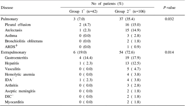
4. Complications
Complications, including myocarditis (2 cases), ar- thritis (3 cases), vasculitis (5 cases), asthma (3 cases), ARDS (1 case), and DIC (2 cases), were observed in 15.0% of the patients (Table 7). We experienced some unusual complications related to M. pneumoniae pneumonia such as asthma, PIGN, coronary aneurysm and arthritis.
Discussion
Extrapulmonary manifestations of M. pneumoniae pneumonia are uncommon, with cases described as single reports or small series3, 4), because M. pneu- moniae invades and multiplies in the bronchial epi- thelial cells1). A variety of mechanisms have been suggested to explain the involvement of distant or- gan systems. Potential mechanisms include metastatic infection, autoimmunity, toxin generation, micro- thrombosis and altered host immunity4, 5).
In our study, we found some characteristics of M.
pneumoniae pneumonia that were similar to those of previous years6∼8). Compared with previous years6, 7), there were more cases requiring admission and the use of medication for longer periods due to pro- longed fever, protracted cough or continuous rales on auscultation. There was also an increased inci- dence of extrapulmonary symptoms involving the skin, pleural cavity, bone marrow, G-I tract and joints, and of complications such as asthma, nephri- tis, arthritis, and vasculitis. Some of the complica- tions that occurred in our study were extremely severe and unique, and to our knowledge, these complications have not been previously reported.
However, there were no deaths in our study. In one
Table 5. Laboratory Findings of Pleural Fluid
Case 1 Case 2 Case 3 Mean
PH Color Protein (g/dL) LDH* (mg/dL) ADA† (mg/dL) WBC (/mm3) Glucose (mg/dL) Gram stain AFB stain Culture Tapping day☨
7.5 Yellow
4.5 4,758
29 210 129
-
-
- 5 days
7.0 Yellow
3.8 1,718
83 1,450 100
-
-
- 3 days
7.5 Clear to yellow
3.5 1,379
38 210 113
-
-
- 6days
7.33
- 3.93 2,618
50 626.66
114
-
-
- 4.6 days
*Lactic dehydrogenase (LDH), †Adenosine deaminase (ADA), ☨Means Hospital days
Table 6. Radiological Findings on Chest X-ray (N=106)
Site Number of patients (%)
Unilateral Right lobe
Upper Middle Lower Parahilum Left lobe
Upper Lower Parahilum Bilateral Atelectasis Pleural effusion
66 (62.2) 41 (38.6)
10 ( 9.4) 7 ( 6.6) 21 (19.8) 3 ( 2.8) 25 (23.6)
5 ( 4.7) 18 (16.9) 2 ( 1.9) 35 (33.0) 15 (14.9) 16 (15.8)
case, a 4-year-old boy was admitted with cough and gross hematuria. Chest X-ray showed pneumonic in- filtration. The Mycoplasma antibody titer was 1 : 10,240 and C3 was 47 mg/dL. Urinalysis revealed blood (+), protein 2 (+), RBC (many/HPF), and WBC (10∼30/HPF). An ultrasonogram of the kid- ney showed swelling and increased echogenicity in both kidneys. In other case, a 3-year-old boy was admitted with a persistent fever, cough and dyspnea.
Chest X-rays showed pneumonic consolidation, ac- companied by a profuse amount of pleural effusion.
The Mycoplasma antibody titer was 1 : 40,960. Mild cardiomegaly was detected on a follow-up chest X- ray, and echocardiogram showed mild coronary dila- tation and pericardial effusion. We excluded a case of concomitant Kawasaki disease because the pa- tient's fever pattern was not remittent, was con- trolled with antipyretics, and other clinical features did not correspond to diagnostic criteria of Kawasaki
disease.
We compared the data collected in late 2003 to data from the preceding year (Table 1). In compari- son with patients diagnosed with M. pneumoniae pneumonia at our hospital in 2002, there were re- markable increases in the incidence of mycoplasmal pneumonia, extrapulmonary symptoms, and complica- tions in late 2003. In addition, clinical courses, in- cluding total febrile period, admission period and treatment period were significantly longer than those in 2002. In our survey, the estimated frequencies of mycoplasma pneumonia, extrapulmonary symptoms and complications were 16.4%, 73.5% and 15.0%, respectively, compared to those of 6.3%, 19.0% and 7.1%, respectively, in 2002.
Compared with previous studies, the incidence of extrapulmonary manifestation was remarkably higher in our study. Pyun et al.7) reported an incidence of 25% in 1997 and Lee et al.8) an incidence of 17%
Table 7. Comparison of Frequencies of Complications in Group 1 vs Group 2
Disease No of patients (%)
P-value Group 1* (n=42) Group 2† (n=106)
Pulmonary Pleural effusion Atelectasis Asthma
Bronchiolitis obliterans ARDS☨
Extrapulmonary Gastroenteritis Hepatitis Vasculitis Hemolytic anemia IDA∫
Arthritis
Aseptic meningitis DIC∥
Myocarditis
3 (7.0) 2 (4.7) 1 (2.3) 0 (0.0) 0 (0.0) 0 (0.0) 6 (19.0)
4 (14.4) 1 ( 2.3) 0 ( 0.0) 0 ( 0.0) 1 ( 2.3) 0 ( 0.0) 0 ( 0.0) 0 ( 0.0) 0 ( 0.0)
37 (35.4) 16 (15.0) 15 (14.9) 3 ( 2.8) 2 ( 1.8) 1 ( 0.9) 54 (72.6)
19 (17.9) 13 (12.5) 5 ( 4.7) 4 ( 3.8) 4 ( 3.8) 3 ( 2.8) 2 ( 1.8) 2 ( 1.8) 2 ( 1.8)
0.032
0.014
*Patients admitted with M. pneumoniae pneumonia in 2002, †Patients enrolled in our study with M. pneu- moniae pneumonia since late 2003, ☨Acute respiratory distress syndrome, ∫Iron defiency anemiam, ∥Dis- seminated intravascular coagulation
P value 0.05 means significant, statistically
in 1993. In our study, the most common extrapul- monary manifestation was gastrointestinal complica- tion, occurring in 30.4% of patients. Elevated liver enzymes were observed in 12.5% of patients. Hepa- tic dysfunction in these patients was transitory, and recovery of normal liver function correlated directly with the resolution of the mycoplasma respiratory disease10). The pathogenesis of self-limiting hepatitis may be attributed to a direct cytolytic effect medi- ated by the infecting mycoplasma, or to an immu- nological autoimmune disorder resulting from the production of heterophil antibodies, etc.4, 10) In pa- tients with persistent rales on auscultation, the inci- dence of extrapulmonary symptoms was higher, but this correlation could not be explained.
In evaluating laboratory results, we found that neither the level of the initial mycoplasma antibody titer at admission, nor the follow-up titer correlated with the duration of fever, length of admission, duration of medication, incidence of extrapulmonary symptoms or associated complications. However, the higher values of CRP and ESR at admission posi- tively correlated with both the duration of fever and length of admission. There were no further positive correlations among the variables we evaluated.
We found that 33.0% of patients showed bilateral plulmonary infiltration on chest X-ray, a percentage that was much higher than 17% and 13% in previ- ous years6, 8). Pulmonary infiltrations in M. pneumo- niae pneumonia patients were shown unilaterally in 70∼90% of cases and bilaterally in 10∼30% of cases and were more likely to occur in the sequence of right lower lobe, left lower lobe, and right upper lobe11, 12). The prevalence of right lower lobe in- volvement can be attributed to exudate shift in the alveoli due to gravity and the anatomical disadvan- tage of the right bronchus, which is straighter and shorter than the left bronchus11).
Chest HRCT was performed in 4 patients who had continuous crackles on auscultation over a 3- month period, even though the severity of crackles
had improved over time. In 2 of these patients, bronchiolitis obliterans was observed and was sub- segmentally localized in small areas in the previous- ly infiltrated lobe. Compared with previous years8, 9), more patients in our study had pleural effusion (15.0
%), though M. pneumoniae pneumonia is generally accompanied by small pleural effusion in approxi- mately 20% of cases11, 13). In analysis of the pleural effusion, the protein level, LDH and WBC counts corresponded to exudate, while the other values cor- responded to transudate. Such findings are similar to that seen in TB pleurisy. The values of ADA in pleural effusions ranged from 29 to 83 in our study, and the value for 1 patient extended into the terri- tory of suspected TB pleurisy. The only way to dif- ferentiate TB pleurisy is by collectively considering the Mantoux test, TB contact history, AFB staining, TB culture, and TB PCR14, 15). Compared to patients without pleural effusion, M. pneumoniae pneumonia patients with accompanying pleural effusion are known to have a longer duration of illness, a higher incidence of complications, and a higher probability of co-infection with viral agents, such as the adeno- virus and parainfluenza virus16∼18). In 9% of patients with persistent cough and severe continuing asthma symptoms, various viral studies were performed to check concomitant viral infection. Respiratory syn- cytial virus was found in one case.
M. pneumoniae infection is known to possibly ex- acerbate asthmatic symptoms and play a pathogenic role in asthma19). In our study, asthma was newly developed in 3 cases. The exacerbation of previous- ly-diagnosed and treated asthma was observed in 5 cases. Possible causal mechanisms of mycoplasma - induced airway inflammation and hyperresponsiveness have been investigated, including increased Th2 re- sponses and inflammatory neuropeptides19, 20).
Complications of M. pneumoniae pneumonia large- ly fall into two categories those involving the res- piratory system and those that are non-respiratory.
Those with respiratory system involvement include
pleural effusion, lung abscess, Swyer-James syn- drome13), emphysema, bronchiolitis obliterans21), bron- chiectasis, etc. Those that are non-respiratory in nature include congestive heart failure, acute myocar- dial infarction, myocarditis22), pericarditis, Stevens- Johnson syndrome, encephalitis, aseptic meningitis, peripheral neuritis, cerebellar ataxia, transverse mye- litis, Guillain-Barré syndrome, cranial nerve palsy, etc13, 23). 33% of patients with CNS complications suffer permanent sequelae23). Among patients with CNS complications in our study, fortunately only 2 patients had aseptic meningitis and both recovered with no sequelae. According to reported studies, the occurrence of coronary aneurysm and PIGN, as complications of M. pneumoniae pneumonia, are rare. We believe that such complications implicate an autoimmune-mediated mechanism, such as that of vasculitis. The recent demonstration of tuberculin anergy and depressed T-and B-cell lymphocyte fuc- tion during the course of M. pneumoniae infection suggests the possibility that immunosuppression may contribute to the pathogenesis of extrapulmonary complications4, 24).
We found a number of characteristics of M. pneu- moniae pneumonia among cases from late 2003 that were different from those of previous years7∼9). Fur- thermore, we propose the following hypotheses to explain why this outbreak was more clinically severe and accompanied by more frequent extrapulmonary symptoms and complications than outbreaks in pre- vious years : First, it is likely that this epidemic outbreak was caused by a different subtype of M.
pneumoniae from that of previous outbreaks. Accord- ing to one report25), Group I (91.7%) was more prevalent than group II (8.3%) with a three-year cycle of epidemic outbreak from 1997 to 2002 in Korea. To date, there is little published data avail- able to compare the immune responses, disease se- verity, and exchange phenomena between the two groups. Second, this epidemic outbreak may have oc- curred concomitantly with other viral infections, such
as adenovirus, influenza, parainfluenza, and rhino- virus. Third, the appearance of serious autoimmune diseases resulted from impaired and altered immunity in the host. That is, such an altered immune re- sponse could allow for escape of M. pneumoniae from the respiratory tract infection, the developement of autoantibodies, prolonged infection, and perhaps the activation of a quiescent infection. Further stud- ies are needed to substantiate the above hypotheses.
The findings in our study are subject to some limitations. First, this study was performed retrospec- tively. Second, case ascertainment was conducted only in our hospital. Third, determination of the be- ginning and end of the outbreak was not possible with the available data.
In conclusion, M. pneumoniae is generally thought to cause mild disease of the respiratory tract and uncomplicated pneumonia in affected children. How- ever, serious pulmonary and extrapulmonary compli- cations such as reported in our study may occur at any times. Knowledge of the less common pulmo- nary and systemic manifestations is important for differential diagnosis and institution of proper, early antimicrobial treatment and appropriate control mea- sures including use of chemoprophylaxis during out- breaks of acute respiratory illness. A multicenter study is needed to verify the changes in clinical characteristics observed during the 2003 outbreak from previous outbreaks.
한 글 요 약
2003년 하반기에 유행한 Mycoplasma pneumoniae 폐렴의
특징에 대한 고찰
권혜옥 박신이 이준호
포천중문의과대학교 분당차병원 소아과
목 적 : 2003년 하반기 우리나라에서 전국적으로 마이코플라즈마 폐렴의 폭발적인 유행을 보였다.그러나 예년과는 달리 심한 임상경과를 밟거나, 합 병증과 폐외 증상을 동반하는 경우가 많이 관찰되 었기에 저자들은 본원의 경험을 토대로 2003년 유 행했던 마이코플라즈마 폐렴의 임상양상에 대해서 고찰해 보고자 한다.
방 법 : 2003년 8월부터 2004년 4월까지 분당차 병원 소아과에 폐렴증상으로 입원한 환아들 중, 입 원 후 검사한 혈청 마이코플라즈마 항체가가 1 : 320 이상이거나 1주 간격으로 시행한 항체가가 4 배 이상 증가가 있었던 환아 106명을 대상으로 후 향적 고찰을 하였다.
결 과 : 총발열기간은 평균 8.2±2.7일, 입원 후 발열기간은 평균 5.3±2.0일이었다. 호흡기 증상으 로는 기침(96.2%), 콧물(48.6%), 인후통(39.4%), 호 흡곤란(12.3%) 등의 순이었으며 지속기간은 각각 평균 22.1±4.8일, 8.4±2.1일, 4.3±1.2일, 3.8±3.4 일 등의 순이었다. 기침이 3개월까지 가는 경우도 소수에서 관찰되었으나, 치료기간과는 상관관계가 없었다. 비호흡기증상으로는 복통(21.9%), 구토(17.1
%), 설사(16.2%), 두통(14.4%), 피부발진(5.9%), 관 절통(4.9%) 등의 순이었다. 입원기간은 평균 7.4±
4.3일이었으며 총치료기간은 21.6±11.1일이었다.
합병증으로 파종성 혈관 내 응고증(2명), 심근염(2 명), 관절염(3명), 혈관염(5명), 천식(3명), 급성호흡 부전(1명) 등이 관찰되었다.
결 론 : 2003년 하반기 우리나라에서 유행했던 마이코플라즈마 폐렴은 예년과는 달리 심한 임상경 과를 보였고, 적지 않게 합병증을 동반하였다. My- coplasma pneumoniae 아형의 종류에 따라 임상경과 가 심해질 수 있는지는 아직 확실하지 않다. 단지, 다른 바이러스 감염과 동시 감염되는 경우 바이러 스성 폐렴의 증세를 악화시키는 것으로 알려져 있 다. 이번 유행과 다른 바이러스와 공동감염 관련여 부는 본 연구에선 확인할 수 없었다. 소아에서 마 이코플라즈마 폐렴이 심한 임상경과를 밟을 수도 있다는 것을 알아야 하겠다.
References
1) Bertman R, Kliegman R, Jenson H. Nelson Textbook of Pediatrics, 17th ed. Philadelphia :
WB Saunders Co. 2004:990-2.
2) Jacobs E. Mycoplasma pneumoniae virulence factors and the immune response. Rev Med Mi- crobiol 1991;2:83-90.
3) Marrie TJ. Community-acquired pneumonia.
Clin Infect Dis 1994;18:501-13.
4) Fernald GW. Immunologic mechanisms sug- gested in the association of M. pneumoniae in- fection and extrapulmonary diease : a review.
Yale J Biol Med 1983;56:475-9.
5) Feigin R, Cherr J. Textbook of Pediatric infec- tious disease, 3rd ed. Philadelphia : WB Saunders Co. 1992:1866-90.
6) Hong JY, Nah SY, Nam SG, Choi EH, Park JY, Lee HJ. Occurrence of Myco plasma pneu- moniae pneumonia in Seoul, Korea, from 1986 to 1995. J Korean Pediatr Soc 1997;40:607-13.
7) Pyun BY, Kim HH, Chung JT, Lee JS. A study as epidemiologic and clinical aspect of mycoplasma pneumoniae pneumonia during the last 5 years. Pediatr Allergy Respir Dis 1998;8:
240-7.
8) Lee EK, Hong YJ, Lee MI, Ahn DH, Sohn KC. A clinical study on mycoplasma pneu- moniae pneumonia. Pediatr Allergy Respir Dis 1993;3:11-9.
9) Kim BY, Lee HS, Kim IK, Choi CH, You KH. Clinical consideration between in type of pneumonia and cold agglutinin titer, and my- coplasma antibody titer caused by mycoplasmal pneumonia in children. J Korean Pediatr Soc 1993;36:959-67.
10) Squadrini F, Lami G, Pellegrino F, Pinelli G, Bavieri M, Fontana A, et al. Acute hepatitis complicating Mycoplasma pneumoniae infection.
J Infect 1988;16:201-2.
11) Putamen CE, Curtis AM, Simenone JF, Jensen P. Mycoplasma pneumonia : Clinical and roent- genographic patterns. AJR 1975;124:417-22.
12) Light RW. Pleural diseases, 4th ed. Philadel- phia : Lippincott Williams and Wilkins. 2001:
42-86.
13) Lind K. Manifestations and complications of Mycoplasma pneumoniae disease : a review.
Yale J Biol Med 1983;56:461-8.
14) Cassell GH, Cole BC. Mycoplasma as agents of human disease. N Engl J Med 1981;304:80- 9.
15) Klockars M, Kleemola M, Leinomen M, Kos- kela M. Serum adenosine deaminase in viral and bacterial pneumonia. Chest 1991;99:623-6.
16) Chan ED. Fulminant mycoplasma pneumonia.
West J Med 1995;162:133-42.
17) Waris ME, Toikka P, Saarinen T, George RB, Zinskind M, Rasch J, et al. Diagnosis of My- coplasma pneumoniae pneumonia in children. J Clin Microbiol 1998;36:3155-9.
18) Shah DC, Muthiah MM. Adult respiratory dis- tress syndrome due to mycoplasma pneumoniae.
Postgrad Med J 1996;72:241-2.
19) Biscardi S, Lorrot M, Marc E, Moulin F, Bou- tonnat-Faucher B, Helibronner C, et al. Myco- plasma pneumoniae and asthma in children.
Clin Infect Dis 2004;38:1341-6.
20) Johnston SL, Pattemore PK, Sanderson G, Mo- gabgab WJ, Forsgren M, Tunevall G, et al.
Community study of role of viral infections in
exacerbations of asthma in 9∼11 year old chil- dren. BMJ 1995;310:1225-9.
21) Prabhu MB, Barber D, Cockcroft DW. Bron- chiolitis obliterans and mycoplasma pneumonia.
Respir Med 1991;85:535-7.
22) Sands MJ, Satz JE. Turner WE. Scoff LA.
Pericarditis and perimyocarditis associated with active mycoplasma pneumoniae Infection. Ann Int Med 1977;86:544-8.
23) Koskiniemi M. CNS manifestations associated with Mycoplasma pneumoniae infections : sum- mary of cases at the University of Helsinki and review. Clin Infect Dis 1993;17:52-7.
24) Sabato AR, Cooper DM, Thong YH. Transitory depression of immune fuction following Myco- plasma pneumoniae infection in children. Pedi- atr Res 1981;15:813-6.
25) Kim SS, Kang H, Ahn BM, Lee WW, Kim ER, Kim SY, et al. Study of exchange pheno- menon of mycoplasma pneumoniae in children from 1997∼2002. Korean J Pediatr 2004;47:24- 30.