Clinical Characteristics of Transplant-associated Encephalopathy in Children
We aimed to analyze characteristics of encephalopathy after both hematopoietic stem cell and solid organ pediatric transplantation. We retrospectively reviewed medical records of 662 pediatric transplant recipients (201 with liver transplantation [LT], 55 with heart transplantation [HT], and 67 with kidney transplantation [KT], 339 with allogeneic hematopoietic stem cell transplantation [HSCT]) who received their graft organs at Asan Medical Center between January 2000 and July 2014. Of the 662 patients, 50 (7.6%) experienced encephalopathy after transplantation. The incidence of encephalopathy was significantly different according to the type of organ transplant: LT, 16/201 (8.0%), HT, 13/55 (23.6%), KT, 5/67 (7.5%), and HSCT, 16/339 (4.7%) (P < 0.001). Drug-induced encephalopathy (n = 14) was the most common encephalopathy for all transplant types, but particularly after HSCT. Hypertensive encephalopathy was the most common after KT and HT, whereas metabolic encephalopathy was the most common after LT. The median time to encephalopathy onset also differed according to the transplant type: 5 days after KT (range 0–491 days), 10 days after HT (1–296 days), 49.5 days after HSCT (9–1,405 days), and 39 days after LT (1–1,092 days) (P = 0.018). The mortality rate among patients with encephalopathy was 42.0% (n = 21/50). Only 5 patients died of neurologic
complications. Transplant-associated encephalopathy presented different characteristics according to the type of transplant. Specialized diagnostic approach for neurologic complications specific to the type of transplant may improve survival and quality of life in children after transplantation.
Keywords: Encephalopathy; Transplantation; Liver; Kidney; Heart; Hematopoietic Stem Cells
Yun-Jeong Lee,1* Mi-Sun Yum,2* Eun-Hee Kim,3 Min-Jee Kim,2 Kyung Mo Kim,2 Ho-Joon Im,2 Young-Hwue Kim,2 Young Seo Park,2 and Tae-Sung Ko2
1Department of Pediatrics, Kyungpook National University Hospital, Daegu, Korea; 2Department of Pediatrics, Asan Medical Center Children’s Hospital, University of Ulsan College of Medicine, Seoul, Korea; 3Department of Pediatrics, CHA Gangnam Medical Center, CHA University, Seoul, Korea
* Yun-Jeong Lee and Mi-Sun Yum contributed equally to this work.
Received: 2 August 2016 Accepted: 30 October 2016 Address for Correspondence:
Tae-Sung Ko, MD
Department of Pediatrics, Asan Medical Center Children’s Hospital, University of Ulsan College of Medicine, 88 Olympic-ro 43-gil, Songpa-gu, Seoul 05505, Korea
E-mail: [email protected]
https://doi.org/10.3346/jkms.2017.32.3.457 • J Korean Med Sci 2017; 32: 457-464
INTRODUCTION
Solid organ and hematopoietic stem cell transplantation (HSCT) have been performed increasingly as a therapeutic modality for end-stage organ failure, a variety of hematologic disorders, and non-malignancy disease. Solid organ transplants performed in the United States in 2015 exceeded 30,000 and continued to in- crease (1). Although remarkable advances in transplantation medicine improved the outcomes of allogeneic graft recipients, neurologic complications are still common with variety inci- dence ranging from 7% to 77% of allograft recipients after HSCT and solid organ transplantation (2-12). One of the most delete- rious neurologic complications is transplant-associated enceph- alopathy, which has various etiologies, including the underly- ing disorder motivating the transplant, immunosuppressant neurotoxicity, metabolic derangement, central nervous system (CNS) infection, and stroke (9,12-15). As the surgical procedure, intensity of immunosuppression, infection prophylaxis regimen, underlying neurologic symptoms, and recipient’s age vary ac- cording to the type of transplant, the presentation of the enceph-
alopathy may differ according to the specific type of organs trans- planted.
To help shape neurological care in pediatric transplantation, we investigated the overall incidence, etiologies and clinical course of encephalopathy after allogeneic HSCT and solid or- gan transplantation in pediatric patients from a single tertiary center in Korea. In addition, we dissected the differences among the transplant types to identify the best peritransplant care ap- proach after transplantation from a neurological perspective.
MATERIALS AND METHODS
We retrospectively reviewed the medical records of all patients younger than 18 years at the time of transplantation who receiv- ed a transplant, including allogeneic hematopoietic stem cell, kidney, heart, and liver transplantation (LT), between January 2000 and July 2014 at Asan Medical Center Children’s Hospital, a single tertiary center in Korea. Before identifying patients with encephalopathy after transplantation, we excluded patients who underwent autologous HSCT or 2 or more organ transplants at
the same time. Encephalopathy was defined as any altered level of consciousness or newly developed brain dysfunction, such as seizures, tremors, visual problems, or personality changes, after transplantation that was not caused by sedative medica- tion. We intended to exclude neurologic deterioration not di- rectly associated with transplantation procedures, preexisting CNS manifestations associated with CNS involvement of un- derlying disease, such as leukemic or lymphohistiocytosis infil- tration into the CNS and hepatic encephalopathy persistent af- ter transplantation, was excluded from the study. The following data were collected from the medical records: patient charac- teristics, underlying disease, preoperative neurologic symptoms, the etiologies of encephalopathy, encephalopathy onset time, clinical course, and survival after transplantation. Neurological examinations, neuroimaging, laboratory studies (blood count, coagulation profile, electrolyte, chemistry, serum trough level of immunosuppressants, cerebrospinal fluid analysis, microbi- ological studies), and electroencephalography findings at the time of events were also reviewed by 2 pediatric neurologists (LYJ, YMS). The etiology of the encephalopathy was determined by reviewing clinical history and symptoms and microbiologi- cal, electrophysiological, and radiological characteristics. En- cephalopathies were categorized into 6 groups: drug-induced, hypertensive, anoxic, metabolic, CNS infection-associated, and unknown etiology. Drug-induced encephalopathy was defined as an acute encephalopathy temporally related to the adminis- tration of any drugs, such as immunosuppressant, chemother- apeutic, and antibiologic agents, that resolved after withdrawal of the drug and based on exclusion of other causes. Identifica- tion of immunosuppressant related encephalopathy was sup- ported in case of high serum trough level of immunosuppres- sant (e.g., tacrolimus > 15 ng/mL or cyclosporine > 400 ng/mL) at that time of neurologic complications without any other causes.
Hypertensive encephalopathy was defined as encephalopathy in the presence of pronounced hypertension. When encepha- lopathy is caused by hypertension secondary to immunosup- pressant, the patient is defined as drug-induced encephalopa- thy. Anoxic encephalopathy was defined as acute symptoms of cerebral perfusion reduction, including ischemic stroke or in- tracranial hemorrhage (ICH). Metabolic encephalopathy was defined as encephalopathy that developed with the occurrence of renal failure, liver failure, or electrolyte and glucose imbalance.
CNS infection-associated encephalopathy was diagnosed by positive identification of a pathogen from the cerebrospinal flu- id, blood, or tissue pathology and/or a cerebrospinal fluid find- ing compatible with infection. Posterior reversible encephalop- athy syndrome (PRES) was defined as an independent clinico- radiological entity involving a posterior lesion in the cerebral hemisphere and characterized by altered consciousness, corti- cal blindness, and/or convulsion (16).
Statistical analysis
Group differences according to the transplanted organ were as- sessed by the χ2 test or Fisher’s exact test for categorical variables and the Kruskal-Wallis test for continuous variables, followed by post hoc testing. All tests of significance were 2-sided, with a significance level of P < 0.05. For multiple comparisons, the sig- nificance level was corrected for the number of comparisons by the Bonferroni method (Bonferroni-adjusted P < 0.008). All sta- tistical analyses were performed using SPSS version 18.0 (SPSS Inc., Chicago, IL, USA).
Ethics statement
The present study protocol was reviewed and approved by the Institutional Review Board of Asan Medical Center (Registry No.
20150724). Informed consent was waived by the board.
RESULTS
Patient characteristics and incidence of encephalopathy From January 2000 through July 2014, 662 pediatric patients underwent organ transplantation at Asan Medical Center Chil- dren’s Hospital: LT in 201 patients, kidney transplantation (KT) in 67, heart transplantation (HT) in 55, and allogeneic HSCT in 339. Of the 662 patients, 50 (7.6%) developed encephalopathy after transplantation at a mean ± standard deviation follow-up of 53.1 ± 46.6 months: 16 of 201 LT recipients (8.0%), 13 of 55 HT recipients (23.6%), 5 of 67 KT recipients (7.5%), and 16 of 339 allogeneic HSCT recipients (4.7%) (Fig. 1). The incidence of encephalopathy was significantly different according to the type of organ transplant (P < 0.001). We compared the characteris- tics of the patients with encephalopathy according to the type of transplant (Table 1). The median age at the time of transplan- tation (P < 0.001) and presence of preoperative neurologic symp- toms (P = 0.002) differed according to the encephalopathy group.
LT patients (n = 8, 50.0%) were the most likely to show preopera-
Fig. 1. The incidence of encephalopathy according to the type of organ transplant.
LT = liver transplantation, HT = heart transplantation, KT = kidney transplantation, HSCT = hematopoietic stem cell transplantation.
%
LT
(n = 16/201) HT
(n = 13/55) KT
(n = 5/67) HSCT (n = 16/339) 100
90 80 70 60 50 40 30 20 10 0
Encephalopathy P<0.001
Fig. 2. The etiology of encephalopathy according to the type of organ transplant.
LT = liver transplantation, HT = heart transplantation, KT = kidney transplantation, HSCT = hematopoietic stem cell transplantation, CNS = central nervous system.
*P = 0.006; †P = 0.004; ‡P = 0.002.
%
Overall LT HT KT HSCT
100 90 80 70 60 50 40 30 20 10 0
Overall P<0.001
Unknown Metabolic Anoxic CNS infection Hypertension Drug
‡
†
*
Table 1. Characteristics of the patients with encephalopathy
Characteristics LT (n = 16) HT (n = 13) KT (n = 5) HSCT (n = 16) P value
Age, yr 1.4 (0.5–11.0)*,†,‡ 8.4 (0.7–17.2)† 14.4 (10.3–15.1)‡ 6.3 (1.2–15.8)* < 0.001§
Male sex 8 (50.0) 7 (53.8) 4 (80.0) 9 (56.3) 0.558
Follow-up duration, mon 36.4 (1.0–116.0) 52.7 (1.0–137.0) 119.8 (19.0–143.0) 37.1 (0.3–129.1) 0.178
Preoperative neurologic symptom 8 (50.0)* 4 (28.6) 0 (0.0) 0 (0.0)* 0.002§
Onset time of encephalopathy, day 39 (1–1,092) 10 (1–296)ll 5 (0–491) 49.5 (9–1,405)ll 0.018§
Values are presented as number (%) or median (range).
LT = liver transplantation, HT = heart transplantation, KT = kidney transplantation, HSCT = hematopoietic stem cell transplantation.
*P = 0.002, comparison of the HSCT group with the LT group; †P = 0.001, comparison of the LT group with the HT group; ‡P = 0.002, comparison of the LT group with the KT group; §P < 0.05; llP = 0.002, comparison of the HSCT group with the HT group.
Table 2. Patients with encephalopathy after LT (n = 16/201) Pt. No. Sex/Age,
yr Diagnosis Neurologic symptom NC onset,
day Etiology MRI/CT Outcome
1 M/0.8 BA Seizure 422 Metabolic Hypoglycemia
(glucose 9 mg/dL)
N Alive
2 F/0.6 BA Mental change 20 Metabolic HE - Death (liver failure)
3 F/1.8 BA Mental change 184 Metabolic HE - Death (liver failure)
4 F/8.0 BA Mental change 124 Metabolic HE - Death (liver failure)
5 M/0.9 BA Mental change 1 Metabolic HE Diffuse brain edema Alive, epilepsy
6 M/0.8 ALF Mental change, seizure 79 Metabolic HE - Death (liver failure)
7 F/1.4 ALF Mental change, seizure 14 Metabolic HE Diffuse brain edema Death (liver failure)
8 M/0.5 ALF Mental change 27 Metabolic HE Diffuse T2 HSI WM Alive, epilepsy
9 F/11.0 Budd-Chiari syndrome Seizure 51 HTN BP 160/110 mmHg Frontal hypodensity Alive
10 F/2.8 Citrullinemia Mental change 1,092 Anoxic ICH ICH, brainherniation Death (ICH)
11 M/5.0 BA Mental change, tremor 59 Drug FK506 16.9 ng/mL
BP 155/80 mmHg
N Alive
12 F/4.0 Alagille syndrome Seizure 16 Drug FK506 32.2 ng/mL
BP 160/100 mmHg Multifocal calcification Death (GI bleeding)
13 M/2.0 PFIC type 1 Mental change 5 Drug FK506 23 ng/mL
BP 133/53 mmHg
Mild ventriculomegaly Alive
14 F/1.4 Factor X deficiency Seizure 3 Drug FK506 30.3 ng/mL
BP 118/80 mmHg Encephalomalacia Alive
15 M/0.8 ALF Seizure 100 Unknown - Alive
16 F/0.8 BA Seizure 22 Unknown - Alive
LT = liver transplantation, NC = neurologic complication, MRI = magnetic resonance imaging, CT = computed tomography, BA = biliary atresia, N = no abnormality, HE = hepatic encephalopathy, ALF = acute liver failure, HSI = high signal intensity, BP = blood pressure, WM = white matter, HTN = hypertensive, ICH = intracranial hemorrhage, FK506 = tacrolimus, GI = gastrointestinal, PFIC = progressive familial intrahepatic cholestasis.
tive neurologic symptoms, followed by HT patients (n = 4, 28.6%).
The median encephalopathy onset time was significantly dif- ferent according to the transplanted organ (P = 0.018). Enceph- alopathy tended to occur in the early postoperative period after KT and HT, whereas it occurred in the late postoperative period after allogeneic HSCT and LT. The encephalopathy onset time was significantly different between HSCT and HT patients (P = 0.002).
Etiology of encephalopathy
In all 50 patients with encephalopathy, the most common etiol- ogy was drug-induced encephalopathy (n = 14, 28.0%) (Fig. 2).
The distribution of etiologies was significantly different accord- ing to the type of transplant (P < 0.001). Post hoc analysis indi- cated that the distribution of etiologies was significantly differ- ent in HSCT, HT, and KT from LT. The most prevalent etiology of encephalopathy differed according to the type of transplant:
metabolic encephalopathy in LT (n = 8), hypertensive enceph- alopathy in KT (n = 4) and HT (n = 6), and drug-induced en-
cephalopathy in allogeneic HSCT (n = 7).
Organ-specific encephalopathy Liver transplantation
Sixteen patients developed encephalopathy after LT at a median follow-up of 36.4 months (range 1–116 months) after the trans- plantation (Table 2). The most common etiology was metabolic encephalopathy (n = 8), followed by drug-induced encepha- lopathy (n = 4). Of the 8 patients with metabolic encephalopa- thy, 7 had hepatic encephalopathy, 5 of whom died because of graft failure. All 4 patients with drug-induced encephalopathy presented with a toxic serum concentration of immunosuppres- sant and elevated blood pressure. One patient (patient 10), who had citrullinemia, Epstein-Barr virus (EBV)-associated post-trans- plant lymphoproliferative disorder (PTLD) and coagulopathy developed ICH around 3 years after transplantation and died of brain herniation.
Heart transplantation
Thirteen patients (7 males) were identified as having encepha- lopathy at a median follow-up of 52.7 months (range 1–137 mon- ths) after transplantation (Table 3). The most common enceph- alopathy in HT patients was hypertensive encephalopathy (n = 6), followed by anoxic encephalopathy (n = 3) and drug-induced encephalopathy (n = 3). Cardiopulmonary resuscitation events occurred in 4 patients (3 in hypertension encephalopathy, 1 in anoxic encephalopathy) and extracorporeal membrane oxy- genation (ECMO) insertion in 1 patient with anoxic encepha- lopathy before neurologic symptom onset. Except for 1 patient with ICH, the 12 other encephalopathic events developed with- in 2 months after transplantation. In particular, 5 cases of hy- pertensive encephalopathy and 2 cases of ischemic encepha- lopathy occurred within 2 weeks after transplantation. Three patients with hypertension (patient 2, 7, and 9) were diagnosed with PRES and 2 of them (patient 7 and 9) also had calcineurin inhibitor neurotoxicity. One patient, who was taking anticoagu-
Table 3. Patients with encephalopathy after HT (n = 13/55) Pt. No. Sex/
Age, yr Diagnosis Neurologic symptom NC onset,
day CPR/ECMO Etiology MRI/CT Outcome
1 F/3.1 DCMP Seizure, mental change 30 −/− HTN BP 147/97 mmHg Small SDH Death (HF)
2 F/0.7 DCMP seizure, mental change 1 +/− HTN BP 150/78 mmHg PRES Alive, epilepsy
3 F/7.8 DCMP Mental change, seizure 7 −/− HTN BP 149/99 mmHg Mild diffuse brain atrophy Alive 4 M/3.5 HF Mental change, seizure 10 +/− HTN BP 170/90 mmHg Multiple calcification Alive, mental retardation
5 F/3.1 DCMP Mental change 11 +/− HTN BP 170/85 mmHg N Alive
6 M/11.0 DCMP Seizure 5 −/− HTN BP 150/96 mmHg - Alive
7 M/8.4 RCMP Seizure, mental change, visual impairment
28 −/− Drug FK506 13.6 ng/mL
BP 135/85 mmHg
PRES Alive
8 M/7.5 RCMP Seizure, mental change 11 −/− Drug FK506 5.6 ng/mL
BP 105/69 mmHg Small SDH Alive
9 M/12.9 HCMP Seizure, mental change, psychosis
59 −/− Drug CsA 1,699 ng/mL
BP 136/82 mmHg
PRES Alive
10 F/11.2 HF Seizure, mental change, delirium
1 −/− Anoxic - Diffuse hypodensity Alive
11 F/14.1 RCMP Mental change 4 −/+ Anoxic - Bilateral putaminal T2 HSI Death (respiratory failure)
12 M/12.6 DCMP Mental change 296 +/− Anoxic ICH ICH, herniation Death (ICH)
13 M/17.2 HCMP Mental change 6 −/− Unknown - Small SDH Death (HF)
HT = heart transplantation, NC = neurologic complication, CPR = cardiopulmonary resuscitation, ECMO = extracorporeal membrane oxygenation, MRI = magnetic resonance imaging, CT = computed tomography, DCMP = dilated cardiomyopathy, HTN = hypertensive, BP = blood pressure, SDH = subdural hemorrhage, HF = heart failure, HCMP = hypertrophic cardiomyopathy, CsA = cyclosporine, PRES = posterior reversible encephalopathy syndrome, N = no abnormality, FK506 = tacrolimus, RCMP = restrictive cardio- myopathy, HSI = high signal intensity, ICH = intracranial hemorrhage.
Table 4. Patients with encephalopathy after KT (n = 5/67) Pt. No. Sex/Age,
yr Diagnosis Neurologic
symptom
NC onset,
day Etiology MRI/CT Outcome
1 M/10.3 Reflux nephropathy Mental change 38 HTN BP 150/90 mmHg N Alive
2 M/14.4 Reflux nephropathy Seizure 0 HTN BP 180/90 mmHg PRES Alive
3 F/15.1 Henoch-Schönlein nephritis Seizure 1 HTN BP 180/110 mmHg - Alive
4 M/15.0 Chronic renal failure, unknown etiology Seizure 5 HTN BP 179/110 mmHg PRES Alive
5 M/10.6 Bilateral dysplastic kidney Fever, mental
change 491 CNS infection EBV encephalitis → PTLD Multifocal enhancing
T2 HSI Alive
KT = kidney transplantation, NC = neurologic complication, MRI = magnetic resonance imaging, CT = computed tomography, HTN = hypertensive, BP = blood pressure, N = no abnormality, PRES = posterior reversible encephalopathy syndrome, CNS = central nervous system, EBV = Epstein-Barr virus, PTLD = post-transplant lymphoproliferative disorder, HSI = high signal intensity.
lant medication after surgical repair of an aortic aneurysm, de- veloped abnormal coagulation function resulting in ICH and died of ICH.
Kidney transplantation
Five patients developed encephalopathy after KT (Table 4). Hy- pertensive encephalopathy developed in 4 patients within 7 days after transplantation. Neuroimaging revealed typical white matter lesionson the occipital lobe that were consistent with PRES in 2 patients with hypertensive encephalopathy (patient 2 and 4). One patient (patient 5) presented with mental change, seizure, headache, and fever and was diagnosed with EBV en- cephalitis 1 year after transplantation and finally with CNS PTLD.
Hematopoietic stem cell transplantation
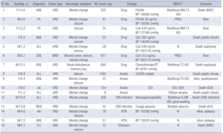
Encephalopathy occurred in 16 patients (9 males) at a median follow-up of 37.1 months (range 0.3–129.1 months) after alloge- neic HSCT (Table 5). Except for 1 patient with a full-matched related donor transplant, 15 patients underwent HSCT from anunrelated donor. The most common etiology was drug-in- duced encephalopathy (n = 7), followed by anoxic encepha- lopathy (n = 4). Three patients with calcineurin inhibitor neu-
rotoxicity (patient 2, 5, and 6) presented coexisting hyperten- sion during neurologic symptoms and 2 of them (patient 2 and 6) were diagnosed with PRES and recovered without any resid- ual neurologic deficit. One patient who had chronic lung graft versus host disease (GVHD) (patient 8) presented with anoxic encephalopathy after cardiopulmonary resuscitation and ap- plication of ECMO due to respiratory failure. Two patients were diagnosed with CNS infection and one of them was confirmed as having aspergillosis. Two patients with ICH had thrombocy- topenia, and in one of them, CNS infection developed before hemorrhage. Two with ICH and one with meningoencephalitis died because of their neurologic symptom.
Outcome of encephalopathy
Of the 50 patients with encephalopathy, 21 (42.0%) died. The mortality rate was 62.5% in allogeneic HSCT patients (n = 10), 43.8% in LT patients (n = 7), 30.8% in HT patients (n = 4) and 0.0% in KT patients (P = 0.070, between 4 groups). Five patients died of neurologic complications, including ICH (2 in the allo- geneic HSCT group, 1 in the HT, 1 in the LT) and meningoen- cephalitis (1 in the allogeneic HSCT group). All patients with ICH presented coagulopathy, or thrombocytopenia and one
Table 5. Patients with encephalopathy after allogeneic HSCT (n = 16/339)
Pt. No. Sex/Age, yr Diagnosis Donor type Neurologic symptom NC onset, day Etiology MRI/CT Outcome
1 F/14.6 AML URD Mental change 523 Drug FK506 -
BP 138/86 mmHg Multifocal WM T2
HSI Death (MOF)
2 M/4.6 FA URD Mental change,
seizure 91 Drug FK506 26 ng/mL
BP 150/90 mmHg PRES Alive
3 F/12.2 FA URD Seizure 34 Drug CsA 439 ng/mL
BP 137/86 mmHg
Multifocal WM T2 HSI
Alive
4 F/2.2 AML URD Mental change,
seizure 22 Drug CsA 302 ng/mL
BP 140/90 mmHg - Death (septic shock)
5 M/1.2 ALL URD Mental change,
seizure 29 Drug CsA 249 ng/mL
BP 160/100 mmHg N Death (asphyxia)
6 M/5.7 CML MRD Blurred vision, seizure, mental change
677 Drug CsA 312 ng/mL
BP 160/110 mmHg
PRES Alive
7 M/15.5 AML URD Visual disturbance,
memory loss 690 Drug Chemotherapy/RT
BP 92/61 mmHg Multifocal T2 HSI Death (asphyxia)
8 F/6.9 ALL URD Seizure 1405 Anoxic cGVHD related - Death (septic shock)
9 F/4.4 AML URD Mental change,
seizure 23 Anoxic - Multifocal T2 HSI Alive, quadriparesis
10 F/9.0 AA URD Mental change 154 Anoxic ICH ICH, SDH Death (ICH)
11 F/1.2 ALL URD Mental change 9 Anoxic - Diffuse atrophy Death (septic shock)
12 M/15.8 ALL URD Mental change 258 CNS infection Meningoencephalitis Multifocal FLAIR
HSI, gyral swelling Death (CNS infection)
13 M/13.8 MDS URD Mental change 42 CNS infection Fungal abscess Multiple abscess Death (ICH)
14 M/4.6 AA URD Mental change,
seizure 18 HTN BP 150/90 mmHg N Alive, epilepsy
15 M/7.2 AML URD Mental change 57 HTN BP 138/83 mmHg N Alive, epilepsy
16 M/2.1 TLL URD Mental change,
seizure 12 Unknown - N Death (MOF)
HSCT = hematopoietic stem cell transplantation, NC = neurologic complication, MRI = magnetic resonance imaging, CT = computed tomography, URD = unrelated donor, FK506 = tacrolimus, BP = blood pressure, WM = white matter, HSI = high signal intensity, MOF = multi-organ failure, FA = Fanconi anemia, PRES = posterior reversible en- cephalopathy syndrome, AML = acute myeloid leukemia, CsA = cyclosporine, ALL = acute lymphoblastic leukemia, CML = chronic myeloid leukemia, cGVHD = chronic graft versus host disease, ICH = intracranial hemorrhage, AA = aplastic anemia, SDH = subdural hemorrhage, CNS = central nervous system, FLAIR = fluid-attenuated inversion re- covery, N = no abnormality, TLL = T-cell acute lymphoblastic leukemia.
had coexisting CNS infection at that time of neurologic symp- tom. The remaining 16 patients died of other transplant-related causes; graft failure, multi-organ failure, septic shock, pulmo- nary hemorrhage, and gastrointestinal bleeding. Of the 29 sur- viving patients, there were residual neurologic symptoms in 7 patients (24.1%) after transplantation. Residual neurologic defi- cits included epilepsy, quadriparesis, dysphagia, and cognitive impairment. All 7 patients with PRES completely recovered from their neurologic symptoms, except for 1 who hadepilepsy be- fore HT.
DISCUSSION
This is the first study to compare the characteristics of encepha- lopathy according to the type of organ transplant from a neuro- logical perspective. In this study, transplant-associated enceph- alopathy was common after pediatric solid organ transplanta- tions and allogeneic HSCTs. Previous studies (2-12) reported an estimated incidence of transplant-related neurologic com- plications that ranged from 7% to 77% according to the type of organ studied and the population. As we disregarded other neu- rological problems, we cannot directly compare the incidence of encephalopathy with previous studies reporting the overall incidence of neurologic complications.
We also showed that the incidence, etiologies and character- istics of encephalopathy differ according to the type of organ transplant. In our study population, the incidence of transplant- related encephalopathy was highest for HT (13/55, 23.6%), fol- lowed by LT (16/201, 8.0%), KT (5/67, 7.5%), and allogeneic HS- CT (16/339, 4.7%). Highest incidence of encephalopathy in HT is probably due to multi-factorial risk factors during pre- and post-transplant period. As previous reports, cardiac arrest, pro- longed cardiopulmonary bypass, use of anticoagulants, com- plications of cardiac catheterization, microemboli during intra- operative period, and hemodynamic instability during peritrans- plant period may lead the HT recipients to be vulnerable to neu- rologic complications (11,17,18). Furthermore, long-standing cerebral hypoperfusion caused by underlying heart disease can increase an intrinsic risk for neurologic complications such as anoxic and hypertensive encephalopathy (19-23). In other or- gan transplantations, previous reports suggested that patients with preexisting neurologic impairment caused by chronic or- gan failure have predisposing factors that lead to neurologic com- plications after transplantation (5,24-26). In an analysis of 101 LT recipients, preoperative hepatic encephalopathy was suggest- ed to be the strongest predictive factor associated with neuro- logic complications (25). In another analysis of brain magnetic resonance imaging of KT and LT recipients, various neurora- diological findings, including brain atrophy, venous thrombo- sis, white matter change, and ICH, were seen to be secondary to chronic liver disease and end stage renal disease (24). Preex-
isting neurologic impairment due to chemotherapy and radia- tion induced neurotoxicity, opportunistic infections, or ICH be- fore transplantation were also prevalent in HSCT candidates (27,28). In our study, the prevalence of pre-transplantation neu- rologic symptoms was significantly different according to the type of organ transplant, with symptoms most commonly seen in LT recipients (3 hepatic encephalopathy, 5 epilepsy, 1 ICH), followed by HT recipients (2 ischemic stroke, 1 delirium, 1 epi- lepsy). This high prevalence of preexisting neurological prob- lems in HT and LT can be the another cause of the higher inci- dence of post-transplant neurological complications.
Our results showed that the attributable etiologies of enceph- alopathy were significantly different according to the type of or- gan transplant. The incidence of hypertensive encephalopathy was particularly high in KT (4/5, 80.0%) and HT (6/13, 46.2%) recipients, similar to previous studies (3,11,15). Both calcineu- rin inhibitor and hemodynamic instability have been reported to be decisive factors for hypertensive encephalopathy in KT and HT (3,11). We defined the patients with hypertensive en- cephalopathy secondary to immunosuppressant neurotoxicity as drug related encephalopathy and most cases of calcineurin inhibitor neurotoxicity were related to hypertension (4 in LT, 2 in HT, 6 in HSCT) showing the close relation between the calci- neurin inhibitor usage and hypertension. In HT, the restoration of perfusion pressure in previously hypoperfused brain after in- creasing cardiac output with impaired cerebral autoregulation are reported to be the cause of the hypertensive encephalopa- thy after HT (20-22,29). In KT, post-transplant hypertension with- in 1 months after transplantation was reported with high inci- dence of 70%–86% in recipients (30,31) and caused by more complicated factors including immunosuppressant usage, graft renal artery stenosis, sequalae of antibody-mediated rejection, allograft dysfunction, and genetic factors (32). Thus, we suggest that early control of hemodynamic instability, particularly in KT and HT, and careful use of calcineurin inhibitor in all types of organ transplant to prevent hypertensive encephalopathy.
The high incidence of the metabolic encephalopathy in LT can be explained by the frequent hepatic encephalopathy of primary liver failure caused by delayed stabilization of graft func- tion or graft rejection. In the LT patients of our study, 7 of the 8 patients with metabolic encephalopathy developed hepatic en- cephalopathy due to graft failure.
In allogenic HSCT recipients with encephalopathy, the most common etiology was drug-related encephalopathy, which in- cluded calcineurin inhibitor neurotoxicity in 6 patients. Simi- larly, calcineurin inhibitor neurotoxicity has been reported as the most common etiology of neurologic complications after HSCT (4,6,33). In allogenic HSCT, severe GHVD and an unre- lated or mismatched donor type have been suggested to be risk factors for neurologic complications of HSCT (6,33,34). These factors may be in part attributable to aggressive immunothera-
peutic treatment of GVHD, which may lead to a higher risk of drug-induced neurotoxicity (14). Moreover, opportunistic CNS infection occurred more frequently in patients with severe GVHD, which was related to chronic immunosuppression (35). Although we could not analyze the risk of encephalopathy in patients with GVHD, 10 of the 16 allogenic HSCT patients with encephalopa- thy suffered from grade III or higher GVHD in our study.
Interestingly, we also found that the encephalopathy onset time was significantly different according to the type of trans- planted organ (P = 0.018). Encephalopathy occurred earlier in HT recipients than in HSCT recipients (median 10 days vs. 49.5 days, P = 0.002). The different etiological distribution of enceph- alopathy in patients with different transplant types could explain this result. Hypertensive encephalopathy, the most common etiology of HT, mainly occurred in the acute period because of hemodynamic instability. In contrast, encephalopathy in HSCT recipients tended to develop in the later period of transplanta- tion due to engraftment failure, chronic GHVD, and other treat- ment related morbidities.
The mortality rate of patients with encephalopathy was not significantly different according to the type of transplant. How- ever, it is notable that all the patients with KT associated enceph- alopathy survived. The high prevalence of hypertensive enceph- alopathy without devastating neurologic complications such as ICH may be the major factor for this high survival rate in KT. The relatively higher mortality rate in HSCT and LT were associated with other transplant-related morbidity including multiorgan failure, graft failure, and septic shock. Further study with larger cohort to compare the predisposing factors of mortality in each type of transplant will be needed.
In our study, the major neurological cause of death was ICH.
ICH is a well-known cause of morbidity and mortality in all types of transplant recipients. It is seen in 1.0% to 7.0% of LT recipi- ents and 0.6% to 5.0% of HSCTs due to coagulopathy, thrombo- cytopenia, and coexisting CNS infection (14,36). Disseminated intravascular coagulopathy, anticoagulation therapy, prolonged extracorporeal circulation, and hyperperfusion due to improved cardiac output have been implicated in intracranial bleeding in HT recipients (11,37). One study reported that ICH was com- paratively rare and responsible for only 1.0% of the deaths after KT (38). This study can explain the higher survival in KT recipi- ents.
The present study had a number of limitations, including a retrospective study carried out in single-center setting and pos- sibility of underestimation of minor neurologic complications in the immediate postoperative period. Yet, this study can be an important guidance on the consultation about the transplanta- tion-related CNS morbidity for the pediatric neurologists.
Transplant-associated encephalopathy in pediatric patients had different characteristics in incidence, etiology, and clinical course according to the type of transplant. Hypertensive enceph-
alopathy was prevalent in acute post-transplant period of HT and KT, and metabolic encephalopathy in LT. All transplant re- cipients including HSCT recipients must be carefully monitored to prevent drug associated encephalopathy. To the best of our knowledge, the present study is the first to assess the clinical features of encephalopathy according to the type of transplant.
Although little evidence is available to correlate perioperative parameters with the definite etiology of encephalopathy, our results indicate that diagnostic approach of neurologic compli- cations should be optimized for each type of transplant.
ACKNOWLEDGMENT
The authors would like to thank Sun-Ok Kim (Asan Medical Cen- ter) for statistical analysis.
DISCLOSURE
The authors have no potential conflicts of interest to disclose.
AUTHOR CONTRIBUTION
Conceptualization: Lee YJ, Yum MS, Kim EH, Kim MJ, Kim KM, Im HJ, Kim YH, Park YS. Data curation: Lee YJ. Investigation: Lee YJ, Yum MS, Kim EH, Kim MJ, Kim KM, Im HJ, Kim YH, Park YS, Ko TS. Supervision: Ko TS. Writing - original draft: Lee YJ. Writ- ing - review & editing: Yum MS, Ko TS.
ORCID
Yun-Jeong Lee http://orcid.org/0000-0003-3472-5336 Mi-Sun Yum http://orcid.org/0000-0003-1311-3955 Eun-Hee Kim http://orcid.org/0000-0003-3143-9545 Min-Jee Kim http://orcid.org/0000-0002-8081-6522 Kyung Mo Kim http://orcid.org/0000-0001-7896-6751 Ho Joon Im http://orcid.org/0000-0001-8799-4068 Young-Hwue Kim http://orcid.org/0000-0001-7960-4752 Young Seo Park http://orcid.org/0000-0001-7653-2036 Tae-Sung Ko http://orcid.org/0000-0002-8213-8964 REFERENCES
1. U.S. Department of Health and Human Services, Health Resources and Services Administration. OPTN/SRTR 2012 annual data report [Internet].
Available at http://srtr.transplant.hrsa.gov/annual_reports/2012/Default.
aspx [accessed on 28 January 2016].
2. Kang JM, Kim YJ, Kim JY, Cho EJ, Lee JH, Lee MH, Lee SH, Sung KW, Koo HH, Yoo KH. Neurologic complications after allogeneic hematopoietic stem cell transplantation in children: analysis of prognostic factors. Biol Blood Marrow Transplant 2015; 21: 1091-8.
3. Ghosh PS, Kwon C, Klein M, Corder J, Ghosh D. Neurologic complications following pediatric renal transplantation. J Child Neurol 2014; 29: 793-8.
4. Faraci M, Lanino E, Dini G, Fondelli MP, Morreale G, Dallorso S, Manzitti C, Calevo MG, Gaggero R, Castagnola E, et al. Severe neurologic compli- cations after hematopoietic stem cell transplantation in children. Neurol- ogy 2002; 59: 1895-904.
5. Lee YJ, Yum MS, Kim EH, Choi HW, Oh SH, Kim DY, Kim KM, Ko TS. Risk factors for neurological complications and their correlation with survival following pediatric liver transplantation. Pediatr Transplant 2014; 18: 177- 84.
6. Koh KN, Park M, Kim BE, Im HJ, Seo JJ. Early central nervous system com- plications after allogeneic hematopoietic stem cell transplantation in chil- dren. Korean J Hematol 2010; 45: 164-70.
7. Ghaus N, Bohlega S, Rezeig M. Neurological complications in liver trans- plantation. J Neurol 2001; 248: 1042-8.
8. Pless M, Zivkovic SA. Neurologic complications of transplantation. Neu- rologist 2002; 8: 107-20.
9. Senzolo M, Ferronato C, Burra P. Neurologic complications after solid or- gan transplantation. Transpl Int 2009; 22: 269-78.
10. Iguchi A, Kobayashi R, Yoshida M, Kaneda M, Watanabe N, Cho Y, Arioka H, Naito H, Shikano T, Ishikawa Y. Neurological complications after stem cell transplantation in childhood. Bone Marrow Transplant 1999; 24: 647- 52.
11. Andrews BT, Hershon JJ, Calanchini P, Avery GJ 2nd, Hill JD. Neurologic complications of cardiac transplantation. West J Med 1990; 153: 146-8.
12. Jeong MY, Cho YK, Kim SY, Kim YO, Kim CJ, Kook H, Woo YJ, Hwang TJ.
Neurologic complications after hematopoietic stem cell transplantation in children. Korean J Pediatr 2004; 47: 978-85.
13. Zivković S. Neuroimaging and neurologic complications after organ trans- plantation. J Neuroimaging 2007; 17: 110-23.
14. Pruitt AA, Graus F, Rosenfeld MR. Neurological complications of trans- plantation: part I: hematopoietic cell transplantation. Neurohospitalist 2013; 3: 24-38.
15. Pruitt AA, Graus F, Rosenfeld MR. Neurological complications of solid or- gan transplantation. Neurohospitalist 2013; 3: 152-66.
16. Hinchey J, Chaves C, Appignani B, Breen J, Pao L, Wang A, Pessin MS, Lamy C, Mas JL, Caplan LR. A reversible posterior leukoencephalopathy syn- drome. N Engl J Med 1996; 334: 494-500.
17. Belvís R, Martí-Fàbregas J, Cocho D, García-Bargo MD, Franquet E, Agu- do R, Brosa V, Campreciós M, Puig M, Martí-Vilalta JL. Cerebrovascular disease as a complication of cardiac transplantation. Cerebrovasc Dis 2005;
19: 267-71.
18. Jarquin-Valdivia AA, Wijdicks EF, McGregor C. Neurologic complications following heart transplantation in the modern era: decreased incidence, but postoperative stroke remains prevalent. Transplant Proc 1999; 31:
2161-2.
19. Burra P, Senzolo M, Pizzolato G, Tursi V, Livi U, Chierichetti F, Dam M. Fron- tal cerebral blood flow is impaired in patients with heart transplantation.
Transpl Int 2002; 15: 459-62.
20. Massaro AR, Dutra AP, Almeida DR, Diniz RV, Malheiros SM. Transcrani- al Doppler assessment of cerebral blood flow: effect of cardiac transplan- tation. Neurology 2006; 66: 124-6.
21. van Mook WN, Rennenberg RJ, Schurink GW, van Oostenbrugge RJ, Mess WH, Hofman PA, de Leeuw PW. Cerebral hyperperfusion syndrome. Lan-
cet Neurol 2005; 4: 877-88.
22. Schwartz RB. Hyperperfusion encephalopathies: hypertensive encepha- lopathy and related conditions. Neurologist 2002; 8: 22-34.
23. Yu S, Liebeskind DS, Dua S, Wilhalme H, Elashoff D, Qiao XJ, Alger JR, Sanos- sian N, Starkman S, Ali LK, et al. Postischemic hyperperfusion on arterial spin labeled perfusion MRI is linked to hemorrhagic transformation in stroke. J Cereb Blood Flow Metab 2015; 35: 630-7.
24. Agildere AM, Başaran C, Cakir B, Ozgül E, Kural F, Haberal M. Evaluation of neurologic complications by brain MRI in kidney and liver transplant recipients. Transplant Proc 2006; 38: 611-8.
25. Dhar R, Young GB, Marotta P. Perioperative neurological complications after liver transplantation are best predicted by pre-transplant hepatic encephalopathy. Neurocrit Care 2008; 8: 253-8.
26. Sotil EU, Gottstein J, Ayala E, Randolph C, Blei AT. Impact of preoperative overt hepatic encephalopathy on neurocognitive function after liver trans- plantation. Liver Transpl 2009; 15: 184-92.
27. Parasole R, Petruzziello F, Menna G, Mangione A, Cianciulli E, Buffardi S, Marchese L, Nastro A, Misuraca A, Poggi V. Central nervous system com- plications during treatment of acute lymphoblastic leukemia in a single pediatric institution. Leuk Lymphoma 2010; 51: 1063-71.
28. Baytan B, Evim MS, Güler S, Güneş AM, Okan M. Acute central nervous system complications in pediatric acute lymphoblastic leukemia. Pediatr Neurol 2015; 53: 312-8.
29. Gruhn N, Larsen FS, Boesgaard S, Knudsen GM, Mortensen SA, Thom- sen G, Aldershvile J. Cerebral blood flow in patients with chronic heart failure before and after heart transplantation. Stroke 2001; 32: 2530-3.
30. Baluarte HJ, Gruskin AB, Ingelfinger JR, Stablein D, Tejani A. Analysis of hypertension in children post renal transplantation--a report of the North American Pediatric Renal Transplant Cooperative Study (NAPRTCS). Pe- diatr Nephrol 1994; 8: 570-3.
31. Tejani A. Post-transplant hypertension and hypertensive encephalopathy in renal allograft recipients. Nephron 1983; 34: 73-8.
32. Büscher R, Vester U, Wingen AM, Hoyer PF. Pathomechanisms and the diagnosis of arterial hypertension in pediatric renal allograft recipients.
Pediatr Nephrol 2004; 19: 1202-11.
33. Uckan D, Cetin M, Yigitkanli I, Tezcan I, Tuncer M, Karasimav D, Oguz KK, Topçu M. Life-threatening neurological complications after bone mar- row transplantation in children. Bone Marrow Transplant 2005; 35: 71-6.
34. Weber C, Schaper J, Tibussek D, Adams O, Mackenzie CR, Dilloo D, Meisel R, Göbel U, Laws HJ. Diagnostic and therapeutic implications of neuro- logical complications following paediatric haematopoietic stem cell trans- plantation. Bone Marrow Transplant 2008; 41: 253-9.
35. Maschke M, Dietrich U, Prumbaum M, Kastrup O, Turowski B, Schaefer UW, Diener HC. Opportunistic CNS infection after bone marrow trans- plantation. Bone Marrow Transplant 1999; 23: 1167-76.
36. Zivković SA. Neurologic complications after liver transplantation. World J Hepatol 2013; 5: 409-16.
37. Alejaldre A, Delgado-Mederos R, Santos MÁ, Martí-Fàbregas J. Cerebro- vascular complications after heart transplantation. Curr Cardiol Rev 2010;
6: 214-7.
38. Wijdicks EF, Torres VE, Schievink WI, Sterioff S. Cerebral hemorrhage in recipients of renal transplantation. Mayo Clin Proc 1999; 74: 1111-2.