• 검색 결과가 없습니다.

Clinical Characteristics of Korean Patients with Lung Cancer Who Have Programmed Death-Ligand 1 Expression

N/A
N/A
Protected

Academic year: 2021

Share "Clinical Characteristics of Korean Patients with Lung Cancer Who Have Programmed Death-Ligand 1 Expression"

Copied!
7
0
0

로드 중.... (전체 텍스트 보기)

전체 글

(1)

Introduction

Although many treatments have led to improved outcomes, lung cancer remains the leading cause of cancer-related mor- tality worldwide

1-3

. Immune checkpoint inhibitors provide new treatment options for many cancer patients. Programmed death-ligand 1 (PD-L1) is a transmembrane protein that binds to the programmed death-1 (PD-1) receptor. This interaction leads to inactivation of cytotoxic T cells and downregulation of the immune response, permitting cancer progression and me- tastasis. Blocking the PD-1/PD-L1 interaction between tumor cells and activated T-cells permits the immune system to re- main active

4-8

. With the introduction of immune checkpoint in-

Clinical Characteristics of Korean Patients with Lung Cancer Who Have Programmed Death-Ligand 1 Expression

Ha-Young Park, M.D.

1

, In-Jae Oh, M.D., Ph.D.

1,2

, Bo Gun Kho, M.D.

1

, Tae-Ok Kim, M.D.

1

, Hong- Joon Shin, M.D.

1

, Cheol Kyu Park, M.D., Ph.D.

1,2

, Yong-Soo Kwon, M.D., Ph.D.

1

, Yu-Il Kim, M.D., Ph.D.

1

, Sung-Chul Lim, M.D., Ph.D.

1

, Young-Chul Kim, M.D., Ph.D.

1,2

and Yoo-Duk Choi, M.D., Ph.D.

2,3

1

Department of Internal Medicine, Chonnam National University Medical School, Gwangju,

2

Lung and Esophageal Cancer Clinic, Chonnam National University Hwasun Hospital, Hwasun,

3

Department of Pathology, Chonnam National University Medical School, Gwangju, Korea

Background: Programmed death-ligand 1 (PD-L1), a transmembrane protein, binds to the programmed death-1 (PD- 1) receptor, and anti–PD-1 therapy enables immune responses against tumors. This study aimed to assess clinical characteristics of PD-L1 expression using immunohistochemistry among Korean patients with lung cancer.

Methods: We retrospectively reviewed the data of patients with pathologically proven lung cancer from a single institution. PD-L1 expression determined by Tumor Proportion Score (TPS) was detected using 22C3 pharmDx (Agilent Technologies) and SP263 (Ventana Medical Systems) assays.

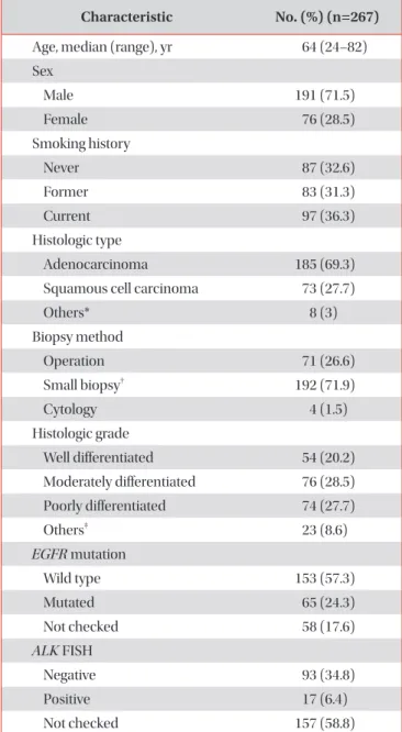
Results: From July 2016 to July 2017, 267 patients were enrolled. The main histologic type was adenocarcinoma (69.3%).

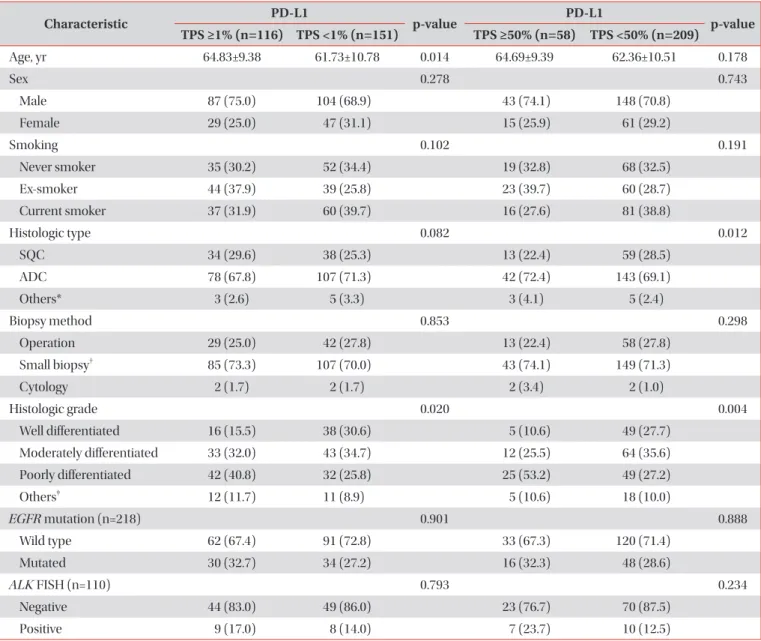
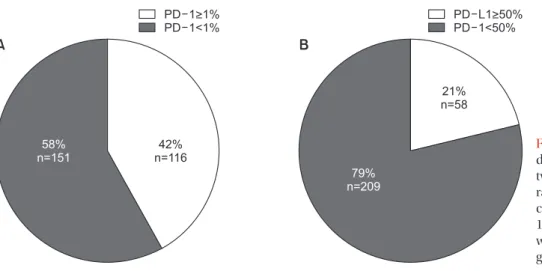
Most participants were smokers (67.4%) and had clinical stage IV disease (60.7%). In total, 116 (42%) and 58 (21%) patients had TPS ≥1% and ≥50%, respectively. The patients were significantly older in TPS ≥1% group than in TPS <1%

group (64.83±9.38 years vs. 61.73±10.78 years, p=0.014), not in TPS ≥50% cutoff value (64.69 ± 9.39 vs. 62.36 ± 10.51, p=

0.178). Regarding histologic grade, higher proportions of poorly differentiated tumor were observed in the TPS ≥1% (40.8%

vs. 25.8%, p=0.020) and TPS ≥50% groups (53.2% vs. 27.2%, p=0.004). Among 34 patients examined with 22C3 and SP263 assays, 27 had positive results in both assays, with a cutoff of TPS ≥1% (r=0.826; 95% confidence interval, 0.736–0.916).

Conclusion: PD-L1 expression, defined as TPS ≥1%, was related to older age and poorly differentiated histology. There was a similar distribution of PD-L1 expression in both 22C3 and SP263 results.

Keywords: Asian Continental Ancestry Group; Patients; Lung Neoplasms; Gene Expression; Carcinoma, Non-Small-Cell Lung

Address for correspondence: In-Jae Oh, M.D., Ph.D.

Department of Internal Medicine, Chonnam National University Hwasun Hospital, 322 Seoyang-ro, Hwasun 58128, Korea

Phone: 82-61-379-7617, Fax: 82-61-379-7619 E-mail: [email protected]

Received: Aug. 25, 2018 Revised: Oct. 21, 2018 Accepted: Jan. 15, 2019 Published online: Feb. 28, 2019

cc It is identical to the Creative Commons Attribution Non-Commercial License (http://creativecommons.org/licenses/by-nc/4.0/).

Copyright © 2019

The Korean Academy of Tuberculosis and Respiratory Diseases.

(2)

hibitors, the treatment for non-small cell lung cancer (NSCLC) is developing rapidly. Anti–PD-1 inhibitors (nivolumab and pembrolizumab) and anti–PD-L1 inhibitors (atezolizumab) have been approved as second-line treatments for patients with PD-L1 expression in advanced NSCLC in the United States, Europe, and Korea. The clinical characteristics of PD- L1 expression must be better understood, to be able to choose effective immune checkpoint inhibitors.

Although there is controversy regarding the use of PD-L1 as a predictive biomarker, until now the expression of PD-L1 had been thought to be the most valuable predictor of the re- sponse to anti‒PD-1/PD-L1 inhibitors

9,10

. Many PD-L1 inhibi- tors have been assessed with different immunohistochemical (IHC) assays using different antibodies, clones, platforms, scoring systems, and cutoff values. For example, the PD-L1 IHC 22C3 pharmDx assay (Agilent Technologies) and SP263 assay (Ventana Medical Systems) are approved as compan- ion diagnostic tests for pembrolizumab. The PD-L1 IHC 28-8 pharmDx assay and SP263 have been used for nivolumab and the PD-L1 IHC SP142 assay for atezolizumab. Because there is a PD-L1 assay for each PD-1/PD-L1 inhibitor, many clinical trials have examined the correlation of PD-L1 expression with clinical outcomes. A blueprint project assessing the analytic comparability among 22C3, 28-8, SP263, and SP142 showed that three assays (excluding SP142) were closely aligned on tumor cell staining

11

.

In previous studies, several clinicopathologic variables have been found to be correlated with PD-L1 expression, such as age, histology, degree of differentiation, and lymph node me- tastasis

12,13

. One study reported that the expression of PD-L1 was significantly higher for women, never smokers, and in ad- enocarcinoma

14

. To clarify these discordant study results, the present study aimed to investigate the clinical and pathologic characteristics of PD-L1 expression in Korean patients with lung cancer who might be candidates for immunotherapy. We also examined the correlation between PD-L1 IHC 22C3 and SP263 data to assess the possibility of using these PD-L1 tests interchangeably.

Materials and Methods

1. Patients and materials

We retrospectively reviewed the clinical and pathologic data of patients with pathologically proven lung cancer from a single institution between July 2016 and July 2017. A total of 267 patients with formalin-fixed, paraffin-embedded tissue samples were included. All data were gathered in accordance with the amended Declaration of Helsinki, following approval of the study by an independent hospital institutional review board (IRB approval number: CNUHH-2018-020). The need for written informed consent was waived because of the retro-

spective design of the study.

2. Immunohistochemistry

PD-L1 expression was detected using qualitative ihc stain-

Table 1. Baseline demographics

Characteristic No. (%) (n=267)

Age, median (range), yr 64 (24–82)

Sex

Male 191 (71.5)

Female 76 (28.5)

Smoking history

Never 87 (32.6)

Former 83 (31.3)

Current 97 (36.3)

Histologic type

Adenocarcinoma 185 (69.3)

Squamous cell carcinoma 73 (27.7)

Others* 8 (3)

Biopsy method

Operation 71 (26.6)

Small biopsy

192 (71.9)

Cytology 4 (1.5)

Histologic grade

Well differentiated 54 (20.2)

Moderately differentiated 76 (28.5) Poorly differentiated 74 (27.7)

Others

23 (8.6)

EGFR mutation

Wild type 153 (57.3)

Mutated 65 (24.3)

Not checked 58 (17.6)

ALK FISH

Negative 93 (34.8)

Positive 17 (6.4)

Not checked 157 (58.8)

*Other types consisting of small cell lung cancer (5 cases) and large cell lung cancer (3 cases).

Small biopsy specimens were col- lected via bronchoscopy, endobronchial ultrasound-transbronchial needle aspiration, or transthoracic needle biopsy of the lung.

“Oth- ers” included specimens for which histologic grade could not be checked.

EGFR: epidermal growth factor receptor; ALK: anaplastic lympho-

ma kinase; FISH: fluorescence in situ hybridization.

(3)

ing with the in vitro diagnostic PD-L1 IHC 22C3 pharmDx test on the Dako Autostainer (Agilent Technologies, Santa Clara, CA; Dako, Carpinteria, CA, USA) and the in vitro diagnostic PD-L1 IHC SP263 test on the Ventana BenchMark platform (Ventana Medical Systems, Tucson, AZ, USA) by one patholo- gist. The 22C3 pharmDx assay was performed for all patients, and both assays were carried out for 34 patients.

For the 22C3 pharmDx assay, sections were stained with anti–PD-L1 22C3 mouse monoclonal primary antibody us-

ing the EnVision FLEX visualization system on a Dako Au- tostainer Link 48 system with negative reagent controls and cell line run controls, as described in the PD-L1 IHC 22C3 pharmDx package insert

11,15

. Deparaffinization, rehydration, and target retrieval was performed with a 3-in-1 procedure using PT Link. Following peroxidase blocking, specimens were incubated with monoclonal mouse primary antibody to PD-L1 or the negative control reagent. Specimens were then incubated with Mouse Linker, followed by incubation with

Table 2. Clinical and pathological characteristics of the 267 patients with PD-L1 expression

Characteristic PD-L1

p-value PD-L1

p-value TPS ≥1% (n=116) TPS <1% (n=151) TPS ≥50% (n=58) TPS <50% (n=209)

Age, yr 64.83±9.38 61.73±10.78 0.014 64.69±9.39 62.36±10.51 0.178

Sex 0.278 0.743

Male 87 (75.0) 104 (68.9) 43 (74.1) 148 (70.8)

Female 29 (25.0) 47 (31.1) 15 (25.9) 61 (29.2)

Smoking 0.102 0.191

Never smoker 35 (30.2) 52 (34.4) 19 (32.8) 68 (32.5)

Ex-smoker 44 (37.9) 39 (25.8) 23 (39.7) 60 (28.7)

Current smoker 37 (31.9) 60 (39.7) 16 (27.6) 81 (38.8)

Histologic type 0.082 0.012

SQC 34 (29.6) 38 (25.3) 13 (22.4) 59 (28.5)

ADC 78 (67.8) 107 (71.3) 42 (72.4) 143 (69.1)

Others* 3 (2.6) 5 (3.3) 3 (4.1) 5 (2.4)

Biopsy method 0.853 0.298

Operation 29 (25.0) 42 (27.8) 13 (22.4) 58 (27.8)

Small biopsy

85 (73.3) 107 (70.0) 43 (74.1) 149 (71.3)

Cytology 2 (1.7) 2 (1.7) 2 (3.4) 2 (1.0)

Histologic grade 0.020 0.004

Well differentiated 16 (15.5) 38 (30.6) 5 (10.6) 49 (27.7)

Moderately differentiated 33 (32.0) 43 (34.7) 12 (25.5) 64 (35.6)

Poorly differentiated 42 (40.8) 32 (25.8) 25 (53.2) 49 (27.2)

Others

12 (11.7) 11 (8.9) 5 (10.6) 18 (10.0)

EGFR mutation (n=218) 0.901 0.888

Wild type 62 (67.4) 91 (72.8) 33 (67.3) 120 (71.4)

Mutated 30 (32.7) 34 (27.2) 16 (32.3) 48 (28.6)

ALK FISH (n=110) 0.793 0.234

Negative 44 (83.0) 49 (86.0) 23 (76.7) 70 (87.5)

Positive 9 (17.0) 8 (14.0) 7 (23.7) 10 (12.5)

Values are presented as mean±SD or number (%).

*Other types included small cell lung cancer (5 cases) and large cell lung cancer (3 cases).

Small biopsy specimens were collected via bron- choscopy, endobronchial ultrasound-trans-bronchial needle aspiration, or transthoracic needle biopsy of the lung.

“Others” included speci- mens for which histologic grade could not be checked.

PD-L1: programmed death-ligand 1; TPS: Tumor Proportion Score; SQC: squamous cell carcinoma; ADC: adenocarcinoma; EGFR: epider-

mal growth factor receptor; ALK: anaplastic lymphoma kinase; FISH: fluorescence in situ hybridization; SD: standard deviation.

(4)

a ready-to-use visualization reagent consisting of secondary antibody molecules and horseradish peroxidase molecules coupled to a dextran polymer backbone. The enzymatic con- version of the subsequently added chromogen results in the precipitation of a visible reaction product at the site of the an- tigen. The color of the chromogenic reaction is modified using a chromogen enhancement reagent; the specimen may then be counterstained and cover slipped. Results were interpreted using a light microscope.

For SP263 assay, sections were stained with anti–PD-L1 SP263 rabbit monoclonal primary antibody and a matched rabbit immunoglobulin G–negative control, using an OptiV- iew DAB IHC Detection Kit (Ventana Medical Systems) on the BenchMark ULTRA automated staining platform

11,16

.

Detection and quantification of the percentage of immuno- reactive tumor cells was performed according to the manu- facturer’s recommendations. Briefly, neoplastic cells were considered positive when any cell membrane staining was present, ignoring pure cytoplasmic immunoreaction. Staining of immune cells was also disregarded

17

. PD-L1 protein expres- sion was determined using the Tumor Proportion Score (TPS), which is the percentage of viable tumor cells showing partial or complete membrane staining

15

. A specimen was consid- ered PD-L1 positive with ≥50% of viable tumor cells exhibiting membrane staining at any intensity (i.e., TPS ≥50%); a weak PD-L1‒positive result is 1%≤TPS<50%. We designated two cat- egories according to cutoffs of TPS 1% or 50%.

3. Statistical analyses

Clinical characteristics and associations with biomarkers were examined by comparing differences using the Fisher exact test or independent sample t test, as appropriate. To evaluate the relationship between PD-L1 expression levels as- certained with the 22C3 and SP263 assays, we investigated 34 samples using precision analysis with Pearson’s concordance correlation coefficient. A p-value of <0.05 was considered sig-

nificant. Statistical analysis was performed using IBM SPSS version 23 (IBM Corp., Armonk, NY, USA).

Results

1. Baseline demographics

A total of 267 patients were included in this study (Table 1);

median age was 64 years (range, 24–82 years). Most patients were male (n=191, 71.5%) and former (n=83, 31.3%) or cur- rent smokers (n=97, 36.3%). The most frequent histologic type was adenocarcinoma (n=185, 69.3%), followed by squamous cell carcinoma (n=73, 27.7%) and small cell lung cancer (n=5, 1.9%). Most patients had clinical stage IV disease (n=162, 60.7%). Some patients were analyzed for the presence of epi- dermal growth factor receptor (EGFR) mutation and anaplas- tic lymphoma kinase (ALK) rearrangement: the result showed 65 patients (24.3%) with EGFR mutation and 17 (6.4%) with ALK rearrangement. Most specimens were obtained by small biopsy (n=192, 71.9%), followed by operation (n=71, 26.6%).

The histologic grade included moderately differentiated (n=76, 28.5%), poorly differentiated (n=74, 27.7%), and well differenti- ated (n=54, 20.2%).

2. Characteristics of patients with PD-L1 expression PD-L1 was evaluated in 267 patients, categorized using cut- offs of TPS 1% or 50% (Table 2, Figure 1). There were 116 (42%) and 58 (21%) patients with TPS ≥1% and ≥50%, respectively.

The correlation between age and PD-L1 expression was not significant (p=0.097). But the patients in the TPS ≥1% group were older than those in the TPS <1% group (64.83±9.38 vs.

61.73±10.78, p=0.014). Furthermore, the patients in the TPS

≥50% group were older than those in the TPS <50% group, but this was not statically significant (64.69±9.39 vs. 62.36±10.51, p=0.178). The group with TPS ≥1% included patients with ad-

Figure 1. Prevalence of programmed

death-ligand 1 (PD-L1) expression using

two cutoff points, in all patients. (A) The

rate of PD-L1 expression was 42% with

cutoff Tumor Proportion Score (TPS)

1%. (B) The rate of PD-L1 expression

was 21% with cutoff TPS 50%. PD-1: pro-

grammed death-1.

(5)

enocarcinoma (n=78, 67.8%) and squamous cell carcinoma (n=34, 29.6%) histology. The TPS ≥50% group also included patients with adenocarcinoma (n=42, 72.4%) and squamous cell carcinoma (n=13, 22.4%). The rate of poorly differentiated pathology was significantly higher in patients with TPS ≥1%

(40.8% vs. 25.8%, p=0.020) and TPS ≥50% (53.2% vs. 27.2%, p=0.004) than their counterparts. There were no differences with respect to smoking, EGFR mutation, ALK rearrangement status, or biopsy site.

3. Relationship between 22C3 and SP263

Among the 34 patients analyzed using both the 22C3 and SP263 assays, 27 patients had positive results in both assays, with a cutoff of 1% or higher (Figure 2A) and 20 patients had positive with a cutoff of 50% or higher (Figure 2B). There was a similar distribution between the 22C3 and SP263 assays as-

says (r=0.826; 95% confidence interval, 0.736–0.916) (Figure 3A). But it didn’t show a complete correlation between two assays for each patient (Figure 3B).

Discussion

In this study, we explored the clinicopathologic character- istics of PD-L1–positive Korean patients with lung cancer. For all enrolled patients, we performed PD-L1 IHC assays using monoclonal mouse anti–PD-1, clone 22C3; PD-L1 expression was described using TPS. A total 42% of patients were PD-L1 positive using a cutoff of TPS ≥1%; this proportion was 21%

with a cutoff of TPS ≥50%. Patients in the PD-L1‒positive (cutoff TPS ≥1%) group were significantly older than those in the PD- L1‒negative group. But in cutoff TPS ≥ 50% group, the patients were older than those in the TPS <50% group, but this was not statically significant. This seems to be due to the small sample size of the TPS ≥50% group to obtain statistical significance.

The most frequent histologic grade was poorly differentiated type in both the TPS ≥1% and TPS ≥50% groups.

In previous studies, several clinicopathologic variables were found to be correlated with PD-L1 expression, but there were discordant results among these studies. Mu et al.

12

evaluated 109 patients with adenocarcinoma and estimated the asso- ciation between expressed PD-L1 and tumor histologic type, degree of differentiation, and lymph node metastasis. Those authors found that higher expression of PD-L1 was related to adenocarcinoma histology and overall survival

12

. Velcheti et al.

18

explored cases of NSCLC including 340 cases in Greece and 204 cases from Yale University in the U.S. They found Figure 2. Prevalence of programmed death-ligand 1 (PD-L1) ex-

pression using the 22C3 pharmDx and SP263 assays. (A) Among the 34 patients analyzed with both the 22C3 and SP263 assays, 27 patients gave positive PD-L1 results for both tests, with a cutoff of Tumor Proportion Score (TPS) ≥1%. (B) Those positive results were found for 20 patients, with a cutoff of TPS ≥ 50%.

Figure 3. Relationship between programmed death-ligand 1 (PD-L1) immunohistochemical 22C3 pharmDx and SP263 assay. (A) Scatter

diagrams illustrating the relationship between expression levels with respect to 22C3 pharmDx and SP263 assays. Each point in the diagram

indicates the percentage of positive tumor cells in the 22C3 (x-axis) and SP263 (y-axis) assays. A significantly high relationship was observed

between the 22C3 and SP263 assays (r=0.826; 95% confidence interval, 0.736–0.916). (B) Analytic comparison of the percentage of tumor

proportion score by case for each assay. Data points expressed the scores on each case (blue points for 22C3 pharmDx and red points for

SP263 assay).

(6)

that advanced stage and squamous cell carcinoma were cor- related with high PD-L1 expression. Cooper et al.

13

analyzed 678 tissue samples from patients with stage I–III NSCLC and 52 paired nodal metastases. The authors showed that younger patients and those with poor differentiation were associated with high PD-L1 expression; there was no association with sex, tumor size, stage, nodal status, or EGFR or KRAS mutation status

13

. Azuma et al.

14

evaluated 164 specimens of surgically resected NSCLC and found that the expression of PD-L1 was significantly higher in women, never smokers, and patients with adenocarcinoma. These inconsistent results suggest that PD-L1 expression might differ by race or residence, and it may be necessary to analyze the clinical characteristics of each group.

We also examined the relationship between IHC assays in 34 patients and found a similar distribution of PD-L1 expres- sion between the 22C3 pharmDx and SP263 assays. Although there has been some controversy regarding use of PD-L1 as a predictive biomarker, the expression of PD-L1 has been thought to be the most valuable predictor of response to anti‒

PD-1/PD-L1 inhibitors

9,10

. Many PD-L1 inhibitors have been assessed with different IHC assays using different antibodies, clones, platforms, scoring systems, and cutoff values

11

. For example, the 22C3 pharmDx and SP263 assay are approved as companion diagnostic tests for pembrolizumab. The 28-8 pharmDx and SP263 assay have been used for nivolumab and the SP142 assay for atezolizumab.

Because there is an PD-L1 assay for each PD-1/PD-L1 in- hibitor, many clinical trials have examined the correlation of PD-L1 expression with clinical outcomes. A blueprint project assessed analytic comparability among the 22C3, 28-8, SP263, and SP142 assays using 39 NSCLC tumor samples. The result showed that three assays (excluding SP142) were closely aligned on tumor cell staining11. In a harmonized trial, 15 lung cancer resection specimens were used to assess interobserver concordance and PD-L1 IHC patterns using the 28-8, 22C3, SP142, and SP263 assays. The result showed that carcinoma cells can be reproducibly scored and there were no differenc- es in interobserver concordance among the tested assays

19

. However, the scoring of immune cells yielded low concor- dance rates and might require specific standardization

19

. A French multicenter harmonization study showed that the 28- 8, 22C3, and SP263 assays were comparable

20

. Another study assessed the concordance between PD-L1 assays using 493 formalin-fixed, paraffin-embedded archival NSCLC samples;

the assays showed similar patterns of tumor membrane stain- ing with high concordance between the SP263, 22C3, and 28-8 assays

21

. And the Italian multicenter comparison study of the 22C3 and SP263 assays found high correlation between PD-L1 IHC expression with the two tests

17

. In short, all these results suggest that the 22C3 and SP263 assays could be used interchangeably. Our study showed a similar distribution but not complete concordance between assays. This might be due

to the small number of subjects who performed both assays simultaneously.

There are some limitations to the present study. First, as mentioned above, this is a retrospective study from a single institution with a small sample size. Second, because most specimens were archival tissue, the PD-L1 IHC results might differ from those of fresh tissue. However, this situation may reflect real world clinical practice in that not all patients are able to undergo rebiopsy before immunotherapy. Third, we could not perform both 22C3 and SP263 assays in all patients;

therefore, the interpretation that there is a high correlation between the two tests should be made with caution. Further large-scale clinical studies should be performed to investigate the clinicopathologic variables of PD-L1 in NSCLC.

In conclusion, this retrospective study demonstrated that Korean patients with lung cancer who had PD-L1 expression (TPS ≥1%) were older and had poorly differentiated histology.

In patients with these characteristics, use of the PD-L1 IHC assay should be actively considered. We also found that the 22C3 pharmDx and SP263 assays could be used interchange- ably. However, a large-scale multicenter study is needed to better understand PD-L1 expression in lung cancer.

Authors’ Contributions

Conceptualization: Oh IJ, Park HY. Methodology: Oh IJ, Choi YD. Formal analysis: Park HY, Oh IJ, Choi YD. Data curation:

Park HY, Oh IJ, Kho BG, Kim TO, Shin HJ, Park CK, Kim YC.

Software: Park HY. Validation: Oh IJ. Investigation: Park HY, Oh IJ, Park CK, Kim YC, Kwon YS, Kim YI, Lim SC. Writing - original draft preparation: Park HY. Writing - review and edit- ing: Oh IJ, Park CK, Kim YC. Approval of final manuscript: all authors.

Conflicts of Interest

No potential conflict of interest relevant to this article was reported.

Acknowledgments

This work was supported by a clinical research grant from Chonnam National University Hwasun Hospital 2017.

References

1. Siegel RL, Miller KD, Jemal A. Cancer statistics, 2017. CA Can- cer J Clin 2017;67:7-30.

2. Park JY, Jang SH. Epidemiology of lung cancer in Korea: re-

(7)

cent trends. Tuberc Respir Dis 2016;79:58-69.

3. Kweon SS. Updates on cancer epidemiology in Korea, 2018.

Chonnam Med J 2018;54:90-100.

4. Pardoll DM. The blockade of immune checkpoints in cancer immunotherapy. Nat Rev Cancer 2012;12:252-64.

5. Francisco LM, Salinas VH, Brown KE, Vanguri VK, Freeman GJ, Kuchroo VK, et al. PD-L1 regulates the development, maintenance, and function of induced regulatory T cells. J Exp Med 2009;206:3015-29.

6. Boussiotis VA. Molecular and biochemical aspects of the PD-1 checkpoint pathway. N Engl J Med 2016;375:1767-78.

7. Chen DS, Irving BA, Hodi FS. Molecular pathways: next- generation immunotherapy: inhibiting programmed death- ligand 1 and programmed death-1. Clin Cancer Res 2012;18:

6580-7.

8. Hall RD, Gray JE, Chiappori AA. Beyond the standard of care:

a review of novel immunotherapy trials for the treatment of lung cancer. Cancer Control 2013;20:22-31.

9. Grigg C, Rizvi NA. PD-L1 biomarker testing for non-small cell lung cancer: truth or fiction? J Immunother Cancer 2016;4:48.

10. Patel SP, Kurzrock R. PD-L1 expression as a predictive bio- marker in cancer immunotherapy. Mol Cancer Ther 2015;

14:847-56.

11. Hirsch FR, McElhinny A, Stanforth D, Ranger-Moore J, Jans- son M, Kulangara K, et al. PD-L1 immunohistochemistry as- says for lung cancer: results from phase 1 of the Blueprint PD- L1 IHC assay comparison project. J Thorac Oncol 2017;12:

208-22.

12. Mu CY, Huang JA, Chen Y, Chen C, Zhang XG. High expres- sion of PD-L1 in lung cancer may contribute to poor progno- sis and tumor cells immune escape through suppressing tu- mor infiltrating dendritic cells maturation. Med Oncol 2011;

28:682-8.

13. Cooper WA, Tran T, Vilain RE, Madore J, Selinger CI, Koho- nen-Corish M, et al. PD-L1 expression is a favorable prog- nostic factor in early stage non-small cell carcinoma. Lung

Cancer 2015;89:181-8.

14. Azuma K, Ota K, Kawahara A, Hattori S, Iwama E, Harada T, et al. Association of PD-L1 overexpression with activating EGFR mutations in surgically resected nonsmall-cell lung cancer. Ann Oncol 2014;25:1935-40.

15. Roach C, Zhang N, Corigliano E, Jansson M, Toland G, Ponto G, et al. Development of a companion diagnostic PD-L1 im- munohistochemistry assay for pembrolizumab therapy in non-small-cell lung cancer. Appl Immunohistochem Mol Morphol 2016;24:392-7.

16. Rebelatto MC, Midha A, Mistry A, Sabalos C, Schechter N, Li X, et al. Development of a programmed cell death ligand-1 immunohistochemical assay validated for analysis of non- small cell lung cancer and head and neck squamous cell car- cinoma. Diagn Pathol 2016;11:95.

17. Marchetti A, Barberis M, Franco R, De Luca G, Pace MV, Staibano S, et al. Multicenter comparison of 22C3 PharmDx (Agilent) and SP263 (Ventana) assays to test PD-L1 expres- sion for NSCLC patients to be treated with immune check- point inhibitors. J Thorac Oncol 2017;12:1654-63.

18. Velcheti V, Schalper KA, Carvajal DE, Anagnostou VK, Syrigos KN, Sznol M, et al. Programmed death ligand-1 expression in non-small cell lung cancer. Lab Invest 2014;94:107-16.

19. Scheel AH, Dietel M, Heukamp LC, Johrens K, Kirchner T, Reu S, et al. Harmonized PD-L1 immunohistochemistry for pulmonary squamous-cell and adenocarcinomas. Mod Pathol 2016;29:1165-72.

20. Adam J, Rouquette I, Damotte D, Badoual C, Danel C, Damio- la F, et al. PL04a.04: multicentric French harmonization study for PD-L1 IHC testing in NSCLC. J Thorac Oncol 2017;12(1 Suppl):S11-2.

21. Ratcliffe MJ, Sharpe A, Midha A, Barker C, Scott M, Scorer P,

et al. Agreement between programmed cell death ligand-1

diagnostic assays across multiple protein expression cutoffs

in non-small cell lung cancer. Clin Cancer Res 2017;23:3585-

91.

수치

Table 1. Baseline demographics
Table 2. Clinical and pathological characteristics of the 267 patients with PD-L1 expression
Figure 1. Prevalence of programmed  death-ligand 1 (PD-L1) expression using  two cutoff points, in all patients
Figure 3. Relationship between programmed death-ligand 1 (PD-L1) immunohistochemical 22C3 pharmDx and SP263 assay

참조

관련 문서

Analysing this research material, The present study were examined that extravasated blood and cancer had interrelationship by comparing cancer patients

Method : : : : We evaluate clinical features of 714 Korean vitiligo patients who lived in the Gwangju chonnam province(315 males and 399 females).. Results

Purpose: This study was designed to evaluate the clinical characteristics and the treatment of colonoscopic perforation and the availability of

In this study, to investigate how applying a critical pathway to stomach cancer patients affects their recovery and treatment, the clinical effect of the critical

Baseline characteristics of genotype 1b chronic hepatitis C patients who treated with daclatasvir and asunaprevir (n=231)... Characteristics of genotype 1b

To assess the clinical usefulness of performing Q-PCR in practice as a diagnostic technique, we compared blindly the Q-PCR results using blood samples of the

(Method) Among 102 patients with metastatic liver carcinoma due to colorectal cancer who were treated at Chosun University Hospital from January 2003 to

Although visceral fat adiposity has known to be associated with clinical, pathologic, and oncologic outcomes in patients with colorectal cancer (CRC), the