Clinical Characteristics and Factors Influencing the Occurrence of Acute Eosinophilic Pneumonia in Korean Military Personnel
Acute eosinophilic pneumonia (AEP) is an uncommon inflammatory lung disease, and limited data exist concerning the clinical characteristics and factors that influence its occurrence. We retrospectively reviewed the records of AEP patients treated at Korean military hospitals between January 2007 and December 2013. In total, 333 patients were identified; their median age was 22 years, and all were men. All patients presented with acute respiratory symptoms (cough, sputum, dyspnea, or fever) and had elevated levels of inflammatory markers including median values of 13,185/μL for white blood cell count and 9.51 mg/dL for C-reactive protein. All patients showed diffuse ground glass opacity/
consolidation, and most had pleural effusion (n = 265; 80%) or interlobular septal thickening (n = 265; 85%) on chest computed tomography. Most patients had normal body mass index (n = 255; 77%), and only 30 (9%) patients had underlying diseases including rhinitis, asthma, or atopic dermatitis. Most patients had recently changed smoking habits (n = 288; 87%) and were Army personnel (n = 297; 89%).The AEP incidence was higher in the Army group compared to the Navy or Air Force group for every year (P = 0.002). Both the number of patients and patients with high illness severity (oxygen requirement, intensive care unit admission, and pneumonia severity score class ≥ III) tended to increase as seasonal temperatures rose. We describe the clinical characteristics of AEP and demonstrate that AEP patients have recently changed smoking habits and work for the Army. There is an increasing tendency in the numbers of patients and those with higher AEP severity with rising seasonal temperatures.
Keywords: Pulmonary Eosinophilia; Clinical Characteristics; Demography; Factors; Army;
Seasonal Chang-gyo Yoon,1 Se Jin Kim,2
Kang Kim,2 Ji Eun Lee,2 and Byung Woo Jhun2
1Department of Preventive Medicine, and 2Division of Pulmonary and Critical Care Medicine, Department of Medicine, The Armed Forces Medical Command, Seongnam, Korea
Received: 9 April 2015 Accepted: 19 October 2015 Address for Correspondence:
Byung Woo Jhun, MD
Division of Pulmonary and Critical Care Medicine, Department of Medicine, The Armed Forces Capital Hospital, 177 Saemaeul-ro, Bundang-gu, Seongnam 13574, Korea Tel: +82.31-725-6298, Fax: +82.31-706-0987 E-mail: [email protected]
http://dx.doi.org/10.3346/jkms.2016.31.2.247 • J Korean Med Sci 2016; 31: 247-253
INTRODUCTION
Acute eosinophilic pneumonia (AEP), first described by Allen et al. in 1989 as a distinct disease entity, is an uncommon in- flammatory lung disease (1). AEP presents with acute-onset re- spiratory symptoms, diffuse radiographic infiltrates, and eosin- ophilic infiltration into the lungs without a known cause of pul- monary eosinophilia, such as drugs, toxins, or parasite infesta- tions (2-4). The clinical severity of respiratory distress associat- ed with AEP varies from mild to severe; however, most patients with AEP exhibit rapid responses to systemic corticosteroid ther- apy without sequelae, even in fatal cases (5-8).
Recent studies concerning the clinical features of AEP have suggested that unidentified inhaled antigens could be partly re- sponsible for the inflammatory process (9-14), and recent mo- lecular studies have revealed that several inflammatory cyto- kines are associated with eosinophil recruitment into the lungs, degranulation, and eosinophil survival (15-19). However, to date, limited data exist concerning the detailed clinical features of AEP and possible factors in its occurrence, limiting insights
into its epidemiology or pathophysiology.
In Korean military hospitals, several cases of AEP are diag- nosed and managed annually, and an abundance of medical records and demographic data regarding the characteristics and outcomes of AEP patients has accumulated (5,6,20,21).
Thus, in this study, we retrospectively investigated the clinico- demographic data of AEP cases managed in Korean military hospitals to evaluate clinical characteristics and associated fac- tors that contribute to the development of, or increased vulner- ability to, AEP.
MATERIALS AND METHODS Study population
We retrospectively reviewed the medical records of consecutive adult patients with newly diagnosed AEP between January 2007 and December 2013 at multiple Korean military hospitals, in- cluding The Armed Forces Capital Hospital, The Armed Forces Busan Hospital, The Armed Forces Cheongpyeong Hospital, The Armed Forces Chuncheon Hospital, The Armed Forces Respiratory Diseases
Daegu Hospital, The Armed Forces Daejeon Hospital, The Armed Forces Gangneung Hospital, The Armed Forces Goyang Hospi- tal, The Armed Forces Hampyeong Hospital, The Armed Forces Hongcheon Hospital, The Armed Forces Ildong Hospital, The Armed Forces Seoul Hospital, The Armed Forces Wonju Hospi- tal, The Armed Forces YangJu Hospital, The Naval Marine Med- ical Center, The Naval Pohang Hospital, and The Air Forces Aero- space Medical Center.
A definitive diagnosis of AEP was based on a modification of the criteria proposed by Philit et al. (2), as reported previously (5): 1) acute onset of febrile respiratory manifestations < 1 month in duration; 2) bilateral diffuse infiltrates on chest radiography;
3) > 25% eosinophils in bronchoalveolar lavage (BAL) or eo- sinophilic pneumonia on lung biopsy; and 4) absence of known causes of pulmonary eosinophilia, including drugs, toxins and infections. If bronchoscopy with BAL or lung biopsy was not available, a clinical diagnosis of AEP was made when a rapid response to systemic corticosteroid therapy was achieved, in addition to definitive diagnostic criteria 1), 2), and 4). All pa- tients with a definitive and clinical diagnosis of AEP were in- cluded in the analysis.
Patient management and data collection
Patients who were clinically suspected of AEP underwent a di- agnostic workup consisting of laboratory examinations, chest radiography, computed tomography (CT), and/or flexible bron- choscopy with BAL. Infectious etiologies were investigated us- ing peripheral blood, sputum, tracheal aspirates and BAL fluid using the following techniques: staining and microbiological culture for bacteria and Mycobacterium tuberculosis; multiplex polymerase chain reaction for respiratory viruses, including in- fluenza virus, parainfluenza virus, adenovirus and respiratory syncytial virus; and serological testing for atypical pathogens, including Mycoplasma pneumoniae, Chlamydia pneumoniae, and parasites using specific antibody tests. Whether diagnostic tests or interventions were performed was decided by the at- tending physician.
Most patients diagnosed with AEP were treated with system- ic corticosteroid. Before May 2007, the dose or duration of cor- ticosteroid tapering was not standardized, while after May 2007, 2- or 4-week tapering protocols were widely used (5). After the publication of a study in May 2012 showing the non-inferiority of 2-week corticosteroid treatments compared to 4-week corti- costeroid treatments in managing AEP patients (5), 2-week ta- pering protocols became widely applied to AEP patients. An example of the 2-week corticosteroid treatment protocol is as follows: the initial dose of corticosteroid is chosen based on the presence of respiratory failure, defined as a partial pressure of arterial oxygen (PaO2)/fraction of inspired oxygen (FiO2) ratio
≤ 300 and/or tachypnea (respiration rate > 30 breaths/min).
Patients with respiratory failure received 60 mg methylprednis-
olone intravenously every 6 hours for 3 days followed by 30 mg oral prednisolone twice daily for 4 days. Patients without respi- ratory failure received 30 mg oral prednisolone twice daily for 7 days, followed by tapering over 2 weeks. However, when oxy- genation was not severely impaired (PaO2/FiO2 ratio > 350) and symptoms were mild, the patient underwent conservative treatment without corticosteroids based on the decision of the attending physician.
The following data were collected retrospectively: demogra- phic characteristics including age, sex, body mass index, un- derlying diseases, history of recent upper-respiratory infections, smoking habits, and working position in military services. Clin- ico-radiological findings and laboratory data at initial presenta- tion were collected, and clinical parameters that reflect disease severity, including oxygen requirement rate, intensive care unit (ICU) admission rate, the need for mechanical ventilation, and pneumonia severity index score, were also collected. Annual incidence rates and seasonal variations were also evaluated.
In calculating annual incidence rates, we used the cited pop- ulation of each military service as a denominator, which is pub- lished biennially by The Ministry of National Defense, Korea, in
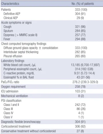
Table 1. Clinical characteristics of patients with AEP
Characteristics No. (%) of patients
Patients Definitive AEP Clinical AEP
333 (100) 304 (91)
29 (9) Acute symptoms or signs
Cough Sputum
Dyspnea ( > MMRC scale II) Fever
321 (96) 284 (85) 257 (77) 294 (88) Chest computed tomography findings
Diffuse ground glass opacity ± consolidation Interlobular septal thickening
Pleural effusion
333 (100) 282 (85) 265 (80) Laboratory findings
White blood cell count, /μL Peripheral eosinophil count, /μL C-reactive protein, mg/dL Eosinophil % in BAL fluid
13,185 (9,700-17,697) 314 (182-538) 9.51 (5.72-14.4)
43 (31-56)
PaO2/FiO2 ratio 276.2 (239.3-329.0)
Oxygen requirement 258 (78)
ICU admission 103 (31)
Mechanical ventilation 8 (2)
PSI classification Class I and II Class III Class IV Class V
242 (72) 86 (26) 4 (1) 1 (1) Diagnostic flexible bronchoscopy 304 (91)
Corticosteroid treatment 306 (92)
Conservative treatment without corticosteroid 27 (8)
PaO2/FiO2 data were missing in 56 cases. Data are presented as medians (interquar- tile ranges) or numbers (%). AEP, acute eosinophilic pneumonia; BAL, bronchoalveolar lavage; PaO2, partial pressure of arterial oxygen; SpO2, oxygen saturation; ICU, inten- sive care unit; MMRC, modified medical research council; MV, mechanical ventilation;
PSI, pneumonia severity index.
the defense white paper. This was used to calculate annual inci- dence rates of AEP among the entire military population and within each service. A Poisson-distribution was applied to esti- mate confidence intervals since most medical events are con- sidered to follow Poisson-distributions, especially rare diseases like AEP. Follow-up data were last obtained on February 1, 2014.
All medical records were anonymized or de-identified.
Statistical analysis
Data are presented as medians and interquartile ranges (IQR) for continuous variables and as numbers and percentages for categorical variables. Data were compared using the Kruskal- Wallis test (more than two independent groups) for continuous variables, and a χ2 or Fisher’s exact test for categorical variables.
Values of P < 0.05 were considered to indicate statistical signifi- cance. All statistical analyses were performed using the PASW software (ver. 18.0; SPSS Inc., Chicago, IL, USA).
Ethics statement
The study protocol was designed in accordance with the Decla- ration of Helsinki and was approved by the institutional review board of the Armed Forces Capital Hospital (AFMC-14-IRB-016) and the Armed Forces Medical Command on behalf of the mil-
itary hospitals that were involved in the current study. Informed consent was exempted by the board.
RESULTS
Clinical characteristics of patients with AEP
Three hundred and thirty-three patients with AEP were identi- fied in the study period, including 304 (91%) definitive and 29 (9%) clinical AEP patients. Clinical characteristics of study pa- tients are shown in Table 1. All patients had acute respiratory symptoms or signs including cough, sputum, dyspnea, or fever.
On initial chest computed tomography scans, all patients had diffuse ground glass opacity with or without consolidation (Fig.
1), 282 (85%) had interlobular septal thickening, and 265 (80%) had pleural effusion. All patients had elevated inflammatory markers, including white blood cell count and C-reactive pro- tein, with median values of 13,185 (IQR, 9,700-17,697)/μL and 9.51 (IQR, 5.72-14.4) mg/dL, respectively, and median periph- eral eosinophil counts of 314 (182-538)/μL. The median value of the PaO2/FiO2 ratio was 276.2 (IQR, 239.3-329.0), 32% of the patients had severe hypoxemia (PaO2/FiO2 < 250), and 258 (78%) patients had an oxygen requirement. Of the study pa- tients, 103 (31%) were admitted to the ICU, 8 (2%) required me-
Fig. 1. Typical chest computed tomography findings in patients with acute eosinophilic pneumonia. Diffuse bilateral ground-glass opacity and interlobular septal thickening are seen in both lungs.
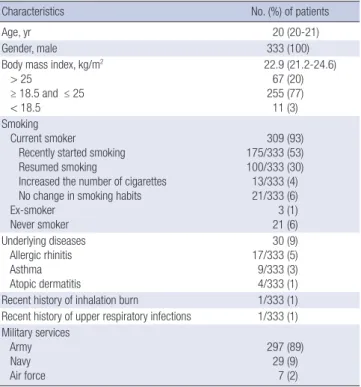
Table 2. Demographics of patients with AEP
Characteristics No. (%) of patients
Age, yr 20 (20-21)
Gender, male 333 (100)
Body mass index, kg/m2 > 25
≥ 18.5 and ≤ 25 < 18.5
22.9 (21.2-24.6) 67 (20) 255 (77) 11 (3) Smoking
Current smoker Recently started smoking Resumed smoking
Increased the number of cigarettes No change in smoking habits Ex-smoker
Never smoker
309 (93) 175/333 (53) 100/333 (30) 13/333 (4) 21/333 (6) 3 (1) 21 (6) Underlying diseases
Allergic rhinitis Asthma Atopic dermatitis
30 (9) 17/333 (5) 9/333 (3) 4/333 (1) Recent history of inhalation burn 1/333 (1) Recent history of upper respiratory infections 1/333 (1) Military services
Army Navy Air force
297 (89) 29 (9)
7 (2)
Data are presented as medians (interquartile ranges) or numbers (%). AEP, acute eo- sinophilic pneumonia.
Table 3. Annual incidence of AEP occurrence according to military service
Years Entire forces Army Navy Air force
2013 11.0 (8.6-13.9) 12.6 (9.8-16.3) 8.8 (3.2-19.2) 0.0 (0.0-5.7) 2012 8.1 (6.1-10.8) 9.5 (7.1-12.7) 2.9 (0.4-10.6) 3.1 (0.4-11.1) 2011 6.3 (4.5-8.6) 7.1 (5.1-10.0) 5.9 (1.6-15.1) 0.0 (0.0-5.7) 2010 6.7 (5.0-9.1) 8.3 (6.1-11.2) 1.5 (0.0-8.2) 0.0 (0.0-5.7) 2009 8.7 (6.7-11.4) 10.0 (7.5-13.2) 7.4 (2.4-17.2) 0.0 (0.0-11.1) 2008 7.8 (5.7-10.0) 8.4 (6.0-11.0) 7.4 (2.4-17.2) 3.1 (0.4-11.1) 2007 2.8 (1.7-4.4) 3.5 (2.1-5.5) 0.0 (0.0-5.4) 0.0 (0.0-5.7) Data are presented as incidence per 100,000 person-year (95% confidence interval).
In each year, the incidence of AEP tended to be higher among Army personnel com- pared to Navy or Air Force personnel, and these differences were statistically signifi- cant (P = 0.0021). AEP, acute eosinophilic pneumonia.
Fig. 2. Incidence of AEP according to military service per year.
Incidence (per 100,000 person-years)
Entire forces Army Navy Air force
15
10
5
0
20132012 20112010 2009 20082007
chanical ventilation support, and approximately one-third of the study patients had more than a pneumonia severity index score (PSI) class II; class III (n = 86; 26%), class IV (n = 4; 1%), and class V (n = 1; 1%). Overall, 306 (92%) AEP patients were treated with systemic corticosteroid and 27 (8%) patients did not receive corticosteroid; all patients survived without sequelae.
Demographics of patients with AEP
The demographics of AEP patients are shown in Table 2. The median age was 22 (IQR, 20-21) years and all patients were male.
The median body mass index was 22.9 (IQR, 21.2-24.6 kg/m2);
67 (20%) patients were overweight ( > 25 kg/m2) and 11 (3%) patients were underweight (< 18.5 kg/m2). Most patients were current smokers (n = 309; 93%); among these, 175 (53%) pa- tients had recently started smoking, 100 (30%) patients had re- sumed smoking, and only 24 (7%) patients were ex-smokers or had never smoked. Only 30 (9%) patients had underlying dis- eases; allergic rhinitis (n = 17; 5%) was the most common, fol- lowed by asthma (n = 9; 3%) and atopic dermatitis (n = 4; 1%).
One patient with a recent history of inhalation burns and one patient with a recent upper respiratory infection were identi- fied. Most of the patients were in the Army (n = 297; 89%), with 29 (9%) in the Navy and only 7 (2%) in the Air Force.
Annual incidences of AEP occurrence according to military service
Because absolute numbers of AEP patients were much lower in the Navy and Air Force than in the Army, as shown by the de-
mographics of study patients, we calculated and compared the annual incidence of AEP occurrence according to military ser- vice (Table 3). Among all forces, in 2008 the incidence of AEP abruptly increased compared to 2007, and by 2013 the incidence of AEP was highest at up to 11.0 per 100,000 person-years (95%
confidence interval, 8.6-13.9; Fig. 2). Interestingly, in each year, the incidence of AEP tended to be higher among Army person- nel compared to Navy or Air Force personnel, and these differ- ences were statistically significant (Kruskal-Wallis, P = 0.002).
However, the differences in other clinico-demographic data between the different military services were not statistically sig- nificant (data not shown).
Seasonal variation in the occurrence of AEP patients Since physicians working at military hospitals generally esti- mate that more cases of AEP are encountered in the summer than in other seasons, we investigated seasonal variations in the occurrence of AEP among all military services to estimate whether occurrence was influenced by season (Table 4). Of all AEP cases, 125 (38%) occurred during the summer, 100 (30%) during the autumn, 58 (17%) during the spring, and only 50 (15%) during the winter. Numbers of AEP patients tended to increase as seasonal temperatures increased (Table 4) and peaked in the summer each year, around August.
Table 4. Numbers of occurrence of AEP patients according to season per year
Years Spring Summer Autumn Winter
Total Mar Apr May Jun Jul Aug Sept Oct Nov Dec Jan Feb
2013 1 2 8 5 11 9 11 10 3 3 5 2 70
2012 5 2 6 4 1 10 9 6 4 3 1 1 52
2011 3 1 5 6 3 6 2 1 6 2 3 2 40
2010 2 1 2 3 7 7 4 7 3 5 1 2 44
2009 2 3 2 4 9 8 7 5 3 4 6 4 57
2008 3 3 6 2 11 8 2 5 6 3 0 2 51
2007 0 0 1 3 3 5 0 4 2 1 0 0 19
Total 16 12 30 27 45 53 35 38 27 21 16 13 333
Data are presented as numbers of AEP patients.
Table 5. Number of AEP patients with a relatively higher severity of illness according to season
Characteristics Spring Summer Autumn Winter
Mar Apr May Jun Jul Aug Sept Oct Nov Dec Jan Feb
Oxygen requirement 12 9 23 22 33 44 27 32 20 16 10 10
ICU admission 4 4 9 7 13 18 9 16 7 5 5 6
Mechanical ventilation 0 0 2 0 1 1 1 1 2 0 0 0
PSI class III 5 2 9 8 9 14 9 10 7 4 4 5
PSI class IV 0 0 0 0 3 0 0 1 0 0 0 0
PSI class V 0 0 1 0 0 0 0 0 0 0 0 0
Data are presented as numbers of AEP patients. AEP, acute eosinophilic pneumonia; ICU, intensive care unit; PSI, pneumonia severity index.
Fig. 3. Levels of C-reactive protein in patients with AEP according to season.
C-reactive protein (mg/dL)
Spring (n = 58)
Summer (n = 125)
Autumn (n = 100)
Winter (n = 50) 35
30 25 20 15 10 5 0
We additionally evaluated the number of AEP patients with a relatively high severity of illness according to season (Table 5).
The number of severe cases tended to increase as seasonal tem- peratures rose and peaked around the summer, from late spring to early autumn. The numbers of patients who needed oxygen (n = 99) and ICU admission (n = 38), and had a PSI score of III (n = 31) or IV (n = 3) tended to be higher in summer than in other seasons. None of the patients who needed mechanical ventilation was seen during the winter. One of the most severe cases of AEP (PSI class V) occurred during late spring, in May.
However, differences in other clinico-demographic data ac- cording to season (spring vs. summer vs. autumn vs. winter) were not statistically significant (data not shown). The initial level of C-reactive protein in AEP patients had a slight tendency to be higher during summer and autumn than during spring and winter (Fig. 3), and median values of C-reactive protein in spring, summer, autumn, and winter were 9.53 (IQR, 5.34-13.1) mg/dL, 9.04 (IQR, 4.90-13.8) mg/dL, 10.10 (IQR, 6.62-16.02) mg/dL, and 7.90 (IQR, 5.96-12.61) mg/dL, respectively; howev- er the differences did not reach statistical significance.
DISCUSSION
In this study, we investigated the clinical characteristics of AEP and evaluated possible demographic factors that may contrib- ute to its development in Korean military personnel. Our data suggest that several environmental factors such as recently chan- ged smoking habits, military service in the Army, and increas-
ing seasonal temperature could be related to the occurrence of AEP. Regarding environmental influences on the development of AEP, previous studies have indicated that inhaled stimulants may trigger the inflammatory processes of AEP (9-12). For ex- ample, an observational study that included 33 AEP patients found that altered smoking habits, not only starting to smoke but also restarting smoking and increasing daily smoking dos- es, were associated with the development of AEP (12). More- over, Rom et al. presented a case study of a firefighter that de- veloped AEP after exposure to dust from the World Trade Cen- ter collapse (10). Similar to these previous studies, the majority of our patients (n = 288; 87%) had recently altered smoking hab- its and the annual incidence of AEP was highest in the Army, which may be because the Army has more ground services, and outdoor activity in mountainous areas of Korea may increase the likelihood of inhaling unidentified antigens.
Interestingly, in this study we found that the occurrence of AEP was related to seasonal variations. The number of AEP pa- tients tended to increase as seasonal temperatures rose and peaked around summer, which was associated with greater dis- ease severity than the other seasons. Moreover, AEP with rela- tively high illness severity was more common in the summer than in other seasons, although seasonal differences in the pa- rameters that indicate disease severity did not reach statistical significance. These phenomena could be partly explained by the rise in temperature as the season changes from winter to summer, possibly increasing the likelihood of inhalation of stim- ulants or unidentified antigens, which in turn may trigger lung
inflammation. One other study has described an association between AEP occurrence and season or climate. When evaluat- ing the clinical features and epidemiology of 18 AEP cases in the United States military personnel deployed in or near Iraq, summer had the highest incidence of AEP (22). However, limit- ed data exist concerning environmental influences on AEP oc- currence, and well-designed studies of its occurrence that ad- just for other confounding factors have not been performed, mainly due to the rarity of the disease, which limits insight into its epidemiology or pathophysiology.
In our study only a small proportion (n = 11; 3%) of patients had a low body mass index (< 18.5 kg/m2) and the majority of patients (n = 255; 77%) had a normal body mass index. This body mass index distribution is uncommon in other pulmonary diseases such as tuberculosis and other inflammatory pulmo- nary diseases. Additionally, only 9% of study patients had un- derlying allergic diseases. These findings suggest that inflam- mation in AEP is not a chronic process and that AEP may have different inflammatory processes than known allergic reac- tions. To date there has not been a comprehensive study of the association of AEP with immunoglobulin E-mediated or aller- gic reactions. The results of a recent study indicated that immu- noglobulin G levels decreased significantly during active dis- ease states and increased during remission, but serum immu- noglobulin E levels did not change significantly, suggesting that the pathogenesis of AEP may negatively impact serum immu- noglobulin G levels but not immunoglobulin E levels (23). How- ever, accurate data concerning the inflammatory process are limited; thus, further pathophysiological studies along with epi- demiological data are needed.
In our study, the incidence of AEP in recent years was similar to previously reported rates. In a study of the characteristics and epidemiology of AEP among the United States military personnel, Shorr et al. (22) reported an AEP incidence of 9.1 per 100,000 person-years (95% confidence interval, 4.3 to 13.3).
However, in our current study the annual AEP incidence rate varied from 2.8 per 100,000 person-years in 2007 to 11.0 per 100,000 person-years in 2013. The lower incidence in 2007 com- pared to other years is likely due to inadequate awareness of AEP in military hospitals in Korea. The peak incidence of AEP in 2013 may be due to improved insight into AEP by military physicians as well as the increased performance of diagnostic flexible bronchoscopies using the BAL procedure in unknown respiratory failure patients. Therefore, more aggressive diagnos- tic evaluation of patients will facilitate determination of accu- rate incidence rates, changes in AEP incidence rates, and aid the investigation of its epidemiology.
This study has some limitations. First, there was no matched normal control group to compare to the AEP group since we performed a retrospective case series study. This type of study was chosen since collecting medical data from healthy military
personnel is difficult as it is not permitted without definite med- ical evidence in any of the Korean armed forces. Thus, based on the results of this study, we evaluated only accurate case series data. Second, because our study was conducted at military hos- pitals only, most patients were previously healthy males with a uniform median age, and thus not representative of the general population. Third, there is a possibility that the incidence or oc- currence of AEP might have been underestimated in our study population because some patients might have been treated at a local military hospital or might have improved spontaneously.
However, we cautiously estimate that this influence is not sig- nificant due to the unique characteristics of the medical system of the Korean military services. According to the military law of Korean military personnel, all soldiers should be treated in mil- itary hospitals first if possible. Accordingly, most patients with suspected AEP were likely transferred to a military hospital, in- cluding the Armed Forces Capital Hospital, which is the largest tertiary referral military hospital in Korea, where bronchoscopy is available.
In conclusion, in this study we investigated the clinical char- acteristics of AEP, demonstrated that patients with AEP frequent- ly have recently changed smoking habits and work for the Army, and found an increasing tendency in the numbers of patients and of patients with higher AEP severity with increasing sea- sonal temperatures. These results highlight the need for future pathophysiological and epidemiological prospective studies to evaluate the impact of external environmental variables on the occurrence and pathophysiology of AEP.
DISCLOSURE
All authors have no potential conflicts of interest to disclose.
AUTHOR CONTRIBUTION
Conception and design of the study: Yoon C, Jhun BW. Data col- lection and analysis: Yoon C, Kim SJ, Kim K, Lee JE, Jhun BW.
Writing the first draft: Yoon C, Jhun BW. Revision of manuscript:
Yoon C, Jhun BW. ICMJE criteria for authorship: Yoon C, Kim SJ, Kim K, Lee JE, Jhun BW.
ORCID
Chang-gyo Yoon http://orcid.org/0000-0002-1543-4659 Se Jin Kim http://orcid.org/0000-0001-7504-812X Kang Kim http://orcid.org/0000-0002-3935-7619 Byung Woo Jhun http://orcid.org/0000-0002-6348-8731 REFERENCES
1. Allen JN, Pacht ER, Gadek JE, Davis WB. Acute eosinophilic pneumonia
as a reversible cause of noninfectious respiratory failure. N Engl J Med 1989; 321: 569-74.
2. Philit F, Etienne-Mastroïanni B, Parrot A, Guérin C, Robert D, Cordier JF. Idiopathic acute eosinophilic pneumonia: a study of 22 patients. Am J Respir Crit Care Med 2002; 166: 1235-9.
3. Hayakawa H, Sato A, Toyoshima M, Imokawa S, Taniguchi M. A clinical study of idiopathic eosinophilic pneumonia. Chest 1994; 105: 1462-6.
4. Daimon T, Johkoh T, Sumikawa H, Honda O, Fujimoto K, Koga T, Ara- kawa H, Yanagawa M, Inoue A, Mihara N, et al. Acute eosinophilic pneu- monia: thin-section CT findings in 29 patients. Eur J Radiol 2008; 65:
462-7.
5. Rhee CK, Min KH, Yim NY, Lee JE, Lee NR, Chung MP, Jeon K. Clinical characteristics and corticosteroid treatment of acute eosinophilic pneu- monia. Eur Respir J 2013; 41: 402-9.
6. Jhun BW, Kim SJ, Kim K, Lee JE. Clinical implications of initial periph- eral eosinophilia in acute eosinophilic pneumonia. Respirology 2014;
19: 1059-65.
7. Lim SY, Suh GY, Jeon K. Acute eosinophilic pneumonia presenting as life-threatening hypoxaemia necessitating extracorporeal membrane oxygenation. Int J Tuberc Lung Dis 2012; 16: 1711-2.
8. Kawayama T, Fujiki R, Morimitsu Y, Rikimaru T, Aizawa H. Fatal idio- pathic acute eosinophilic pneumonia with acute lung injury. Respirolo- gy 2002; 7: 373-5.
9. Imokawa S, Sato A, Hayakawa H, Toyoshima M, Taniguchi M, Chida K.
Possible involvement of an environmental agent in the development of acute eosinophilic pneumonia. Ann Allergy Asthma Immunol 1996; 76:
419-22.
10. Rom WN, Weiden M, Garcia R, Yie TA, Vathesatogkit P, Tse DB, McGuin- ness G, Roggli V, Prezant D. Acute eosinophilic pneumonia in a New York City firefighter exposed to World Trade Center dust. Am J Respir Crit Care Med 2002; 166: 797-800.
11. Watanabe K, Fujimura M, Kasahara K, Yasui M, Myou S, Kita T, Wata- nabe A, Nakao S. Acute eosinophilic pneumonia following cigarette smok- ing: a case report including cigarette-smoking challenge test. Intern Med 2002; 41: 1016-20.
12. Uchiyama H, Suda T, Nakamura Y, Shirai M, Gemma H, Shirai T, Toyo- shima M, Imokawa S, Yasuda K, Ida M, et al. Alterations in smoking hab- its are associated with acute eosinophilic pneumonia. Chest 2008; 133:
1174-80.
13. Kitahara Y, Matsumoto K, Taooka Y, Moritani C, Nakamura K, Ohashi
N, Daido K, Arita K. Cigarette smoking-induced acute eosinophilic pneu- monia showing tolerance in broncho-alveolar lavage findings. Intern Med 2003; 42: 1016-21.
14. Shiota Y, Kawai T, Matsumoto H, Hiyama J, Tokuda Y, Marukawa M, Ono T, Mashiba H. Acute eosinophilic pneumonia following cigarette smoking. Intern Med 2000; 39: 830-3.
15. Katoh S, Matsumoto N, Matsumoto K, Fukushima K, Matsukura S. Ele- vated interleukin-18 levels in bronchoalveolar lavage fluid of patients with eosinophilic pneumonia. Allergy 2004; 59: 850-6.
16. Mato N, Bando M, Kusano A, Hirano T, Nakayama M, Uto T, Nakaya T, Yamasawa H, Sugiyama Y. Clinical significance of interleukin 33 (IL-33) in patients with eosinophilic pneumonia. Allergol Int 2013; 62: 45-52.
17. Miyazaki E, Nureki S, Fukami T, Shigenaga T, Ando M, Ito K, Ando H, Sugisaki K, Kumamoto T, Tsuda T. Elevated levels of thymus- and acti- vation-regulated chemokine in bronchoalveolar lavage fluid from pa- tients with eosinophilic pneumonia. Am J Respir Crit Care Med 2002;
165: 1125-31.
18. Allen JN, Liao Z, Wewers MD, Altenberger EA, Moore SA, Allen ED. De- tection of IL-5 and IL-1 receptor antagonist in bronchoalveolar lavage fluid in acute eosinophilic pneumonia. J Allergy Clin Immunol 1996; 97:
1366-74.
19. Okubo Y, Horie S, Hachiya T, Momose T, Tsukadaira A, Takashi S, Suzu- ki J, Isobe M, Sekiguchi M. Predominant implication of IL-5 in acute eo- sinophilic pneumonia: comparison with chronic eosinophilic pneumo- nia. Int Arch Allergy Immunol 1998; 116: 76-80.
20. Lee JE, Rhee CK, Lim JH, Lee SM, Shim YS, Lee CT, Lee SW. Fraction of exhaled nitric oxide in patients with acute eosinophilic pneumonia. Chest 2012; 141: 1267-72.
21. Jhun BW, Kim SJ, Kim K, Lee JE, Hong DJ. Clinical implications of corre- lation between peripheral eosinophil count and serum levels of IL-5 and tryptase in acute eosinophilic pneumonia. Respir Med 2014; 108: 1655- 62.
22. Shorr AF, Scoville SL, Cersovsky SB, Shanks GD, Ockenhouse CF, Smoak BL, Carr WW, Petruccelli BP. Acute eosinophilic pneumonia among US Military personnel deployed in or near Iraq. JAMA 2004; 292: 2997-3005.
23. Matsuno O, Takenaka R, Hiroshige S, Ono E, Nishitake T, Ueno T, Mi- yazaki E, Kumamoto T. Reduced IgG levels found during acute eosino- philic pneumonia, which normalize during recovery from disease. Respir Med 2008; 102: 899-903.