Korean Circulation Journal
Introduction
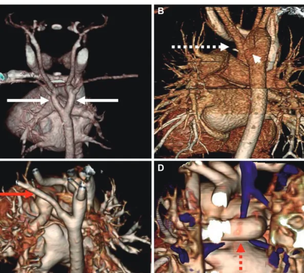
Vascular rings are rare congenital anomalies that primarily result from an embryological derangement of the paired aortic arches or branching pulmonary arteries.
1)2)The symptoms and physical find- ings produced by vascular rings are related to the structure(s) they
Print ISSN 1738-5520 • On-line ISSN 1738-5555
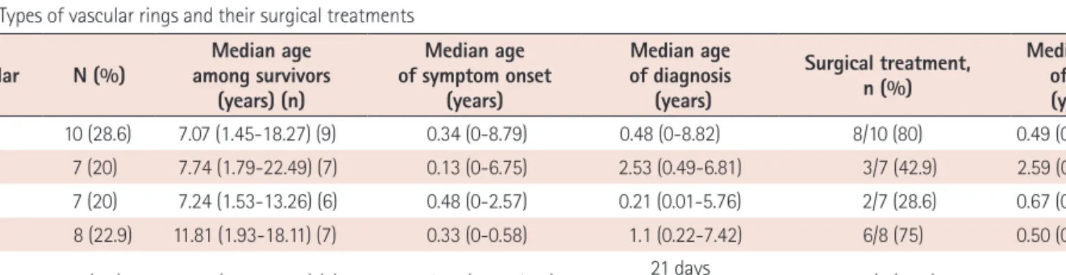
Clinical Course of Vascular Rings and Risk Factors Associated With Mortality
Yoon Jung Suh, MD 1 , Gi Beom Kim, MD 1 , Bo Sang Kwon, MD 1 , Eun Jung Bae, MD 1 , Chung Il Noh, MD 1 , Hong Gook Lim, MD 2 , Woong Han Kim, MD 2 , Jeong Ryul Lee, MD 2 , and Yong Jin Kim, MD 2
1