Osteosarcomatous Transformation in
Mazabraud Syndrome:
A Case Report
Mazabraud 증후군에서 발생한 골육종으로 악성 변환:
증례 보고
Yang Il Park, MD , Ji Young Yoo, MD* , Joon Seog Kong, MD
Department of Radiology, Korea Cancer Center Hospital, Korea Institute of Radiological and Medical Sciences, Seoul, Korea
Mazabraud syndrome is a rare benign disease that is accompanied by polyostotic fibrous dys- plasia and intramuscular myxoma. Malignant transformation of fibrous dysplasia occurs in ap- proximately 1% of cases. To date, only eight cases of malignant transformation, of fibrous dys- plasia to osteosarcoma, in Mazabraud syndrome have been reported worldwide. The authors report the first case of osteosarcomatous transformation in a patient with Mazabraud syn- drome in the Republic of Korea, focusing on imaging findings.
Index terms Mazabraud Syndrome; Transformation, Neoplastic Cell; Fibrous Dysplasia of Bone;
Myxoma; Osteosarcoma; Magnetic Resonance
서론
Mazabraud 증후군은 1926년에 Henschen이 처음으로 보고하고 1967년 Mazabraud에 의해 정립되었으며, 다골성(polyostotic) 섬유성 이형성(fibrous dysplasia)과 근육 내 점액 종(intramuscular myxoma)이 함께 나타나는 드문 양성질환으로(1) 대한민국에서의 보 고는 1예가 있다(2).
섬유성 이형성에서 악성 변환이 일어난 경우는 약 1%로 알려져 있고, Mazabraud 증후군 의 섬유성 이형성에서 발생한 골육종으로 악성 변환에 대한 보고는 아직까지 전 세계에 8예 에 불과하다(3).
저자들은 Mazabraud 증후군 환자가 통증과 만져지는 종괴를 주소로 내원하여 시행한 영 상검사에서 섬유성 이형성의 악성 변환이 골육종으로 진단된 증례를 경험하였기에 영상 소
Received December 16, 2020 Revised December 31, 2020 Accepted January 5, 2021
*Corresponding author Ji Young Yoo, MD
Department of Radiology, Korea Cancer Center Hospital, Korea Institute of Radiological and Medical Sciences, 75 Nowon-ro, Nowon-gu, Seoul 01812, Korea.
Tel 82-2-970-1576 Fax 82-2-970-2433 E-mail [email protected] This is an Open Access article distributed under the terms of the Creative Commons Attribu- tion Non-Commercial License (https://creativecommons.org/
licenses/by-nc/4.0) which permits unrestricted non-commercial use, distribution, and reproduc- tion in any medium, provided the original work is properly cited.
ORCID iDs Yang Il Park https://
orcid.org/0000-0002-9557-9371 Ji Young Yoo
https://
orcid.org/0000-0002-2115-0441 Joon Seog Kong
https://
orcid.org/0000-0003-4151-4617
견을 중심으로 보고하고자 한다.
증례 보고
50세 남자가 내원 1달 전부터 발생한 좌측 정강이 통증, 압통과 만져지는 종괴를 주소로 내원했 다(연구 대상자로부터 서면으로 동의를 취득하였다. 또한, 헬싱키 1975 선언에 따라 연구를 진행하 였다). 내원 12년 전부터 외부 병원에서 좌측 하지의 다골성 섬유성 이형성으로 경과 관찰 중이었다.
내원 당시 혈액검사에서 alkaline phosphatase (이하 ALP)는 1402 U/L, lactate dehydroge- nase (이하 LDH)는 264 U/L로 증가하였다.
내원 당시 촬영한 단순 X선 영상에서 좌측 경골 근위부에 팽창성의 방사선투과 병변이 있고 골 내막에 조개모양의 잠식(endosteal scalloping), 피질골 파괴, 골 기질 석회화, 근위부 경골의 앞, 뒤로 수직 선상(perpendicular) 골막반응을 포함하고 있는 연조직 음영이 있었다(Fig. 1A). 내원 5 개월 전 촬영한 좌측 경골 단순 X선 영상에서는 연조직 음영, 피질골 파괴 및 악성 골막 반응은 보 이지 않았다(Fig. 1B).
자기공명영상(이하 MRI)에서 좌측 근위부 경골의 골단에서부터 골간에 걸쳐 약 19 cm 크기의 병변과 앞, 뒤, 외측으로 경골을 둘러싸는 연조직 종괴가 관찰되었다(Fig. 1C). 이 병변은 T1 강조 영상[repetition time/echo time (이하 TR/TE), 600/10]에서 저신호강도, T2 강조영상(TR/TE, 3600/73)에서 불균질한 저신호 및 고신호강도를 보이며 지방 억제 조영증강 T1 강조영상(TR/TE, 560/10)에서 불균질하게 조영증강되었다.
또한 좌측 대퇴부의 내측광근(vastus medialis)과 중간광근(vastus intermedius)에 걸쳐 7.5 cm × 4 cm × 4.5 cm의 경계가 뚜렷하고 지방억제 T2 강조영상(TR/TE, 3500/103)에서 고신호 를 보이고 지방 억제 조영증강 T1 강조영상(TR/TE, 655/9.5)에서 종괴 변연부에 조영증강을 보이 는 낭성 종괴가 있었다(Fig. 1D).
전신 골스캔에서 좌측 대퇴골, 슬개골, 경골에 섭취증가가 있었고, 이와 일치하는 부위의 단순 X 선 촬영 영상에서 섬유 이형성증을 시사하는 영상 소견을 보였다(Fig. 1E).
위의 소견들을 종합하면 좌측 하지의 다골성(polyostotic) 섬유성 이형성과 좌측 대퇴부의 근육 내 점액종을 동반하는 Mazabraud 증후군 환자에서 경골의 섬유성 이형성이 악성 변환을 한 것으 로 보였다. 종양 제거를 위해 경골 및 대퇴부 낭성 종괴의 광범위 절제 및 종양 대치물을 이용한 재 건술을 시행하였다.
적출된 좌측 대퇴부의 낭성 종괴는 현미경 소견에서 내부에 풍부한 점액성 물질이 있어(Fig. 1F, left) 점액종으로 진단되었다.
적출된 좌측 경골의 종양은 육안적으로 근위부 경골의 골단에서부터 골간까지 침범하는 종괴이
고 주변 연조직을 침범하고 있다. 현미경 소견에서 좌측 경골의 종괴는 미네랄화되지 않은 레이스
모양의 유골들과 종양성 과색소성 세포들이 관찰되어(Fig. 1F, middle) 골아세포성 골육종으로 확
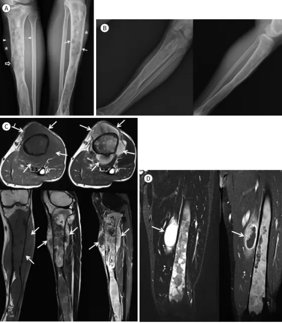
Fig. 1. A case of a 50-year-old male with malignant transformation of Mazabraud syndrome on the left tibia.
A. Internal oblique and lateral radiographs of the left lower leg show expansile lesions of the proximal tibia with endosteal scalloping (arrows), cortical destruction (blank arrow), perpendicular periosteal reaction (ar- rowheads), and soft tissue mass formation (asterisks).
B. Anteroposterior and lateral radiographs of the left lower leg, obtained 5 months before (A), show expans- ile lesion with ground glass appearance in the left tibia from the proximal epiphysis to distal metaphysis.
C. Approximately 19 cm size lesion involves the left tibia from the proximal epiphysis to middle metaphysis, showing cortical destruction, osteoid matrix calcification, periosteal reaction and soft tissue mass (arrows).
The lesion shows low SI on T1WI, mixed low to high SI on T2WI, and heterogeneous enhancement on en- hanced fat saturated T1WI (left upper: axial T1WI, right upper: axial T2WI, left lower: coronal T1WI, middle lower: sagittal T2WI, right lower: sagittal enhanced T1WI with fat saturation).
D. Coronal T2WI and enhanced T1WI with fat saturated images show a well-defined soft tissue mass with T2 high SI and peripheral enhancement at the vastus medialis and intermedius muscles of the left proximal medial thigh represents myxoma (arrows).
SI = signal intensity, T1WI = T1-weighted imaging, T2WI = T2-weighted imaging A
C
D B
고찰
Mazabraud 증후군은 1960년대에 처음 알려진 이후로 매우 드물게 보고되는 양성 질환으로 근 육 내 점액종과 섬유성 이형성이 함께 있는 경우 진단한다(1).
Mazabraud 증후군의 진단에 있어 MRI는 가장 정확한 영상검사이다. MRI에서 점액종은 경계 가 분명하고 물과 유사한 신호강도를 보이며 다양한 정도로 조영증강된다. 섬유성 이형성은 T1 강 조영상에서 경계가 명확하고 균질한 저신호강도, T2 강조영상에서 등신호에서 고신호강도를 보 이고, 경계부에 저신호강도의 두꺼운 경화성 띠를 보일 수 있다(4). 이 두 가지 질환이 영상검사에
Fig. 1. A case of a 50-year-old male with malignant transformation of Mazabraud syndrome on the left tibia.E. Plain radiographs of the pelvis and left thigh show expansion of the medullary cavity (arrows) with ground-glass lesions (blank arrows) in the left ilium, entire femur, and distal tibia, representing typical findings of polyostotic fibrous dysplasia.
F. Photomicrograph images show soft tissue myxoma with a paucity of cells, abundance of mucoid materials, and almost a complete absence of vascular structures (left, H&E stain, × 200). Image shows osteoblastic type of osteosarcoma with unmineralized lace-like osteoids (arrows) and neoplastic hyperchromatic cells (middle, H&E stain, × 100). The image shows typical fibrous dysplasia consisting of small, principally non- mineralized, trabeculae of woven bone (arrows) in bland cellular and collagenous matrix (right, H&E stain, × 200).
H&E = hematoxylin and eosin E
F
에 가장 많으며 절제 후 재발하는 경우는 드물다. Mazabraud 증후군에서 점액종은 주로 섬유성 이형성의 주변에 발생하고 다발성이 많으며 자주 재발하나, 섬유성 이형성의 악성 변환에 영향을 미치지 않는다(1, 5). 본 증례에서도 근육 내 점액종은 대퇴골 주변에 있지만 골육종은 경골에서 발생하여 악성 변환과 관련이 없었다.
섬유성 이형성은 섬유결합조직의 비정상적인 증식으로 정상 골수와 해면골이 미숙골과 섬유성 간질로 대치되는 골질환으로 단골성(monostotic)과 다골성의 형태로 올 수 있다(6). Mazabraud 증후군에서는 주로 다골성으로 나타나고, 골스캔에서 방사성추적자(radiotracer)의 섭취가 강하 게 증가되어 보이므로 다골성 섬유성 이형성의 분포를 확인할 수 있다(7).
섬유성 이형성의 약 1%에서 악성 변환을 보이고, 악성 섬유성 조직구종(malignant fibrous histiocytoma), 섬유육종(fibrosarcoma), 골육종, 연골육종 등으로 변할 수 있다(8, 9). 다골성 섬 유성 이형성이 단골성 섬유성 이형성보다 악성 변환이 발생하는 빈도가 더 높다(6). 새로 발생한 통증이나 종창 등의 임상적인 변화가 있을 때 악성 변환을 의심할 수 있다(5).
악성 변환 시 급격한 성장, 피질골 파괴, 골막반응, 주변 연조직 종괴 형성과 병적 골절을 영상검 사에서 보일 수 있다(5). 골육종은 혈액검사에서 암표지자인 ALP와 LDH가 상승하는 것으로 알려 져 있다. 본 증례에서도 혈액검사에서 ALP와 LDH가 상승되어 골육종으로 악성 변환을 확인할 수 있다(10).
결론적으로, 저자들은 좌측 하지의 다골성 섬유성 이형성과 좌측 대퇴부의 근육 내 점액종을 동 반한 Mazabraud 증후군 환자의 경골에서 골육종으로 변환한 증례를 경험하였다. Mazabraud 증 후군 환자가 통증이 발생하거나 새로 만져지는 종괴를 호소할 경우 악성 변환의 가능성을 고려하 고 추가적인 영상검사를 시행할 필요가 있다.
Author Contributions
Conceptualization, all authors; investigation, P.Y.I., K.J.S.; methodology, Y.J.Y., P.Y.I.; project admin- istration, Y.J.Y.; resources, P.Y.I., K.J.S.; supervision, Y.J.Y.; visualization, P.Y.I.; writing—original draft, P.Y.I.; and writing—review & editing, Y.J.Y.
Conflicts of Interest
The authors have no potential conflicts of interest to disclose.
Funding None
REFERENCES
1. Cabral CE, Guedes P, Fonseca T, Rezende JF, Cruz Júnior LC, Smith J. Polyostotic fibrous dysplasia associat- ed with intramuscular myxomas: Mazabraud’s syndrome. Skeletal Radiol 1998;27:278-282
2. Chun KA, Kim KT, Kim YJ, Maeng LS, Lee EJ. Fibrous dysplasia associated with intramuscular myxoma (Maz- abraud’s syndrome): a case report. J Korean Radiol Soc 1999;40:975-978
3. Multani I, Popovic S, Parasu N, Ghert M. Osteosarcomatous transformation in the setting of Mazabraud’s syndrome: a case report and review of the literature. Case Rep Orthop 2019;2019:2638478
4. Iwasko N, Steinbach LS, Disler D, Pathria M, Hottya GA, Kattapuram S, et al. Imaging findings in Mazabraud’s syndrome: seven new cases. Skeletal Radiol 2002;31:81-87
5. Majoor BCJ, van de Sande MAJ, Appelman-Dijkstra NM, Leithner A, Jutte PC, Vélez R, et al. Prevalence and clinical features of Mazabraud syndrome: a multicenter European study. J Bone Joint Surg Am 2019;101:
160-168
6. Ruggieri P, Sim FH, Bond JR, Unni KK. Malignancies in fibrous dysplasia. Cancer 1994;73:1411-1424 7. Fitzpatrick KA, Taljanovic MS, Speer DP, Graham AR, Jacobson JA, Barnes GR, et al. Imaging findings of fi-
brous dysplasia with histopathologic and intraoperative correlation. AJR Am J Roentgenol 2004;182:1389- 1398
8. Huvos AG, Higinbotham NL, Miller TR. Bone sarcomas arising in fibrous dysplasia. J Bone Joint Surg Am 1972;54:1047-1056
9. Kumar R, Madewell JE, Lindell MM, Swischuk LE. Fibrous lesions of bones. Radiographics 1990;10:237-256 10. Liu PP, Leung KS, Kumta SM, Lee KM, Fung KP. Bone-specific alkaline phosphatase in plasma as tumour
marker for osteosarcoma. Oncology 1996;53:275-280