1
책임저자: 육정환, 서울시 송파구 풍납2동 388-1
138-736, 울산대학교 의과대학 서울아산병원 외과 Tel: 02-3010-3496, Fax: 02-474-9027
E-mail: [email protected]
접수일:2009년 8월 25일, 게재승인일:2009년 10월 19일 본 연구는 Ethicon, Inc., Johnson and Johnson company의 연구비지 원에 의하여 이루어졌음.
Vicryl and PDS Plus Antibacterial 봉합사의 항균력에 대한 생체 외 연구
울산대학교 의과대학 서울아산병원 외과학교실, 1진단검사의학교실
육정환ㆍ성흥섭
1In Vitro Antibacterial Efficacy of Vicryl and PDS Plus Antibacterial Suture
Jeong Hwan Yook, M.D., Heungsup Sung, M.D.1
Departments of Surgery and 1Laboratory Medicine, Asan Medical Center, University of Ulsan College of Medicine, Seoul, Korea
Purpose: Surgical site infection (SSI) is the most common nosocomial infection in surgical patients, and this accounts for approximately 17% of all hospital-acquired infections. Suture materials are possibly significant sources of SSI. This study aims to evaluate the in vitro antibacterial efficacy of Vicryl and PDS plus antibacterial suture coating with triclosan against bacteria.
Methods: Vicryl and PDS plus antibacterial suture coating with and without triclosan were tested for in vitro efficacy against methicillin-susceptible Staphylococcus aureus, methicillin-resistant S. aureus, methicillin-resistant Staphylococcus epidermidis, Escherichia coli by a zone of inhibition assay and test of bacterial adhesion and viability.
Results: Vicryl and PDS plus antibacterial suture coating with triclosan demonstrated activity against all tested bacteria in vitro. Evaluations by a zone of inhibition assay and test of bacterial adhesion and viability show the antibacterial activity compared with untreated sutures. Pretreatment of surgical sutures with fetal bovine serum did not diminish antibacterial activity of the triclosan-coated sutures compared with non-coated sutures (P<0.01).
Conclusion: Vicryl and PDS plus antibacterial suture reduced in vitro colonization of several strains of bacteria compared with untreated control sutures. (J Korean Surg Soc 2010;78:1-6)
Key Words: Surgical site infection, Suture, Triclosan, Antibacterial 중심 단어: 수술부위 감염, 봉합사, 트리클로산, 항균성
서 론
수술부위 감염(surgical site infection, SSI)은 모든 병원내 감염 중에서 약 17%를 차지한다.(1) 이러한 수술 부위 감염 은 수술 후의 합병증 발생으로 인한 입원기간의 연장을 가
져오며 이에 따른 치료 비용의 상승을 가져오고 심할 경우 에는 사망에 이르는 경우도 있기 때문에 수술부위 감염을 줄이려는 노력이 외과의와 병원들에서 우선시 되고 있다.(2) 수술실의 공기 정화 시설이나 수술 도구 및 수술 부위의 살균 방법의 향상, 수술기법의 발달, 예방적 항균제의 적절 한 사용 등이 있으며 이런 노력들 가운데, 수술 중에 사용된 기구나 봉합사 등에 의한 감염을 줄이는 것도 한가지 방법 이 될 수 있다. 살균된 수술 환경에서 일어나는 감염은 괴사 조직이나 신체에 이식된 의료용 삽입물에 오염된 세균이 증식하여 생긴다.(3) 동물 실험에서 봉합사, 금속, 합성 물질 등이 적은 수의 병독성이 낮은 포도상구균에 의해서도 쉽 게 감염이 발생하는 것을 관찰할 수 있다.(4-9) 봉합사는 의
료용 삽입물처럼 외과적 수술 부위에 이물질로 작용하여 세 균의 번식처가 될 수 있다. 이는 특정 봉합사의 모세관 현상 에 의해서 일어난다.(10,11) 이식된 의료용 삽입물과 연관된 감염은 잘 치료되지 않으며 추가적인 외과적 수술이 필요하 기도 하고 패혈증으로 사망에 이르게 하기도 한다.(12) 아직 까지 논란이 있지만 수술부위 감염을 막기 위한 전신적인 항균제 사용은 임상적으로 많이 실시되고 있으며, 또 다른 방법으로는 의료용 삽입물이나 봉합사 등이 세균에 노출되 더라도 감염이 덜 일어나도록 처치할 수도 있다. 즉, 세균이 삽입물이나 봉합사 등에 부착되어 번식하는 것을 막을 수 있도록 하여 이물질과 연관된 감염을 줄이는 것이다. 이물 질에 이미 감염을 일으킨 세균을 항균제로 박멸하는 것은 더욱 어려우므로 봉합사 자체에 항균제를 코팅해서 봉합사 가 세균에 감염되는 것을 미리 막으려 하는 방법이 고안되 었다. 외과 영역에서 봉합사와 연관된 감염을 가장 잘 일으 키는 세균은, Staphylococcus aureus, Staphylococcus epidermi- dis, Escherichia coli, Klebsiella pneumoniae 등이다.(1) 이번 연구에서는 항균제를 처리한 봉합사인 Vicryl Plus 항균 봉 합사(Ethicon, Inc., Johnson and Johnson company, Somerville, NJ, USA)를 S. aureus, S. epidermidis, E. coli 등의 세균에 감 염시켜서 실제로 항균효과가 있는지를 생체 외에서 평가하 였다.(13)
방 법
1) 봉합사
평판배지에서의 세균 성장의 억제대를 평가하기 위해서 2-0 Vicryl, 2-0 Vicryl Plus Antibacterial, 2-0 PDS II, 2-0 PDS Plus Antibacterial, 4-0 Vicryl, 4-0 Vicryl Plus Antibacterial, 4-0 PDS II, 4-0 PDS Plus Antibacterial 봉합사(Ethicon, Inc., Johnson and Johnson company, Somerville, NJ, USA)를 각각 5 cm 길이로 무균적으로 잘랐다. 세균이 점착(adhesion)되 는 정도와 세균의 생존력(viability)을 평가하기 위해서 2-0 Vicryl, 2-0 Vicryl Plus Antibacterial, 4-0 Vicryl, 4-0 Vicryl Plus Antimicrobial 봉합사를 각각 1 cm 길이로 무균적으로 잘랐다. 봉합사는 실험에 사용하기 전까지 실온에 보관하 였다.(13-15)
2) 세균
메티실린 감수성 S. aureus (methicillin-susceptible S. aureus, MSSA) American Type Culture Collection (ATCC) 25923, 메티
실린 내성 S. aureus (methicillin-resistant S. aureus, MRSA) ATCC 43300, 카테터 연관 혈류 감염증 환자의 카테터에서 분리된 메티실린 내성 S. epidermidis (904C1223) 균주 및 소 변에서 분리된 extended-spectrum β-lactamase (ESBL) 생성 E. coli (906E2411)를 사용하였다. 4균주를 혈액한천배지에 획선한 후 5% CO2 배양기에서 35oC, 하룻밤 배양하였다. 순 수 분리된 집락을 tryptic soy broth 20 ml에 접종하여 35oC에 서 18시간 배양하였다. 배양된 균액을 3,000 rpm에서 10분간 원심분리하여 균 침사를 얻은 후 Mueller-Hinton broth에 0.5 McFarland 탁도(1.5×10⁸ CFU/ml)로 희석하였다.(14)
3) 평판배지에서의 세균 성장의 억제대(zone of in- hibition) 평가
0.5 McFarland 탁도의 MSSA, MRSA, S. epidermidis, E.
coli 균액을 각각 Mueller-Hinton 배지에 면봉으로 잔디 도말 하였다. 5 cm 길이로 자른 4가지 2-0 봉합사를 세균이 도말 된 평판배지 한 개에 무균적으로 놓았다. 같은 방법으로 4 가지 4-0 봉합사를 세균이 도말된 평판배지 한 개에 무균적 으로 놓았다. 35oC 대기 상태로 24시간 배양 후 봉합사 주위 의 억제대를 봉합사 길이 방향과 직각으로 측정하였다.(13- 15) 모든 실험은 5회 반복하였다.
4) 세균 점착력(adhesion)과 생존력(viability) 평가
0.5 McFarland 탁도의 MRSA, S. epidermidis, E. coli 균액 10μl를, 15 ml의 0.5% dextrose가 첨가된 phosphate buffered saline (PBS)에 각각 희석하여 최종 균 농도를 1×10⁵ CFU/ml (5.0 log10 CFU/ml)가 되도록 하였다. 희석된 균액 1 ml에 1 cm 길이의 2-0 Vicryl, 2-0 Vicryl Plus Antibacterial, 4-0 Vicryl 및 4-0 Vicryl Plus Antimicrobial 봉합사를 각각 넣고 35oC에 서 100 rpm으로 교반하면서 1시간 동안 두었다. 각각의 봉 합사를 PBS 1 ml에 3회 부드럽게 세척한 후 0.5% dextrose가 첨가된 PBS 1 ml에 넣고 35oC에서 24시간 배양하였다. 각각 의 봉합사를 PBS 1 ml에 3회 부드럽게 세척한 후 0.5% dex- trose가 첨가된 PBS 1 ml에 넣고 20 kHz의 초음파에 2분간 처리하였다. 초음파 처리한 봉합사 용액을 혈액한천배지에 200μl, 20μl, 2μl씩 접종하여 35oC, 대기 중에서 24시간 배양한 후 집락의 수를 세었다.(13-15) 모든 실험은 5회 반 복하였다.
단백질이 코팅되었을 때 봉합사에 MRSA와 E. coli가 점 착되는 정도와 세균의 생존력을 평가하기 위해 2-0 Vicryl, 2-0 Vicryl Plus Antibacterial 봉합사를 fetal bovine serum
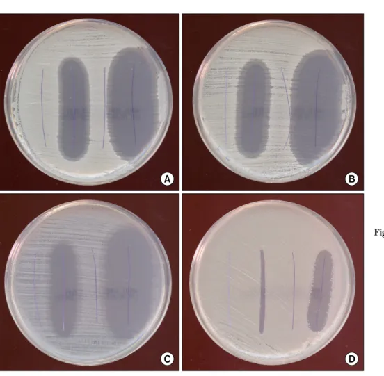
Fig. 1. Zone of inhibition of 2-0 Vicryl, 2-0 Vicryl Plus An- tibacterial, 2-0 PDS II, and 2-0 PDS Plus Antibacterial sutures (left-to-right) again- st (A) methicillin-suscepti- ble Staphylococcus aureus (MSSA) ATCC 25923, (B) methicillin-resistant S. au- reus (MRSA) ATCC 43300, (C) Staphylococcus epider- midis, and (D) extended-spec- trum β-lactamase (ESBL) producing Escherichia coli.
Antibacterial suture materials showed the inhibition of bac- terial growth.
(FBS, CAMBREX. Bio Science, Walkersville, Inc., Walkers- ville, MD, USA)에 30분간 담근 후 위와 동일한 방법으로 실험하였다.(14) 모든 실험은 5회 반복하였다.
5) 통계
균 집락수의 차이는 unpaired Wilcoxon singed rank test를 이용하여 비교하였으며, SPSS version 13.0 (SPSS, Chicago, IL, USA) 통계 프로그램을 사용하였다.
결 과
1) 평판배지에서의 세균 성장의 억제대
MSSA, MRSA, S. epidermidis, E. coli 균 모두 triclosan이 코팅된 봉합사인 Viryl Plus Antimicrobial, PDS Plus Antimi- crobial에서만 억제대를 보였다(Fig. 1, 2). 각각의 균에 대한 억제대는 Table 1과 2에 정리하였다. MSSA, MRSA, S. epi- dermidis에 대한 triclosan 코팅 봉합사의 평균 균 성장 억제 대는 2-0, 4-0 모두 Vicryl Plus Antimicrobial의 경우 10 mm
이상, PDS Plus Antimicrobial의 경우 20 mm 이상의 억제대 를 보였다. 그러나 ESBL 생성 E. coli에 대한 triclosan 코팅 봉합사의 평균 균 성장 억제대는 2-0 Vicryl Plus의 경우 5.4 mm, 4-0 Vicryl Plus의 경우 2.6 mm, 2-0 PDS Plus의 경우 12.6 mm, 4-0 PDS Plus의 경우 10.2 mm로 작았다.
2) 세균 점착력(adhesion)과 생존력(viability)
2-0 굵기의 Vicryl과 Vicryl Plus Antimicrobacterial 봉합사 에 점착된 후 생존한 균수는 MRSA의 경우 1 cm의 봉합사 당 각각 평균 364 CFU (310∼425 CFU)와 평균 14 CFU (5∼
25 CFU), S. epidermidis의 경우 1 cm 봉합사당 각각 평균 289 CFU (245∼360 CFU)와 평균 10 CFU (5∼15 CFU), E.
coli의 경우 1 cm 봉합사당 각각 평균 406 CFU (290∼490 CFU)와 평균 22 CFU (10∼35 CFU)로 모두 통계적으로 유 의한 차이를 보였다(P<0.01)(Fig. 3).
4-0 굵기의 Vicryl과 Vicryl Plus Antimicrobacterial 봉합사 에 점착된 후 생존한 균수는 MRSA의 경우 1 cm의 봉합사 당 각각 평균 352 CFU (295∼445 CFU)와 평균 12 CFU (5∼
Table 1. In vitro diffusion from 2-0 Vicryl with triclosan and 2-0 PDS with triclosan
Zone of inhibition (mm)
Vicryl Vicryl Plus PDS II PDS Plus
Staphylococcus aureus ATCC* 25923, methicillin-susceptible 0 16.0 (15∼17) 0 23.2 (23∼24)
S. aurues ATCC 43300, methicillin-resistant 0 12.2 (11∼13) 0 21.4 (20∼22)
Staphylococcus epidermidis 0 18.4 (18∼19) 0 23.4 (23∼24)
ESBL† producing Escherichia coli 0 5.4 (5∼6) 0 12.6 (12∼13)
*ATCC = American type culture collection; †ESBL = extended-spectrum β-lactamase.
Table 2. In vitro diffusion from 2-0 Vicryl with triclosan and 2-0 PDS with triclosan
Zone of inhibition (mm)
Vicryl Vicryl Plus PDS II PDS Plus
Staphylococcus aureus ATCC* 25923, methicillin-susceptible 0 12.4 (12∼13) 0 21.2 (20∼22)
S. aurues ATCC 43300, methicillin-resistant 0 12.6 (12∼13) 0 21.4 (21∼22)
Staphylococcus epidermidis 0 10.6 (10∼11) 0 23.0 (22∼24)
ESBL† producing Escherichia coli 0 2.6 (2∼3) 0 10.2 (9∼11)
*ATCC = American type culture collection; †ESBL = extended-spectrum β-lactamase.
Fig. 2. Zone of inhibition of 4-0 Vicryl, 4-0 Vicryl Plus An- tibacterial, 4-0 PDS II, and 4-0 PDS Plus Antibacterial sutures (left-to-right) again- st (A) MSSA ATCC 25923, (B) MRSA ATCC 43300, (C) S. epidermidis, and (D) ESBL producing E. coli. An- tibacterial suture materials showed the inhibition of bac- terial growth.
Fig. 3. Mean microbial recovery from Vicryl and Vicryl Plus Antimicrobial sutures exposed to bacterial inoculum for 60 minutes. There were significant differences in bacterial adherence and viability (P<0.01).
Fig. 4. Impact of 20% feta bovine serum (FBS) on MRSA ATCC 43300 and E. coli mean microbial recovery from Vicryl and Vicryl Plus Antimicrobial sutures. There were sig- nificant differences in bacterial adherence and viability (P
<0.01).
15 CFU), S. epidermidis의 경우 1 cm 봉합사당 각각 평균 247 CFU (200∼305 CFU)와 평균 11 CFU (5∼15 CFU), E.
coli의 경우 1 cm 봉합사당 각각 평균 438 CFU (385∼485 CFU)와 평균 27 CFU (20∼35 CFU)로 모두 통계적으로 유 의한 차이를 보였다(P<0.01)(Fig. 3).
2-0 굵기의 Vicryl과 Vicryl Plus Antibacterial 봉합사를 FBS에 30분간 담근 후 동일 실험을 반복했을 때 점착된 후 생존한 균 수는 MRSA의 경우 1 cm의 봉합사당 각각 평균 487 CFU (395∼575 CFU)와 평균 23 CFU (15∼35 CFU), E.
coli의 경우 1 cm 봉합사당 각각 평균 559 CFU (435∼685 CFU)와 평균 32 CFU (25∼45 CFU)였다(P<0.01)(Fig. 4).
고 찰
수술부위 감염은 많은 외과의들의 노력에도 불구하고 여 전히 수술 후 합병증과 사망률의 중요한 원인이며, 이는 항 균제 내성균주의 발생, 수술환자의 고령화, 만성질환 환자 나 장기이식으로 인한 면역억제제 사용 환자의 증가 및 의 료용 삽입물 증가 등에 의한 것으로 생각된다. 이 중에서 봉합사는 신체에 이물질이므로 세균이 감염될 수 있으며, 이로 인해 수술부위 감염을 일으킬 수 있다. 실제로 MRSA, 반코마이신 내성 장구균과 같은 항균제에 내성을 갖는 세 균이나 그람음성 세균들에 의한 수술부위 감염이 증가하고 있는 실정이다. 수술부위에 감염된 Staphylococcus의 50%가 MRSA이고, 분리된 균의 11%는 gentamicin 내성의 E. coli와 Klebsiella라는 보고도 있다.(16) 현재까지 논란이 있지만 수
술부위 감염을 막기 위해서는 전신적인 항생제를 임상적으 로 많이 사용하고 있으며, 또 다른 방법으로는 의료용 삽입 물이나 봉합사 등이 세균에 노출되더라도 감염이 덜 일어 나도록 하는 방법을 고안하여 사용하고 있다. 즉, 감염이 일 어나려면 세균이 삽입물이나 봉합사 등에 부착되거나 감염 되어 번식되어야 하는데, 이것을 막을 수 있도록 하여 이물 질과 연관된 감염을 줄이는 것이다. 이물질에 이미 감염된 세균을 항생제로 박멸하는 것은 더욱 어려우므로 봉합사 자체에 항생제를 덮어 씌워서 봉합사가 세균에 감염되는 것을 미리 막으려하는 방법이다. 세균 감염에 저항성을 갖 는 항균제가 처리된 봉합사는 이물질과 연관된 수술부위 감염을 예방하는 유용한 방법이 될 수 있다. 생체 외 실험으 로 항균제 처리 봉합사가 세균들에 저항하는 항균효과를 가지며, 이런 효과는 3주 이상 또는 봉합사가 흡수되어 없 어지는 기간까지도 지속된다고 보고하고 있다.(15) 저자들 은 실험을 통해 triclosan이 코팅된 Vicryl Plus Antimicrobial 봉합사가 평판배지에서의 세균 성장을 억제할 뿐만 아니라 세균의 점착(adhesion)과 생존(viability)을 저하시킴을 관찰 할 수 있었다. Triclosan이 코팅된 Vicryl Plus Antimicrobial 봉합사가 MRSA, S. epidermidis, ESBL 생성 E. coli 등의 세 균을 억제하며, 수술부위 감염을 줄일 수 있는 가능성을 보 여 주었다. 이런 결과들이 임상적으로 유용하게 사용되려면 실제적으로 신체 내에서도 감염을 줄이면서 다른 합병증을 유발하지는 않는지에 대한 임상연구가 필요하다고 하겠다.
결 론
생체 외 실험이지만 항균 물질인 triclosan이 처리된 Vi- cryl Plus Antibacterial 봉합사와 PDS Plus Antibacterial 봉합 사는 항균 처리되지 않은 일반 봉합사에 비교하여 S. aur- eus, S. epidermidis, E. coli에 의한 감염을 줄이거나 방지하는 효과가 있음을 보였으며, 향후 봉합사와 연관된 수술부위 감염을 예방하는데 이용할 수 있을 것으로 기대된다.
REFERENCES
1) Mangram AJ, Horan TC, Pearson ML, Silver LC, Jarvis WR.
Guideline for prevention of surgical site infection, 1999. Hos- pital Infection Control Practices Advisory Committee. Infect Control Hosp Epidemiol 1999;20:250-80.
2) Weaving P, Cox F, Milton S. Infection prevention and control in the operating theatre: reducing the risk of surgical site in- fections (SSIs). J Perioper Pract 2008;18:199-204.
3) Vadaux P, Francois P, Lew DP, Waldvogel FA. Host factors predisposing to and influencing therapy of foreign body in- fections. In: Waldvogel FA, Bisno AL, editors. Infections As- sociated with Indwelling Medical Devices. 3rd ed. Washing- ton, DC: ASM Press; 2000. p.1-26.
4) Kaiser AB, Kernodle DS, Parker RA. Low-inoculum model of surgical wound infection. J Infect Dis 1992;166:393-9.
5) Chuard C, Lucet JC, Rohner P, Herrmann M, Auckenthaler R, Waldvogel FA, et al. Resistance of Staphylococcus aureus recovered from infected foreign body in vivo to killing by antimicrobials. J Infect Dis 1991;163:1369-73.
6) Zimmerli W, Waldvogel FA, Vaudaux P, Nydegger UE. Pa- thogenesis of foreign body infection: description and character- istics of an animal model. J Infect Dis 1982;146:487-97.
7) Christensen GD, Simpson WA, Bisno AL, Beachey EH.
Experimental foreign body infections in mice challenged with slime-producing Staphylococcus epidermidis. Infect Immun 1983;40:407-10.
8) Vaudaux P, Grau GE, Huggler E, Schumacher-Perdreau F, Fiedler F, Waldvogel FA, et al. Contribution of tumor necrosis factor to host defense against staphylococci in a guinea pig model of foreign body infections. J Infect Dis 1992;166:58-64.
9) Zimmerli W. Experimental models in the investigation of de- vice-related infections. J Antimicrob Chemother 1993;31(Suppl D):97-102.
10) Chu CC, Williams DF. Effects of physical configuration and chemical structure of suture materials on bacterial adhesion.
A possible link to wound infection. Am J Surg 1984;147:
197-204.
11) Katz S, Izhar M, Mirelman D. Bacterial adherence to surgical sutures. A possible factor in suture induced infection. Ann Surg 1981;194:35-41.
12) Anderson JM, Marchant RE. Biomaterials: factors favoring colonization and infection. In: Waldvogel FA, Bisno AL, editors. Infections Associated with Indwelling Medical De- vices. 3rd ed. Washington, DC: ASM Press; 2000. p.89-110.
13) Ming X, Rothenburger S, Yang D. In vitro antibacterial effi- cacy of MONOCRYL plus antibacterial suture (Poliglecaprone 25 with triclosan). Surg Infect (Larchmt) 2007;8:201-8.
14) Edmiston CE, Seabrook GR, Goheen MP, Krepel CJ, Johnson CP, Lewis BD, et al. Bacterial adherence to surgical sutures:
can antibacterial-coated sutures reduce the risk of microbial contamination? J Am Coll Surg 2006;203:481-9.
15) Ming X, Rothenburger S, Nichols MM. In vivo and in vitro antibacterial efficacy of PDS plus (polidioxanone with triclo- san) suture. Surg Infect (Larchmt) 2008;9:451-7.
16) Esuvaranathan K, Kuan YF, Kumarasinghe G, Bassett DC, Rauff A. A study of 245 infected surgical wounds in Singa- pore. J Hosp Infect 1992;21:231-40.