The Hepatoprotective Activity of Spatholobi Caulis Water Extract against Cadmium-Induced Toxicity in Rats
Won-Mook Park1, Hong-Sik Choi1, Seung-Mo Kim1, Chang-Hoon Woo2
1Department of Oriental Internal Medicine, Daegu Haany University
2Department of Oriental Rehabilitation Medicine, Daegu Haany University
Objectives: This study was investigated the protective effect of Spatholobi Caulis water extract against cadmium (CdCl2, Cd)-induced hepatic toxicity in rats.
Methods: To induce acute hepatic toxicity, Cd (4 mg/kg body weight) was dissolved in normal saline and intravenously injected into rats. Then, the rats received either a vehicle or silymarin (100 mg/kg) or Spatholobi Caulis water extract (30, 50 mg/kg/day) for 3 days, and were exposed to a single injection of Cd 24 h after the last Spatholobi Caulis/vehicle treatment.
Results: Alanine aminotransferase (ALT), aspartate aminotransferase (AST) and lactate dehydrogenase (LDH) were significantly increased by Cd treatment. In contrast, pretreatment with Spatholobi Caulis reduced ALT, AST and LDH. Cd-intoxicated liver damage was significantly inhibited by treatment of Spatholobi Caulis 30 and 50 mg/kg at histopathological observations in the present study.
Conclusions: These results can be considered as direct evidence that Spatholobi Caulis has favorable inhibitory effects on the Cd-intoxicated liver damages. The efficacy of Spatholobi Caulis 30 mg/kg shows similar effects to that of silymarin 100 mg/kg, and more favorable hepatoprotective effects were observed in Spatholobi Caulis 50 mg/kg as compared with silymarin 100 mg/kg against Cd-intoxicated hepatopathies in the present study.
Key Words : Spatholobi Caulis, cadmium, hepatic toxicity
계혈등 물추출물의 간세포 보호효과
박원묵1, 최홍식1, 김승모1, 우창훈2
1대구한의대학교 한의학과 간계내과학교실,
2대구한의대학교 한의학과 한방재활의학과교실
Original Article
⋅Received:21 June 2010 ⋅Revised:2 September 2010 ⋅Accepted:9 September 2010
⋅Correspondence to:최홍식(Hong-Sik Choi)
경상북도 문경시 문경읍 하리 360번지 시립문경요양병원 한방1과
Tel:010-5116-1461, Fax:+82-54-572-3200, E-mail:[email protected] 서 론
계혈등 (Spatholobi Caulis)은 중국의 복건, 광동, 광서, 운남 등지에 분포하는 콩과 (Leguminosae)에 속한 落葉 木質大藤本인 密花豆 (Spatholobus suberectus DUNN)의 근경을 건조한 것으로, 性은 溫 無毒하고 味는 苦微甘하다. 주로 肝, 腎經에 작용하
며 行血補血, 活血去瘀, 舒筋活絡하는 효능이 있어 月經不調, 痛經, 血虛經閉, 關節痠痛, 手足麻木, 風濕 痺痛 등을 치료하며1), 또한 혈액에 대한 항응고작용 및 혈압강하작용이 있어서 고혈압, 심장질환 등에 응용되고 있다2).
계혈등의 성분으로는 formononetin, daidzein, isoliquiritigenin, licochalcone A, (2R)-7-hydroxy-6-
methoxy-flavanone, friedelin, friedelan-3β-ol, sativan, β- sistosterol, stigmasterol, campesterol, (3R,4R)- 2’,4’-dihydroxy-6,7-methylene- dioxyisoflavan-4-ol, genistein, medicagol, pseudobaptigenin, (3R,4R)-7’, 2’- dihydroxy-4’-methoxyisoflavan-4-ol 등이 알려져 있으며3,4), 계혈등에 대한 연구로는 항균작용5), 항산 화작용6,7), 항노화작용7), 항염증작용8-10), 뇌혈류개선
작용8,11), 세포주기억제 및 세포사멸유도작용12-17), 지
질대사조절작용18) 등과 같이 다양한 방면에서 연구 가 이루어지고 있다.
카드뮴 (Cd, Cadmium)은 중금속 중의 하나로 산 업화의 발달에 따라 생활환경에 널리 분포하게 되었
으며19,20), 급성 카드뮴중독은 dysuria, polyuria, 흉
통, 피로, 두통을 유발하는 것으로 알려져 있고21), 음식물 및 공기를 통한 만성적인 카드뮴의 체내 유 입은 폐질환, 간질환 및 신장질환을 유발하는 것으 로 알려져 있다22). 카드뮴은 1차적으로 간에 축적되 므로, 간은 카드뮴독성시 매우 중요한 장기로 알려 져 있다23-25).
본 연구에서는 계혈등 물추출물 (SCE; Spatholobi Caulis Extract)의 카드뮴 중독에 대한 간세포보호효 과를 평가하고자, in vitro에서 카드뮴에 의해 유도 되는 H4IIE cell의 세포독성에 대한 SCE의 세포독 성억제효과를 평가하고, in vivo에서 카드뮴에 의해 유발되는 rat의 간손상에 대한 SCE의 간보호효과를 평가하여 유의한 결과를 얻었기에 보고하는 바이다.
재료 및 방법
1. SCE의 제조
계혈등은 대원약업사 (대구, 한국)에서 구입하여 관능평가를 한 후, 200 g을 물 1.5 L에 넣고 3시간 전탕한 후 추출물을 거어즈로 1차 여과하고 3000×g 에서 3분간 원심분리하였다. 원심분리 후의 상층액 만을 취하여 0.2 μm filter (Nalgene, New York, USA)로 여과하였다. 이 여과액을 rotary evaporator (EYELA, Tokyo, Japan)로 농축하고, 이 농축액을
Ultra-Low temperature freezer (Nihon freezer, Japan) 에서 동결시켰다. 동결된 계혈등 추출물을 동결건조 기 (Labconco, USA)로 동결 건조하여 추출물 15.36 g을 얻었으며, 사용 때까지 -20℃에서 보관하였다.
SCE의 수율은 7.68%였으며 in vitro 처치시에는 Dulbecco’s modified Eagle’s medium (DMEM)에 녹여 사용하였고, in vivo 실험에서는 H2O에 녹여 사용하였다.
2. 시약
3-(4,5-dimethylthiazol-2-yl)-2,5-diphenyltetrazoleu m (MTT)은 Sigma (St. Louis, MO, USA)에서 구입 하였고, DMEM과 fetal bovine serum (FBS)는 BioWhittaker사 (Walkersville, MD, USA)와 Life Technologies (Gaithersburg, MD, USA)로부터 구입 하였으며, penicillin 및 streptomycin은 Gibco/BRL (Eggenstein, Germany)로부터 구입하였으며, silymarin 은 Sigma (St. Louis, MO, USA)로부터 구입하여 사 용하였다.
3. 세포배양
Rat 유래 hepatocyte cell line인 H4IIE cell은 American Type Culture Collection (St. Louis, MO, USA)에서 구입하였으며, DMEM에 10% FBS, 100 U/ml penicillin 및 100 μg/ml streptomycin을 혼합한 배지를 사용하여 37℃, 5% CO2 incubator (Sanyo, Japan)에서 배양하였다. 실험과정의 모든 세포는 80~90%의 confluence에서 실험하였고, 20 passages 를 넘기지 않은 세포만 사용하였다.
4. 세포 생존율 측정
H4ⅡE cell을 96 well plate에 5×10⁴ cells/well로 분주한 다음 SCE를 농도별로 처치하여, 세포의 생 존율을 구하였다. 세포에 10, 30, 100, 300 μg/ml의 농도로 SCE를 12시간 전처치한 후, 카드뮴 10 uM 을 처치하여 12, 24 시간 동안 37℃, 5% CO2의 환 경이 유지되는 배양기 (Sanyo, Japan)에서 배양하였
: SCE or silymarin or water (p.o.) : assay
: Cadmium or normal saline (i.v.)
1 2 3 4 (day)
1h
Scheme 1. Schedule of the in vivostudy 다. 배양 후 생존세포에 MTT (0.1 mg/ml)를 50 ㎕
넣고 4시간 배양한 후 배지를 조심스럽게 제거하고 생성된 formazan crystals를 dimethylsulfoxide (DMSO) 에 녹여 Titertek Multiskan Automatic ELISA microplate reader (Model MCC/340, Huntsville, AL)를 사용하여 570 nm에서 흡광도를 측정하였다.
세포의 생존율은 control cell에 대한 백분율로 나타 내었다. [i.e. cell viability (% control) = 100×
(absorbance of treated sample)/(absorbance of control)].
5. 실험동물의 사육
실험동물은 6주령 Sprague-Dawley계 수컷 흰쥐 (140-160 g)를 효창사이언스 (대구, 한국)로부터 공 급받아 1주일 동안 동물실험실환경에 적응시킨 후 실험에 사용하였으며, 사육실 환경은 온도 20-23℃, 습도 60%, 12시간 light/dark cycle을 유지하고, 사료 (Nestle Purina Petcare Korea, Seoul, Korea)와 음료 는 자유롭게 섭취하도록 하였다.
6. 실험동물의 분류 및 처치
실험동물은 각 군당 5마리씩 아무런 처치를 하지 않은 군을 Normal군으로, 카드뮴으로 간독성을 유 발한 군은 Cd군으로, silymarin을 투여하고 카드뮴 을 처치한 군을 Sm군으로, SCE를 30, 50 mg/kg로 투여하고 카드뮴를 처치한 군을 각각 SCE 30군, SCE 50군으로, 카드뮴 대신 꼬리 정맥으로 생리식 염수를 주입하고 silymarin이나 SCE 대신 물을 투여 한 군을 Control군으로 하여 진행하였다.
7. 실험동물의 처치
간독성을 유발하기 위하여 카드뮴을 saline에 녹 인 후 0.2 μm filter로 여과하여 rat의 꼬리정맥을 통 해 카드뮴 4 mg/kg을 주입하였으며, 양성대조군인 Sm군은 카드뮴 처치 전 Srivastava 등26)과 같이 silymarin을 100 mg/kg로 투여하였다. SCE의 효능 을 평가하기 위해서는 카드뮴 처치 전 SCE를 30, 50 mg/kg로 투여하고 카드뮴을 처치하였고, Control 군은 카드뮴 대신 꼬리 정맥으로 생리식염수를 주입 하고, silymarin이나 SCE 대신 물을 투여 하였다.
(Scheme. 1)
8. 혈액생화학적 검사
혈액은 실험동물의 복대정맥으로부터 3 ml이상을 채취한 후 3000×g, 4℃에서 10분간 원심분리하여 혈청을 얻었다. 혈청중 alanine amino- transferase (ALT), aspartate aminotransferase (AST), lactate dehydrogenase (LDH)는 Analysis kits (Pointe Scientific Inc., Canton, MI, USA)와 Automated blood chemistry analyzer (Photometer 5010, Robert Riele GmbH & Co KG, Berlin, Germany)를 사용하 여 분석하였다.
9. 조직학적 평가
간 실질조직의 일부를 샘플로 채취하여 10% 중 성포르말린에 고정시킨 다음, 일반적인 방법으로 탈 수 및 파라핀 포매를 실시하고, 3~4 μm의 절편을 제작하여 Hematoxylin-eosin 염색을 실시한 후 광학
Nor 140
Cell Viability (% of Control)
120
100 80
60 40
20 0
0 10 30 100 300 (㎍/ml)
Cd 10 μM
Nor 120
Cell Viability (% of Control)
100
80
60
40
20
0
0 10 30 100 300 (㎍/ml)
Cd 10 μM
(A) (B)
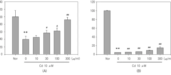
Fig. 1.Increase in cell viability of H4IIE cells exposed to Cd by SCE.
H4IIE cells pre-treated with SCE for 12 and further incubated with SCE+Cd (10 μM) for the next 12 hr (A) 24 hr (B). Cell viability was assessed by MTT assay. Data represent the mean±SD of eight separate experiments. (*, significant as compared to normal,
**P<0.01; #, significant as compared to Cd alone, #P<0.05, ##P<0.01)
현미경 (Nikon, Japan)하에서 관찰하였다. 간 실질 중 변성부위의 비율 (%/mm2 of hepatic parenchyma) 및 변성 간세포의 수 (N/1000 hepatocytes)를 자동영 상분석장치 (DMI-300 Image Processing; DMI, Korea)를 이용하여 각각 평가하였다.
10. 통계적 검증
실험 결과는 mean±S.D.로 나타내었으며, 처치군 간 의 유의성은 one way analysis of varience (ANOVA) 로 검정한 후 Newman-Kleuls test로 검정하였다. 통 계적 유의성 검정은 p<0.05 또는 p<0.01로 하였다.
실험결과
1. SCE가 카드뮴으로 유도된 세포독성에 미치는 영향
카드뮴으로 유발되는 세포독성에 대한 SCE의 보 호효과를 MTT assay로 관찰하였다. 12시간 동안 serum을 제거한 배지에서 H4IIE cell을 배양한 후, SCE를 12시간 동안 농도별로 처치하고, 이후 카드 뮴을 10 μM로 12, 24시간 동안 처치하였다.
12시간에서 Normal군은 100.00±16.71%였으며, Cd군은 39.99±8.36%로 유의한 세포독성이 유도되 었다. 그러나 이러한 세포독성은 SCE 30, 100, 300 μg/ml을 처치한 군에서는 각각 57.39±6.82, 62.68±
7.21, 92.40±5.98 (%)로 유의하게 억제되었다.
24시간에서 Cd군은 4.62±0.07%로 유의하게 세포 독성이 유도되었으며, SCE를 10, 30, 100, 300 μ g/ml을 처치한 군에서는 각각 5.28±0.03, 6.49±0.15, 9.25±0.65, 15.11±2.43 (%)를 나타내어 실험에 사용 된 SCE 전 농도에서 유의하게 세포독성이 억제되었 다(Fig. 1A, 1B).
2. SCE가 카드뮴으로 유도된 혈청 ALT에 미치는 영향
SCE가 카드뮴으로 유도되는 세포독성을 억제함 을 확인하고, 카드뮴 유도 간독성에 대한 SCE의 효 과를 동물모델에서 평가하였다.
본 연구에서 Normal군과 Control군은 각각 50.18
±12.53, 42.42±4.35 (U/L)를 나타내었으며, Cd군은 1597.77±374.48 U/L을 나타내어 유의한 간독성이 유발되었다. 그러나 이러한 ALT의 증가는 Sm군에 서 345.59±217.24 U/L로 유의하게 감소하였으며,
Nor 5000
AST (U/L)
4000
3000
2000
1000
0
Con Cd Sm 30 50 (mg/kg; SCE)
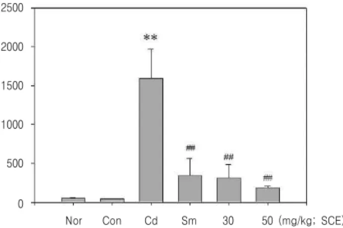
Fig. 3.Effect of SCE on AST levels in Cd-induced liver toxicity.
AST activities were monitored in the serum of rats that had been orally pretreated with SCE (30 or 50 mg/kg for 3 days, injected with Cd (CdCl2, 4 mg/kg body weight, i.v.) treatment. Blood samples were obtained at 24 hr after last drug treatment. Values represent mean±SD (n = 5) (*, significant as compared to normal, **P<0.01; #, significant as compared to Cd alone, ##P<0.01) Nor : non treated group, Con : treated with Normal saline i.v and water p.o group, Cd : treated with Cadmium 4 mg/ml i.v and water p.o group, Sm : treated with Cadmium 4 mg/ml i.v and Silymarin 100 mg/kg p.o group, SCE 30 : treated with Cadmium 4 mg/ml i.v and SCE 30 mg/kg p.o group, SCE 50 : treated with Cadmium 4 mg/ml i.v and SCE 50 mg/kg p.o group.
Nor 2500
ALT (U/L)
2000
1500
1000
500
0
Con Cd Sm 30 50 (mg/kg; SCE)
Fig. 2.Effect of SCE on ALT levels in Cd-induced liver toxicity.
ALT activities were monitored in the serum of rats that had been orally pretreated with SCE (30 or 50 mg/kg for 3 days, injected with Cd (CdCl2, 4 mg/kg body weight, i.v.) treatment. Blood samples were obtained at 24 hr after last drug treatment. Values represent mean±SD (n = 5) (*, significant as compared to normal, **P<0.01; #, significant as compared to Cd alone, ##P<0.01) Nor : non treated group, Con : treated with Normal saline i.v and water p.o group, Cd : treated with Cadmium 4 mg/ml i.v and water p.o group, Sm : treated with Cadmium 4 mg/ml i.v and Silymarin 100 mg/kg p.o group, SCE 30 : treated with Cadmium 4 mg/ml i.v and SCE 30 mg/kg p.o group, SCE 50 : treated with Cadmium 4 mg/ml i.v and SCE 50 mg/kg p.o group.
SCE 30, 50 군에서는 각각 315.27±166.25, 182.48
±25.85 U/L로 유의하게 감소하여 양성대조군인 SM 군과 유사한 감소를 나타내었다(Fig. 2).
3. SCE가 카드뮴으로 유도된 혈청 AST에 미치는 영향
본 연구에서 Normal군과 Control군은 각각 58.37
±14.44, 54.49±15.56 (U/L)를 나타내었으며, Cd군은 3559.17±617.62 U/L을 나타내어 유의한 간독성이 유발되었다. 그러나 이러한 AST의 증가는 Sm군에 서 309.94±192.05 U/L로 유의하게 감소하였으며, SCE 30, 50 군에서는 각각 227.12±175.76, 308.91
Nor 4000
LDH (U/L)
3000
2000
1000
0
Con Cd Sm 30 50 (mg/kg; SCE)
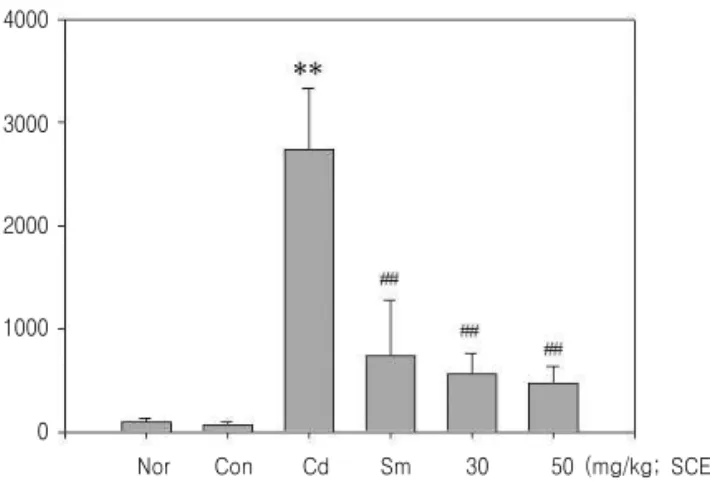
Fig. 4.Effect of SCE on LDH levels in Cd-induced liver toxicity.
LDH activities were monitored in the serum of rats that had been orally pretreated with SCE (30 or 50 mg/kg for 3 days, injected with Cd (CdCl2, 4 mg/kg body weight, i.v.) treatment. Blood samples were obtained at 24 hr after last drug treatment. Values represent mean±SD (n = 5) (*, significant as compared to normal, **P<0.01; #, significant as compared to Cd alone, ##P<0.01) Nor : non treated group, Con : treated with Normal saline i.v and water p.o group, Cd : treated with Cadmium 4 mg/ml i.v and water p.o group, Sm : treated with Cadmium 4 mg/ml i.v and Silymarin 100 mg/kg p.o group, SCE 30 : treated with Cadmium 4 mg/ml i.v and SCE 30 mg/kg p.o group, SCE 50 : treated with Cadmium 4 mg/ml i.v and SCE 50 mg/kg p.o group.
±243.46 (U/L)로 유의하게 감소하여 양성대조군인 SM군과 유사한 감소를 나타내었다(Fig. 3).
4. SCE가 카드뮴으로 유도된 혈청 LDH에 미치는 영향
본 연구에서 Normal군과 Control군은 각각 96.50
±31.37, 75.75±17.21 (U/L)를 나타내었으며, Cd군은 2744.33±591.79 U/L을 나타내어 유의한 간독성이 유발되었다. 그러나 Sm군에서 738.60±537.92 U/L 로 유의하게 감소하였으며, SCE 30, 50 군에서는 각각 563.67±201.35, 475.17±165.05 (U/L)로 유의하 게 감소하여 양성대조군인 Sm군과 유사한 감소를 나타내었다(Fig. 4).
5. SCE가 카드뮴으로 유도된 간의 조직학적 변화에 미치는 영향
카드뮴에 의해 유도된 간독성에 미치는 SCE의 효과를 평가하기 위하여 조직학적 평가를 하였다.
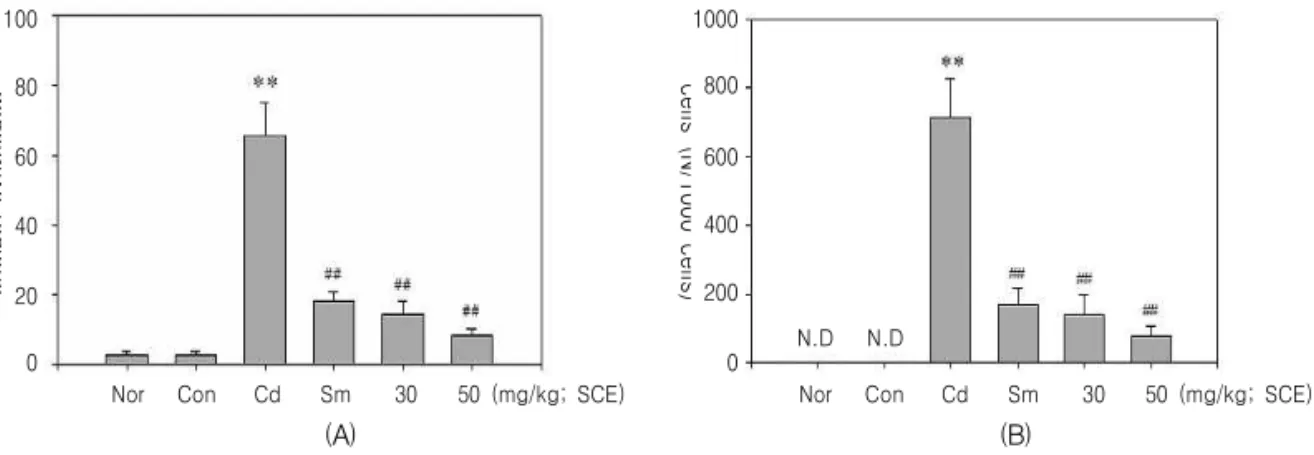
간 변성율은 Normal군과 Control군에서 각각 2.69±1.03, 2.77±1.23%를 나타내었으며, 카드뮴에
의해서는 간 변성율이 65.53±9.54%로 유의하게 증 가되었다. 그러나 이러한 간 변성율의 증가는 Sm군 에서 18.25±2.52% (-72.15%)로 유의하게 감소했으 나 SCE 30, 50 mg/kg에서도 14.62±3.74, 8.37±2.10 (-77.69 및 -87.22%)로 유의하게 감소하였으며, 이는 양성대조군인 Sm군보다 더 낮은 변성율을 나타낸 것이다.
변성 간세포의 수는 Normal군과 Control군에서 잘 나타나지 않았으며, 카드뮴에 의해서 변성된 간 세포의 수가 711.80±116.11개로 유의하게 증가되었 다. 그러나 이러한 변성 간세포 수의 증가는 Sm군 에서 169.00±49.61개 (-76.26%)로 유의하게 감소되 었고, SCE 30, 50 mg/kg는 142.00±55.64, 77.40±
31.10개 (-80.05 및 -89.13%)로 유의하게 감소되었 으며, 이는 양성대조군인 Sm군보다 더 적은 변성 간세포의 수를 나타낸 것이다.
조직학적 변화로는 Cd군과 control군에서는 괴사 조직 주변의 충혈과 출혈을 동반한 소엽중심성 괴사 가 관찰되지만 SCE 30, 50 군에서는 괴사억제효과 가 관찰되었다(Fig. 5A, 5B, Fig. 6).
Nor 100
Percentages of degerative regions
80
60
40
0
Con Cd Sm 30 50 (mg/kg; SCE) 20
Nor 1000
Number of degenerativecells (N/1000 cells)
800
600
400
0
Con Cd Sm 30 50 (mg/kg; SCE) 200
N.D N.D
(A) (B)
Fig. 5.Effect of SCE on hepatopathies in Cd-induced liver toxicity.
Hepatopathies were observed in the liver of rats that had been orally pretreated with SCE (30 or 50 mg/kg for 3 days, injected with Cd (CdCl2, 4 mg/kg body weight, i.v.) treatment. The liver samples were obtained at 24 hr after last drug treatment. (A) Percentage of hepatic degenerative regions (%), (B) Number of degenerative hepatocytes (N/1000 cells) Values represent mean±SD (n = 5) (*, significant as compared to normal, **P<0.01; #, significant as compared to Cd alone, ##P<0.01) Nor : non treated group, Con : treated with Normal saline i.v and water p.o group, Cd : treated with Cadmium 4 mg/ml i.v and water p.o group, Sm : treated with Cadmium 4 mg/ml i.v and Silymarin 100 mg/kg p.o group, SCE 30 : treated with Cadmium 4 mg/ml i.v and SCE 30 mg/kg p.o group, SCE 50 : treated with Cadmium 4 mg/ml i.v and SCE 50 mg/kg p.o group.
(A) (B)
(C) (D)
(E) (F)
(G) (H)
(I) (J)
(K) (L)
Fig. 6.Changes on Histological Profiles of the Liver.
Note that centrolobular necrosis with peripheral hemorrhages/congestions were detected in Cd control, but they were dramatically and dose-dependently inhibited by treatment of both dosages of SCE in the present study. these are considered as direct evidences that SCE has favorable inhibitory effects on the Cd-intoxicated liver damages. All HE stain; Scale bars = 160μm.
Nor (a, b) : non treated group, Con (c, d) : treated with Normal saline i.v and water p.o group, Cd (e, f) : treated with Cadmium 4 mg/ml i.v and water p.o group, Sm (g, h) : treated with Cadmium 4 mg/ml i.v and Silymarin 100 mg/kg p.o group, SCE 30 (i, j) : treated with Cadmium 4 mg/ml i.v and SCE 30 mg/kg p.o group, SCE 50 (k, l) : treated with Cadmium 4 mg/ml i.v and SCE 50 mg/kg p.o group.
고 찰
계혈등 (Statholobi Caulis)은 密花豆 (Spatholobus suberectus DUNN)의 근경으로 行血補血, 活血去瘀, 舒筋活絡하는 효능이 있어 月經不調, 痛經, 血虛經 閉, 關節痠痛, 手足麻木, 風濕痺痛 등을 치료하며1), 또한 항균작용5), 항산화작용6), 뇌혈류개선 및 항염
증작용5,7,11) 등이 있는 것으로 보고되고 있다.
카드뮴은 19세기 초에 발견되었으며, 산업의 발 전과 더불어 그 사용범위와 사용량이 증가되어 왔 다. 카드뮴은 부식되지 않는 내식성 때문에 전기도 금, 용접, 합금, 염화비닐의 안정제, 형광등, 반도체, 보석, 자동차와 항공기의 제작, 축전지, 광전지, 색 소, 살충제, 유리제조 및 사진술 등과 같은 산업분야 에 광범위하게 사용되어져 왔다29).
일반적으로는 환경오염으로 인해 물과 농축산물 등의 식품을 통해 소화기계 경로로 카드뮴에 노출될 수 있고, 이외에도 공기 혹은 흡연으로도 노출될 수 있으며, 장기간 노출 시에는 체내에 축적되어 장애 를 유발한다30-32). 우리나라에서 전국적인 카드뮴 식 이 섭취량에 대한 정확한 보고는 없으나, 문33)에 의 하면 여주, 서울, 부산, 천안, 함안의 여성 231명을 대상으로 카드뮴의 섭취량 및 혈중 카드뮴농도를 조 사한 결과, 카드뮴의 섭취량은 기하 평균치로 볼 때 16.7 μg/day에서 21.2 μg/day로 나타났으며, 혈중 카 드뮴은 1.27-2.74 μg/L를 나타낸 것으로 보고되었다.
또한 이러한 섭취량과 혈중 농도를 동아시아 국가들 과 비교해볼 때, 카드뮴 섭취량의 경우는 일본에 비 해 2/3수준을 나타내었고, 중국이나 타이완에 비해 서는 약 2배 정도 높게 섭취되고 있는 것으로 나타 났다. 혈중 카드뮴농도 역시 카드뮴섭취량에 대응하 는 농도를 나타내어, 비교된 동아시아 국가 7개국 중 일본에 이어서 2위를 나타내었다.
섭취 또는 흡입된 카드뮴은 대부분은 간, 신장, 정소 및 뇌 등에 장기간 축적되어34) 헤모글로빈 농 도 저하35), 지질과산화 (lipid peroxidation) 촉진36), 항산화 방어기전 저해37), 생식능력 감소38) 등의 심 각한 장애를 야기하는 것으로 보고되어 있다.
이러한 카드뮴 독성의 완화에 대한 연구로는 녹 두나물39), 杜仲40), 茵蔯蒿41), 魚腥草메탄올 추출물42), 天花粉43), 녹차 catechin44), 五味子45), 葛根46), 키틴 및 키토산47), Vitamin E48), 대두 isoflavone49), 우룡 차, 홍차, 보이차50), 셀레늄51), 甘草 및 liquiritigenin
52-54)
등의 일부 약재에 관해 이루어지고 있다.
이에 본 연구에서는 계혈등 물추출물 (SCE)의 카 드뮴 독성에 대한 간세포 보호효과를 평가하고자 in
vitro에서 카드뮴에 의해 유도되는 세포독성에 대한
SCE의 억제효과를 평가하고, in vivo에서 카드뮴에 의해 유발되는 간손상에 대한 SCE의 간보호 효과를 평가하고자 하였다.
본 연구의 결과, in vitro에서 카드뮴을 12시간 처 치한 경우 Normal군은 100.00± 16.71%이었으나 Cd 군은 39.99±8.36%로 유의한 세포독성이 유도되었 고, SCE는 30, 100, 300 μg/ml에서 유의하게 세포독 성을 억제하였다. 또 카드뮴을 24시간 처치한 경우 Cd군은 4.62±0.07%로 유의한 세포 독성이 유도되 었고, SCE는 10, 30, 100, 300 μg/ml에서 유의하게 세포독성을 억제하였다.
일반적으로 카드뮴은 다른 중금속인 수은의 경우 처럼 포유동물 세포에서 노출조건 및 실험모델에 따 라서 necrosis와 apoptosis 모두를 유발할 수 있는 것 으로 알려져 있다55). Habeebu 등27)의 연구에서는 카 드뮴을 5-60 μmol Cd/kg ip로 실험동물에 처치하였 을 때, 카드뮴처치 후 9-14시간에서는 간세포의 apoptotic cell death가 최대치를 나타내고, necrotic cell death는 14-48시간에 최대치를 나타내는 것으로 보고되어 있다27,55).
본 연구에서는 SCE가 카드뮴으로 유도된 세포독 성을 억제하는 in vitro에서의 연구결과를 바탕으로, in vivo에서 rat에서의 간손상에 대한 SCE의 보호효 과를 평가하였다. 일반적으로 카드뮴 중독성 간손상 은 조직병리학적으로 소엽 중심성 괴사와 함께 괴사 조직 주변성 충․출혈이 인정되므로56,57), 이러한 조 직병리학적 변화를 통해 다양한 해독물질들의 약리 작용이 평가되어 왔다57-60).
카드뮴에 의한 간독성을 유발하기 위하여 Lee 등
53)과 같이 카드뮴을 4 mg/kg 으로 rat의 꼬리정맥을 통해 주입하여 간독성을 유발하였으며, 양성대조군 인 Sm은 Srivastava 등26)과 같이 silymarin을 100 mg/kg로 투여하였다.
본 연구에서 카드뮴으로 유도되는 간독성은 이전 의 연구들과28,39,61,62)
같이 유의한 ALT, AST, LDH 의 상승을 동반한 간손상을 유발하였으며, 카드뮴에 의하여 유의성 있게 증가된 ALT는 SCE의 전처치 로 유의성 있게 감소되었다. ALT는 aminotransferase 중의 하나로, 혈청 ALT는 여러 장기들 중에서도 간 세포변성과 괴사를 가장 민감하게 반영하기 때문에 간담도 질환의 특징적 지표로 다용되고 있다28,63).
AST는 ALT와 함께 각종 간장 질환에서 일정한 변화경향을 보이는 경우가 많으므로, 급성 간염의 진단 및 간염의 경과 관찰 등과 같은 간장 관련 질 환에서 유력한 지표가 된다64). 카드뮴에 의해 AST 역시 Normal군에 비해 유의하게 증가하였으며, SCE는 카드뮴에 의해 증가된 AST의 수준을 유의하 게 감소시켰다.
LDH는 해당계 효소로서 급성 간염에서는 amino- transferase와 동일하게 간세포로부터 빠져나와서 초 기에 상승을 나타낸다57). 본 연구에서도 카드뮴은 LDH의 수준을 유의하게 증가시켰으며, 이러한 증 가는 SCE에 의하여 유의하게 감소하였다. 특히 SCE는 조추출물 (crude extract)로서, 30 또는 50 mg/kg를 투여하였음에도 양성대조군으로 사용된 silymarin 100 mg/kg 처치군인 Sm군과 유사한 정도의 감소를 나타내어, 뛰어난 간보호효과를 나타내었다.
카드뮴에 의해 유도된 간독성에 미치는 SCE 효 과의 조직학적 평가에서도 혈액화학적 검사와 유사 한 간보호효과를 나타내었다. 간 변성율은 이전의 연구57)와 같이 카드뮴에 의해 65.53±9.54%로 유의 하게 증가되었고, Sm군에서는 이러한 증가가 18.25
±2.52%로 72.15% 감소되었다. 그리고 SCE 30, 50 mg/kg군에서는 각각 14.62±3.74% (77.69% 감소) 및 8.37±2.10% (87.22% 감소)로 유의하게 감소되었 으며, 이는 양성대조군인 Sm군보다 더 낮은 변성율 을 나타낸 것이다.
변성 간세포의 수에 있어서도, Normal군이나 Control군에서는 잘 나타나지 않았으나 카드뮴에 의 해서는 711.80±116.11개로 유의하게 증가되었고, Sm군에서는 169.00±49.61개로 76.26% 감소하였다.
그리고 SCE 30군, SCE 50군에서는 각각 142.00
±55.64개 (80.05% 감소) 및 77.40±31.10 (89.13%
감소)로 유의하게 감소되어, 양성대조군인 Sm군보 다 더 낮은 변성 간세포의 수를 나타내었다.
이러한 in vitro 및 in vivo의 결과는 계혈등이 카 드뮴 독성에 대한 간세포 보호효과가 있음을 객관적 으로 제시하는 결과이다. 따라서 향후 계혈등을 활 용한 카드뮴 독성 완화에 대한 방제의 개발 및 임상 연구, 치료 기전의 연구, 효과적인 치료물질의 개발 등이 함께 수행되어야 할 것으로 생각된다.
결 론
본 연구에서는 계혈등 물추출물의 카드뮴 독성에 대한 간세포 보호효과를 평가하고자 in vitro에서 카 드뮴에 의해 유도되는 세포독성에 대한 SCE의 억제 효과를 평가하고, in vivo에서 카드뮴에 의해 유발되 는 간손상에 대한 SCE의 간보호 효과를 평가하였다.
그 결과 in vitro에서는 계혈등물추출물 30, 100, 300 μg/ml의 농도에서 유의한 세포독성 억제를 나 타내었으며, in vivo에서는 계혈등 30 mg/kg에서 silymarin 투여군과 유사한 효과를 나타내었고, 계혈 등 50 mg/kg에서 silymarin 투여군보다 더 우수한 간보호효과를 나타내었다. 이러한 결과는 계혈등이 카드뮴 독성에 대한 간세포 보호효과가 있음을 말해 준다고 볼 수 있으며, 향후 계혈등의 간세포 보호 기전에 대한 연구와 유효 치료 물질 개발에 대한 연 구가 있어야 할 것으로 생각된다.
참고문헌
1. 전국한의과대학 본초학교수. 본초학. 서울:영림 사. 1992:445-6.
2. 鄭虎占. 中藥現代硏究與應用. 北京:學苑出版社.
1998:2539-46.
3. 윤정선. 계혈등의 플라보노이드 성분[석사학위 논문]. 서울: 서울대학교대학원: 2003.
4. Zhu YP. Chinese materia medica. The Netherlands : Harwood academic publisher. 1998:468-70.
5. Choi HS, Kim YC, Lee JS, Jo MR, Seo CH, Park SI. Antibacterial Activities of Hot-water and Ethyl Alcohol Extracts of Medicinal Herbs on Fish Pathogenic Bacteria. J. Fish Pathol. 2004;
17(1):39-55.
6. Cha BC, Lee EH, Noh MA. Antioxidant Activity of Spatholobus suberectus Dunn. Kor. J.
Phamacogn 2005;36(1):50-5.
7. Sim GS, Kim JH, Lee DH, Park SM, Pyo HB, Zhang YH, et al. Effects of the Spatholobi calulis extract on Antioxidation and Inhibition of Matrix Metalloproteinase in Human Skin Fibroblasts.
Korean J. Biotechnol. Bioeng. 2005;20(1):40-5.
8. Lee SL, Choi CH, Baek JU, Youn DH, Jeong SH, Han U, et al. Experimental Study of Patholobi Caulis on the Transient Cerebral Ischemia in Rats. Korean J. Oriental Phyology
& Pathology. 2007;21(5):1127-34.
9. Choi SI, Park SJ, Byun SH, Lee JR, Park MK, Kim SC. Effects of Spatholobi Caulis MeOH Extract on the Production of NO and Pro-inflam- matory Cytokines in LPS-activated Raw264.7 Cells. Korean J. Herbology. 2009;24(2):21-7.
10. Choi JS, Song TW, Kim DH. A Study on the Effects of Spatholubus suberectus Dunn on the Inhibition of Arthritis Induced by Collagen on the Mouse. Korean J. Herbology. 2003;18(3):
79-99.
11. Lee SL, Jeong HW. Experimental Effects of SPATHOLOBI CAULIS on the Cerebral Blood Flow and Lactate Dehydrogenase Activity.
Korean J. Oriental Phyology & Pathology. 2006;
20(1):25-30.
12. 이화경. 계혈등이 자궁근종세포의 증식억제와 세포자멸사 관련 유전자의 발현에 미치는 영향
[석사학위논문]. 대구: 대구한의대학교대학원:
2004.
13. 김병회. 계혈등이 자궁경부암세포의 세포자멸사 유도와 관련유전자 발현에 미치는 영향[석사학 위논문]. 대구: 대구한의대학교대학원: 2005.
14. 전용지. 계혈등이 자궁내막암세포의 증식억제 및 세포자멸사에 미치는 영향[석사학위논문]. 대 구: 대구한의대학교대학원: 2005.
15. Kim DC. Induction of cell cycle arrest and p21cip1/waf1 expression in human uterine leioma cells by isoliquiritigenin one of the chemical constituents of Spatholobus suberectus Dunn[박사학위논문]. 대구: 대구한의대학교대학 원: 2005.
16. 박선영. 계혈등의 Beta-sistosterol 성분이 자궁근 종세포 증식억제와 세포자멸사의 유도에 미치는 영향[석사학위논문]. 대구: 대구한의대학교대학 원: 2005.
17. Hwang SG, Lee HC, Kim CK. Kim DG, Song HJ, Park YJ, et al. Spatholobus suberectus Water Extract induces Apoptotic Cell Death via Inhibition of Cell Cycle in p53-Dificient Human Leukemia Cell Line Jurkat Korean J. Oriental Phyology & Pathology. 2001;15(6):887-92.
18. Wang W, Wang J, Zhao D, Liu H, Zhou W, Chen K. Comparison of Spatholobus suberectus Dunn Euonymus alatus (Thunb.) Sied. and Eupolyphage sinensis Walkeron regulation of plasma lipid.
Zhongguo Zhoug Yao Za Zhi. 1991;16:299- 301,320.
19. Buchet JP, Lauwerys R, Roels H, Bernard A, Bruaux P, Claeys F, et al. Renal effects of cadmium body burden of the general population.
Lancet 1990;336:699-702.
20. Hare L, Tessier A. Predicting animal cadmium concentrations in lakes. Nature 1996;380:430-2.
21. Wittman R, Hu H. Cadmium exposure and nephropathy in a 28-year-old female metals worker. Environ Health Perspect. 2002;110:1261-6.
22. Patrick L. Toxic metals and antioxidants: part II.
The role of antioxidants in arsenic and cadmium toxicity. Altern Med Rev. 2003;8:106-28.
23. Swiergosz-Kowalewska R. Cadmium distribution and toxicity in tissues of small rodents. Microsc Res Tech. 2001;55:208-222.
24. Theocharis S, Margeli A, Fasitsas C, Loizidou M, Deliconstantinos G. Acute exposure to cadmium causes time-dependent liver injury in rats. Comp Biochem Physiol C. 1991;99:127-30.
25. Theocharis SE, Margeli AP, Giannakou N, Drakopoulos DS, Mykoniatis MG. Cadmium- induced hepatotoxicity in three different rat strains. Toxicol Lett. 1994;70:39-48.
26. Srivastava RK, Sharma S, Verma S, Arora B, Lal H. Influence of diabetes on liver injury induced by antitubercular drugs and on silymarin hepatoprotection in rats. Methods Find Exp Clin Pharmacol. 2008 Dec;30(10):731-7.
27. Habeebu SS, Liu J, Klaassen CD. Cadmium- induced apoptosis in mouse liver. Toxicolgy and applied pharmacology. 1998;149;203-9.
28. 김순호, 손한철, 이은엽, 장철훈. 최신임상검사진 단학. 서울:계축문화사. 1996:150-7.
29. 유승흠. 예방의학. 서울:계축문화사. 2004:520-2.
30. Lee KJ, Kim JJ, Hwang JH, Kim KY.
Background Exposure of Healthy Office Workers to Cadmium in Selected Korean Women. J.
Occupational and Environmental Hygiene. 2008;
18(2)108-14.
31. Page AL, Chang AC. Cadmium, Springer-Verlag.
Berlin Heodelberg. 1986:33.
32. Hwang YO, Kim SU, Ryu SH, Ham HJ, Park GY, Park SG. Contents of mercury, lead, cadmium, and arsenic in dried marine products.
Analytical Science & Technology. 2009;22(4):
336-44.
33. Moon CS. Evaluation of Cd and Pb Intake and Exposure Routes in Some Korean Women.
Korean J. Env. Hlth. 2007;33(5):353-8.
34. Samarawickrama GP. Biological effects of
cadmium in mammals, In Webb M(ed): “The chemistry, biochemistry and biology of cadmium.”
Amsterdam:Elsevier-North Holland Biomedical Press. 1979:341-422.
35. Kunimoto M, Miysaka K, Miura T. Changes in membrane properties of rat blood cells induced by cadmium accumulating in the membrane fraction. J. Biochem. 1986;99:397-406.
36. Manca D, Ricard AC, Trottier B, Chevalier G.
Studies on lipid proxidation in rat tissues following administration of low and moderate doses of cadmium chloride. Toxicology. 1991;67 :303-23.
37. Sarkar S, Yadav P, Trivedi R, Bansal AK, Bhatnagar D. Cadmium-induced lipid peroxi-dation and the status of the antioxidant system in rat tissues, J. Trace Element Med. Biol. 1995;9:
144-9.
38. Dalton T, Fu K, Enders GC, Palmiter RD, Andrews GK. Analysis of the effects of overexpression of metallothionein-I in transgenic mice on the reproductive toxicology of cadmium.
Environ Health Persp. 1996;104:68-76.
39. Choi IH, Kim SO, Kim KS, Lee MY. Effect of Mungbean Sprouts Juice on Cadmium-Induced Hepatotoxicity in Rats. J. Korean Soc. Food Sci.
Nutr. 1998;27(5):980-6.
40. Lee IK, Kim JG. Effects of Extract of Eucommia ulmoides Oliv on the Reduction of Lead and Cadmium in Organs of Rats. J. KPHA. 2000;
26(1):22-8.
41. Lee CH, Cho JK, Choe IS, Han KH, Kim CY.
Effects of Mugwort - water Extracts on Cadmium Toxicity in Rats. Korean J. Food Science of Animal Products. 1999;19(2):188-97.
42. Lee JH, Jeong SI, You IS, Kim SK, Lee KN, Han DS, et al. . The Inhibitory Effects of the Methanol Extract of Houttuynia cordata THUNB against Cadmium Induced Cytotoxicity (V). Kor.
J. Pharmacogn. 2001;32(1):61-7.
43. Lee JH, You IS, Kim SK, Lee KN, Chung WY, Han DS, et al. The Inhibitory Effects of Trichosanthes kirilowii Root against Cadmium Induced Cytotoxicity (III) Kor. J. Pharmacogn.
2001;32(1):15-21.
44. Choi JH, Rhee SJ. Effects of Green Tea Catechin on Cadimium Accumulation in Chronic Cadmium Poisoned Rats. Korean J. Nutr. 2001;34(4):384- 92.
45. Han SH, Shin MK, Chung YH. Effects of the Omija (Schizandra chinensis Baillon) Extract on the Metabolism and Renal Cadmium Contents in Cadmium Administered Rats. J. Korean Soc.
Food Sci. Nutr. 2002;31(6):1102-6.
46. Chung YH, Han SH, Shin MK. Effects of Pueraria radix in Water Extract on the Detoxifi- cation in Rat administered with Cadmium.
KOREAN J. DIETARY CULTURE. 2002;17(4):
456-64.
47. Yu IS, Lyu MH. A Study on the Effect of Chitin, Chitosan on the Cadmium Accumulation and the Enzyme Activity in Rat Kidney. EnvScience.
2002;11:55-60.
48. Kim MJ, Hong JH, Lee SJ. Effect of Vitamin E on Cadmium Accumulation and Excretion in Chronic Cadmium Poisoned Rats. Korean J.
Nutr. 2003;36(7):691-8.
49. Shim JY, Cho YM, Kim YK, Lee HO, Om AS.
Effects of soy isoflavones on detoxification of cadmium. Korean J. Soybean Society. 2005;22 (1):19-24.
50. Kim TK, Park BH, Lee SI, Kim SD. Effects of Tea Beverages on Cadmium Accumulation and Excretion in Rats Given Cadmium. Korean Journal of Food Preservation . 2008;15(1):161-8.
51. Rhim TJ. The Effects of Selenium on Cadmium- Induced Toxicity and Lipid Peroxidation in Rat Hepatocyte Primary Culture. Korean J. Society of Environmental Agriculture . 2003;22(2):94-9.
52. Kim SC, Byun SH, Yang CH, Kim CY, Kim JW,
Kim SG. Cytoprotective effects of Glycyrrhizae radix extract and its active component liquiritigenin against cadmium-induced toxicity (effects on bad translocation and cytochrome c-mediated PARP cleavage). Toxicology. 2004;197(3):239-51.
53. Lee JR, Park SJ, Lee HS, Jee SY, Seo J, Kwon YK, et al. Hepatoprotective Activity of Licorice Water Extract against Cadmium-induced Toxicity in Rats. Evid Based Complement Alternat Med.
2009;6(2):195-201.
54. Park EY. Hepatoprotective and renoprotective effects of liquiritigenin, a component of licorice, on cadmium-induced toxicity in rats[석사학위논 문]. 대구: 대구한의대학교 대학원: 2008.
55. Pram TN, Marion M, Denizeau F, Jumarie C.
Cadmium-induced apoptosis in rat hepatocytes does not necessarily involve caspase-dependent pathways. Toxicology in vitro. 2006;20:1331-42.
56. Thophon S, Kruatrachue M, Upatham ES, Pokethitiyook P, Sahaphong S, Jaritkhuan S.
Histopathological alterations of white seabass, Lates calcarifer, in acute and subchronic cadmium exposure. Environ Pollut. 2003;121(3):307-20.
57. Tzirogiannis KN, Panoutsopoulos GI, Demonakou MD, Hereti RI, Alexandropoulou KN, Mykoniatis MG. Effect of hepatic stimulator substance (HSS) on cadmium-induced acute hepatotoxicity in the rat liver. Dig Dis Sci. 2004;49(6):1019-28.
58. Horiguchi H, Oguma E, Kayama F, Sato M, Fukushima M. Dexamethasone prevents acute cadmium-induced hepatic injury but exacerbates kidney dysfunction in rabbits. Toxicol Appl Pharmacol. 2001;174(3):225-34.
59. Shibasaki T, Matsumoto H, Gomi H, Ohno I, Ishimoto F, Sakai O. Effects of a hepato-protective agent and a hepato-secreting chelator on cadmium- induced nephrotoxicity in Syrian hamsters. Biol Trace Elem Res. 1996;52(1):1-9.
60. El-Ashmawy IM, Youssef SA. The antagonistic effect of chlorpromazine on cadmiumtoxicity.
Toxicol Appl Pharmacol. 1999;161(1):34-9.
61. Jeong YG, Choe MG, Park ST, Kim TY, Jeong YT. Toxic Effects of Cadmium(CdCl2) on the Liver and Heart in the Rat. EnvScience. 2003;12:
51-64.
62. Sauer J-M, Waalkes MP, Hooser SB, Kuester RK, McQueen CA, Sipes IG. Suppression of Kupffer cell function prevents cadmium induced
hepatocellular necrosis in the male Sprague- Dawley rat. Toxicology 1997;121:155-64.
63. Rej, R. Aspartate aminotransferase activity and isoenzyme proportions in human liver tissues.
Clin Chem. 1978;24(11):1971-9.
64. Sherlock S., Dooley J. Diseases of the Liver and Biliary System, 11th ed. London:Blackwell Science.
2002:23.