ARTICLE
Int J Thyroidol 2015 November 8(2): 129-136 http://dx.doi.org/10.11106/ijt.2015.8.2.129Received March 18, 2015 / Revised May 18, 2015 / Accepted June 15, 2015
Correspondence: Ju Won Seok, MD, PhD, Department of Nuclear Medicine, College of Medicine, Chung-Ang University, 102 Heukseok-ro, Dongjak-gu, Seoul 156-755, Korea
Tel: 82-2-6299-2897, Fax: 82-2-6299-2899, E-mail: joneseok@cau.ac.kr
Copyright ⓒ 2015, the Korean Thyroid Association. All rights reserved.
This is an open-access article distributed under the terms of the Creative Commons Attribution Non-Commercial License (http://creative- commons.org/licenses/by-nc/4.0/), which permits unrestricted non-commercial use, distribution, and reproduction in any medium, provided the original work is properly cited.
핵의학적 관점에서 본 미국갑상선학회 진료권고안
중앙대학교 의과대학 핵의학과
석주원
ATA Guideline in a View Point of Nuclear Medicine
Ju Won Seok
Department of Nuclear Medicine, College of Medicine, Chung-Ang University, Seoul, Korea
Since the American Thyroid Associationʼs guidelines for the management of thyroid nodule and differentiated thyroid cancer were published in 1996 and revised in 2006 and 2009, significant scientific advances have occurred in the field. The new revised guideline for informing clinicians, patients, researchers, and health policy makers on updated published evidence relating to the diagnosis and management of thyroid nodules and differentiated thyroid cancer is recently announced. We reviewed the part which is related the nuclear medical diagnosis and treatment in the new guideline and expected it will be made the new guideline for nuclear medicine physician based on the consensus among nuclear medicine physicians and the verification through further research.
Key Words: Thyroid nodule, Differentiated thyroid cancer, Guideline, Nuclear medicine
서 론
1996년에 미국갑상선학회(American Thyroid Associ- ation: ATA)에서 갑상선결절과 분화갑상선암이 있는 환자들을 위한 진료권고안이 처음 발표되었고, 2006년 에 이어 2009년에 그 내용이 개정된 이후 새로운 진료 권고안이 만들어졌다.1-3) 지난 15-20년간 세계적으로 갑상선결절 및 암의 빈도가 증가하면서 많은 수의 갑 상선암에 대한 임상연구 논문이 발표되었고 그에 따른 갑상선결절과 분화갑상선암의 진단과 치료에서 많은 발전이 있었지만, 여러 분야에서 임상적인 논란거리들 또한 대두되었다. 그런 문제점들을 반영하여 새로운 진료권고안이 필요하다는 필요성이 증가하여 이번에 새로운 진료권고안이 발표되었지만, 미국갑상선학회 내에서도 이견이 많았고, 특히 우리나라의 임상적인
의료나 문화적인 환경 등을 고려해볼 때 다소 이견을 낼만한 요소들이 있다는 점을 확인할 수 있었다.
대한핵의학회에서는 대한갑상선학회와 함께 대한갑 상선학회 갑상선결절 및 암 진료권고안 개정안이나 갑 상선암 방사성요오드 치료에 관한 환자 안내서 등을 발표하였으나,4,5) 핵의학 부문의 특수성 등을 고려한 핵의학 전문의들을 위한 갑상선 환자 진료권고안이 필 요하다는 점에 많은 이들이 공감해왔다. 이번에 개정 된 진료권고안에서 동위원소 치료에 대한 내용 또한 많은 수정이 이루어져서 대한핵의학회 내의 갑상선연 구회를 중심으로 우리나라 실정에 맞는 핵의학 전문의 를 위한 갑상선 환자 진료권고안의 제정에 나서게 되 었고, 그에 대한 준비 과정으로 이번에 개정된 미국갑 상선학회의 진료권고안을 살펴보고, 핵의학적인 면에 서 논란이 될만한 점들을 검토해보고자 한다.
본 론
치료반응에 대한 평가
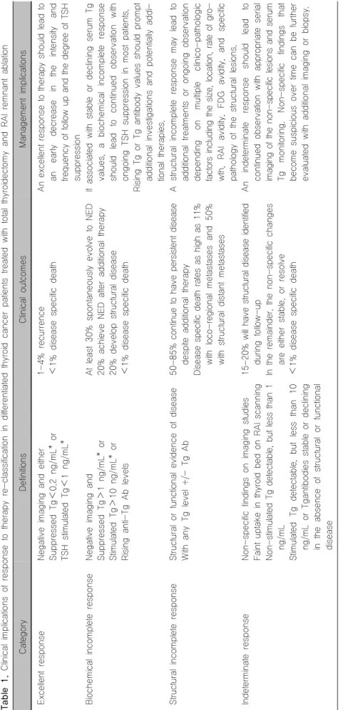
추적 기간 중 얻어지는 모든 임상적, 생화학적, 영상 학적, 그리고 조직병리학적인 결과들은 환자의 치료에 대한 결과를 평가하고 임상적인 상태를 재분류하는 데 사용된다. 이런 재분류 체계는 초기 치료를 한 후의 분 화갑상선암 환자들을 임상적으로 관리하는 데 매우 중 요하게 사용될 수 있다. 치료 반응에 따른 4가지 분류 를 하는 개념은 Tuttle 등6,7)과 Vaisman 등8)에 의해 서 술되었는데, 임상적인 영향과 치료에 대한 반응을 우 수한 치료반응, 생화학적 불완전 치료반응, 구조적 불 완전 치료반응, 미결정 치료반응으로 분류하고 있다 (Table 1).
우수한 치료반응: 질환에 대한 임상적이나 생화학적 또는 구조적인 증거가 없는 경우
생화학적 불완전 치료반응: 병변이 국소화된 증거는 없으나 티로글로불린 수치의 이상이나 항 티로글로불 린 항체의 증가가 있는 경우
구조적 불완전 치료반응: 국소적 또는 원격전이가 지속되거나 새로 발견된 경우
미결정 치료반응: 확실하게 양성이나 악성으로 분류 할 수가 없는 비특이적인 생화학적 또는 구조적 증상 이 있는 경우로 명백한 구조적 병변의 증거가 없이 항 티로글로불린 항체가 안정적이거나 감소하는 환자들 도 포함된다.
동위원소 치료 시행에 대한 권고
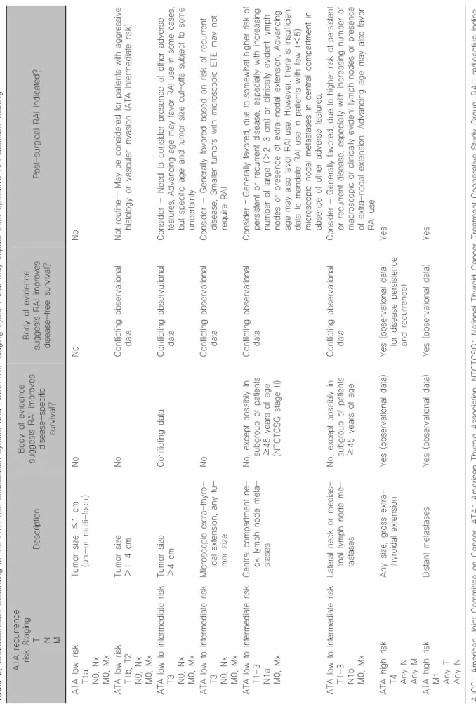
갑상선전절제 이후 잔여조직 제거를 위한 동위원소 치료는 분화갑상선암의 저위험군 환자에게는 권고되 지 않는다.9-19) 또한 엽절제를 한 경우나 전절제를 한 단발성의 미세유두암 환자에서 다른 나쁜 조건들이 없 다면 잔여조직 제거를 위한 동위원소 치료는 권고되지 않는다. 전절제를 한 다발성의 미세유두암 환자에서도 다른 나쁜 조건들이 없다면 잔여조직 제거를 위한 동 위원소 치료는 권고되지 않는다. 갑상선전절제를 시행 한 분화갑상선암의 중간 위험군 환자에서는 동위원소 치료가 권고된다. 갑상선전절제를 시행한 분화갑상선 암의 고위험군 환자에서는 잔여조직 제거를 위한 동위 원소 치료가 강력하게 권고된다(Table 2).20-23)
인체재조합 갑상선자극호르몬
인체재조합 갑상선자극호르몬을 사용하는 경우 잔 여조직 제거의 효율을 낮추지 않으면서 단기간의 삶의 질을 높일 수 있고, 장기간의 추적관찰 결과에서도 의 미 있는 차이를 보여주지 않아서, 광범위한 림프절 전 이가 관찰되지 않는 저위험군과 중간위험군 분화갑상 선암 환자들(예: T1-T3, N0/Nx/N1a, M0)에서 잔여조 직 제거를 위한 동위원소 치료가 계획되어 있다면, 갑 상선호르몬을 중단하는 대신에 인체재조합 갑상선자 극호르몬을 사용한 준비과정으로 충분히 대체할 수 있 다고 권고하고 있다. 원격전이는 없으나 광범위한 림 프절 전이가 관찰되는 중간위험군의 분화갑상선암 환 자에서도 동위원소 치료에 앞선 갑상선호르몬의 중단 을 인체재조합 갑상선자극호르몬을 사용한 준비과정 으로 대체할 수 있다고 권고하고 있다.24-37)
고위험군 분화갑상선암 환자에서는 장기추적 관찰 결과에 대한 연구자료들이 충분하지 않아 동위원소 치 료 전에 인체재조합 갑상선자극호르몬의 사용은 권고 되고 있지 않다.38-40) 다만, 동위원소 투여 이전의 갑상 선호르몬 중단을 불가능하게 만들 수 있는 중요한 동 반질환이 있는 분화갑상선암 환자들에서는 위험도와 관계없이 인체재조합 갑상선자극호르몬이 고려되어야 한다고 권고했다.
동위원소 치료용량의 결정
갑상선전절제를 시행한 저위험군 또는 저위험의 양 상을 보이는 중간위험군의 갑상선암 환자에서 잔여조 직 제거를 위한 동위원소 치료를 시행할 때는 고용량 보다는 약 30 mCi (1.11 GBq)의 저용량 사용을 강하게 권고하고 있다.41-48) 중간위험군이나 고위험군 환자에 서 원격전이는 없으나 현미경적으로 병변이 발견되거 나 의심되어 동위원소로 초기치료를 할 경우에는 30-150 mCi의 용량을 권고하고 있고, 그 이상의 용량을 사용하는 것이 재발병변을 더 감소시키지는 못하는 것 으로 보고하고 있다.49-52)
국소전이나 원격전이가 있는 환자에서 경험적인 동 위원소 치료를 권고하고 있으나, 150 mCi 이상의 동위 원소를 투여하는 경우에는 조직 내로의 최고 허용용량 을 넘는 경우가 종종 발생하므로 70세가 넘는 환자에 서는 허용하지 않도록 권고하고 있다.53-55) 폐 전이나 골 전이가 있는 환자에서는 경험적으로 100-200 mCi (70세 이상에서는 100-150 mCi) 또는 선량측정계로 계 산하여 투여 후 48시간에 전신에 80 mCi가 남도록 하
Table 1. Clinical implications of response to therapy re-classification in differentiated thyroid cancer patients treated with total thyroidectomy and RAI remnant ablation CategoryDefinitionsClinical outcomesManagement implications Excellent responseNegative imaging and either Suppressed Tg<0.2 ng/mL* or TSH stimulated Tg<1 ng/mL*
1-4% recurrence <1% disease specific deathAn excellent response to therapy should lead to anearlydecreaseintheintensityand frequency of follow up and the degree of TSH suppression Biochemical incomplete responseNegative imaging and Suppressed Tg>1 ng/mL* or Stimulated Tg>10 ng/mL* or Rising anti-Tg Ab levels At least 30% spontaneously evolve to NED 20% achieve NED after additional therapy 20% develop structural disease <1% disease specific death
If associated with stable or declining serum Tg values, a biochemicalincompleteresponse shouldlead tocontinued observation with ongoing TSH suppression in most patients. Rising Tg or Tg antibody values should prompt additional investigations and potentially addi- tional therapies. Structural incomplete responseStructural or functional evidence of disease With any Tg level +/- Tg Ab50-85% continue to have persistent disease despite additional therapy Disease specific death rates as high as 11% withloco-regional metastases and50% with structural distant metastases
Astructuralincomplete response maylead to additional treatments orongoing observation depending on multiple clinico-pathologic factors including the size, location, rate of gro- wth, RAI avidity, FDGavidity, andspecific pathology of the structural lesions. Indeterminate responseNon-specific findings on imaging studies Faint uptake in thyroid bed on RAI scanning Non-stimulated Tg detectable, but less than 1 ng/mL StimulatedTgdetectable, but lessthan10 ng/mL or Tgantibodies stable or declining in the absence of structural or functional disease
15-20% will have structural disease identified during follow-up In the remainder, the non-specific changes are either stable, or resolve <1% disease specific death
Anindeterminateresponse shouldlead to continued observation with appropriate serial imaging of the non-specific lesions and serum Tgmonitoring. Non-specific findings that become suspicious over time can be further evaluated with additional imaging or biopsy. *In the absence of anti-Tg antibodies FDG: flourodeoxyglucose, RAI: radioactive Iodine, Tg: thyroglobulin, Tg Ab: thyroglobulin antibody, TSH: thyroid stimulating hormone
Table 2. Characteristics according to the ATA risk stratification system and AJCC/TNM staging system that may impact post-operative RAI decision-making ATA recurrence risk Staging T N M
Description
Body of evidence suggests RAI improves disease-specific survival?
Body of evidence suggests RAI improves disease-free survival? Post-surgical RAI indicated? ATA low risk T1a N0, Nx M0, Mx
Tumor size ≤1 cm (uni-or multi-focal) NoNoNo ATA low risk T1b, T2 N0, Nx M0, Mx
Tumor size >1-4 cmNoConflicting observational dataNot routine – May be considered for patients with aggressive histology or vascular invasion (ATA intermediate risk) ATA low to intermediate risk T3 N0, Nx M0, Mx
Tumor size >4 cmConflicting data Conflicting observational dataConsider - Need to consider presence of other adverse features. Advancing age may favor RAI use in some cases, but specific age and tumor size cut-offs subject to some uncertainty ATA low to intermediate risk T3 N0, Nx M0, Mx Microscopic extra-thyro- idal extension, any tu- mor size
NoConflicting observational dataConsider - Generally favored based on risk of recurrent disease. Smaller tumors with microscopic ETE may not require RAI ATA low to intermediate risk T1-3 N1a M0, Mx
Central compartment ne- ck lymph node meta- stases No, except possibly in subgroup of patients ≥45 years of age (NTCTCSG stage III)
Conflicting observational dataConsider – Generally favored, due to somewhat higher risk of persistent or recurrent disease, especially with increasing number of large (>2-3 cm) or clinically evident lymph nodes or presence of extra-nodal extension. Advancing age may also favor RAI use. However, there is insufficient data to mandate RAI use in patients with few (<5) microscopic nodal metastases in central compartment in absence of other adverse features. ATA low to intermediate risk T1-3 N1b M0, Mx
Lateral neck or medias- tinal lymph node me- tastases No, except possibly in subgroup of patients ≥45 years of age
Conflicting observational dataConsider - Generally favored, due to higher risk of persistent or recurrent disease, especially with increasing number of macroscopic or clinically evident lymph nodes or presence of extra-nodal extension. Advancing age may also favor RAI use ATA high risk T4 Any N Any M
Any size, gross extra- thyroidal extension Yes (observational data)Yes (observational data for disease persistence and recurrence)
Yes ATA high risk M1 Any T Any N
Distant metastasesYes (observational data) Yes (observational data) Yes AJCC: American Joint Committee on Cancer, ATA: American Thyroid Association, NTCTCSG: National Thyroid Cancer Treatment Cooperative Study Group, RAI: radioactive Iodine, TNM: tumor node metastasis
거나, 적색골수에 200 cGy가 되도록 투여하도록 권고 하고 있다.56-59)
방사성요오드 저항성 분화갑상선암
방사성요오드 저항성 분화갑상선암은 종양세포나 전이세포가 방사성요오드를 축적하지 않아서 처음 진 단 시나 치료 후 전신스캔에서 갑상선 자리 주위에 섭 취가 없는 경우, 이전에 방사성요오드 섭취가 관찰되 었으나 종양세포가 방사성요오드를 축적하는 능력을 상실한 경우, 방사성요오드가 어떤 병변에서는 축적되 나 다른 병변에서는 축적되지 않는 경우, 그리고 의미 있는 방사성요오드의 축적이 관찰됨에도 전이병변이 진행되는 경우로 정의하였다.60) 방사성요오드에 저항 성이 있는 분화갑상선암을 가진 환자에서는 더 이상의 방사성요오드 치료는 권고하지 않았다.
Fluorodeoxyglucose Positron Emission Tomo- graphy (FDG-PET)
FDG-PET 검사는 갑상선결절의 미세침흡인생검의 세포학적인 검사결과가 불충분할 때 권고될 수 있는 영상검사라는 데는 동의하지 않았으나,61-63) FDG- PET-CT 검사에서 갑상선결절의 국소적인 섭취가 관 찰될 때는 약 35% 정도까지 악성종양이 발생될 가능성 이 있어 미세침흡인생검이 필요함을 강하게 권고하고
있다.64-66) 또한 갑상선암의 수술 전에 필수적으로 시행
되어야 할 검사로는 권고하지 않았으나, FDG-PET 검 사가 목과 종격동 전이, 그리고 원격전이를 진단하는 데 있어서 민감도가 높은 검사임에는 동의하고 있다.
FDG-PET 검사는 저분화성 갑상선암이나 허슬세포 암의 초기 병기설정이나, 전이 병소를 찾거나 고위험 도 환자를 위한 예후평가를 위해서, 전이 등에 대한 국 소 또는 전신치료의 치료반응 평가를 위해서 필요하다 고 권고하고 있다.67,68)
Single-Photon Emission Computerized Tomo- graphy (SPECT-CT)
갑상선호르몬 중단 후의 진단영상 또는 동위원소 치 료 후 전신영상에서 SPECT-CT를 사용한 검사가 전신 스캔에 의한 평면영상보다 더 나은 해부학적인 위치를 알려주고, 재발이나 전이 소견과 비특이적인 섭취 소 견을 감별하는 데 더 많은 정보를 제공한다는 점에 동 의하고 SPECT-CT에 의한 검사를 권고하고 있다.69-79)
결 론
갑상선결절과 분화갑상선암에 대한 미국갑상선학회 의 새로운 진료권고안의 내용 중 핵의학적 진단과 치 료에 밀접한 관련이 있는 부분들을 살펴보았다. 이 중 치료반응에 대한 새로운 평가방법이나 인체재조합 갑 상선자극호르몬의 시행, 방사성요오드 저항성 분화갑 상선암에 관련된 내용은 그동안의 많은 연구와 지식이 축적됨에 따른 결과물로 여겨지고 있어 핵의학적인 측 면에서도 충분히 수용 가능할 것으로 여겨지고 있고, 특히나 FDG-PET과 SPECT-CT와 관련된 내용들이 진료권고안에 포함된 것은 충분히 고무적인 일로 받아 들여지고 있다.
다만, 핵의학적인 측면에서 기존의 권고안과 상당히 다른 내용들이 포함되어 있는 동위원소 치료 시행에 대한 권고와 동위원소 치료용량의 결정에 관련된 내용 들은 대한핵의학회 내에서도 찬반이 엇갈리고 있고, 우리나라와 같이 평상시 요오드 섭취가 많은 지역에서 사용되는 동위원소 치료용량은 그렇지 않은 지역과는 확실한 차이가 있을 것이라는 반론들이 제시되고 있다. 따라서 미국갑상선학회의 새로운 진료권고안에 대한 내용들을 바탕으로 하여 향후 대한핵의학회 내의 갑상 선연구회를 중심으로 여러 논의 과정을 거치고 수정 보완하여 핵의학전문의를 위한 갑상선암의 진료권고 안이 제정될 것으로 예상된다. 그러나 동위원소 치료 를 포함한 여러 부분에서 향후의 추가 연구를 통한 검 증과 핵의학 전문의들 내의 합의가 필요할 것으로 생 각된다.
중심 단어: 갑상선결절, 분화갑상선암, 방사성요오드 치료.
References
1) Singer PA, Cooper DS, Daniels GH, Ladenson PW, Green- span FS, Levy EG, et al. Treatment guidelines for patients with thyroid nodules and well-differentiated thyroid cancer. American Thyroid Association. Arch Intern Med 1996;156(19):2165-72.
2) Cooper DS, Doherty GM, Haugen BR, Kloos RT, Lee SL, Mandel SJ, et al. Management guidelines for patients with thyroid nodules and differentiated thyroid cancer. Thyroid 2006;16(2):109-42.
3) American Thyroid Association Guidelines Taskforce on Thyroid Nodules and Differentiated Thyroid Cancer, Cooper DS, Doherty GM, Haugen BR, Kloos RT, Lee SL, et al.
Revised American Thyroid Association management guidelines for patients with thyroid nodules and differentiated thyroid
cancer. Thyroid 2009;19(11):1167-214.
4) Yi KH, Park YJ, Koong SS, Kim JH, Na DG, Ryu JS, et al. Revised Korean Thyroid Association Management Guidelines for patients with thyroid nodules and thyroid cancer. J Korean Thyroid Assoc 2010;3(2):65-96.
5) Kim WB, Seok JW, Kim MH, Kim BI, Park YJ, Lee KE, et al. Korean Thyroid Association Guidelines for patients undergoing radioiodine therapy for differentiated thyroid cancers (first edition, 2012). J Korean Thyroid Assoc 2013;6(1):12-25.
6) Tuttle RM, Leboeuf R. Follow up approaches in thyroid cancer:
a risk adapted paradigm. Endocrinol Metab Clin North Am 2008;37(2):419-35, ix-x.
7) Tuttle RM, Tala H, Shah J, Leboeuf R, Ghossein R, Gonen M, et al. Estimating risk of recurrence in differentiated thyroid cancer after total thyroidectomy and radioactive iodine remnant ablation: using response to therapy variables to modify the initial risk estimates predicted by the new American Thyroid Association staging system. Thyroid 2010;20(12):1341-9.
8) Vaisman F, Shaha A, Fish S, Michael Tuttle R. Initial therapy with either thyroid lobectomy or total thyroidectomy without radioactive iodine remnant ablation is associated with very low rates of structural disease recurrence in properly selected patients with differentiated thyroid cancer. Clin Endocrinol (Oxf) 2011;75(1):112-9.
9) Jonklaas J, Sarlis NJ, Litofsky D, Ain KB, Bigos ST, Brierley JD, et al. Outcomes of patients with differentiated thyroid car- cinoma following initial therapy. Thyroid 2006;16(12):1229-42.
10) Jonklaas J, Cooper DS, Ain KB, Bigos T, Brierley JD, Haugen BR, et al. Radioiodine therapy in patients with stage I differentiated thyroid cancer. Thyroid 2010;20(12):1423-4.
11) Sacks W, Fung CH, Chang JT, Waxman A, Braunstein GD.
The effectiveness of radioactive iodine for treatment of low-risk thyroid cancer: a systematic analysis of the peer-reviewed lite- rature from 1966 to April 2008. Thyroid 2010;20(11):1235-45.
12) Sawka AM, Brierley JD, Tsang RW, Thabane L, Rotstein L, Gafni A, et al. An updated systematic review and commentary examining the effectiveness of radioactive iodine remnant ablation in well-differentiated thyroid cancer. Endocrinol Metab Clin North Am 2008;37(2):457-80, x.
13) Ross DS, Litofsky D, Ain KB, Bigos T, Brierley JD, Cooper DS, et al. Recurrence after treatment of micropapillary thyroid cancer. Thyroid 2009;19(10):1043-8.
14) Kim HJ, Kim NK, Choi JH, Kim SW, Jin SM, Suh S, et al. Radioactive iodine ablation does not prevent recurrences in patients with papillary thyroid microcarcinoma. Clin Endocrinol (Oxf) 2013;78(4):614-20.
15) Baudin E, Travagli JP, Ropers J, Mancusi F, Bruno-Bossio G, Caillou B, et al. Microcarcinoma of the thyroid gland: the Gustave-Roussy Institute experience. Cancer 1998;83(3):553-9.
16) Creach KM, Siegel BA, Nussenbaum B, Grigsby PW.
Radioactive iodine therapy decreases recurrence in thyroid papillary microcarcinoma. ISRN Endocrinol 2012;2012:816386.
17) Lin HW, Bhattacharyya N. Survival impact of treatment options for papillary microcarcinoma of the thyroid. Laryn- goscope 2009;119(10):1983-7.
18) Kuo EJ, Roman SA, Sosa JA. Patients with follicular and Hurthle cell microcarcinomas have compromised survival: a
population level study of 22,738 patients. Surgery 2013;154(6):
1246-53; discussion 1253-4.
19) Mallick U, Harmer C, Hackshaw A, Moss L; IoN Trial Management Group. Iodine or Not (IoN) for low-risk differentiated thyroid cancer: the next UK National Cancer Research Network randomised trial following HiLo. Clin Oncol (R Coll Radiol) 2012;24(3):159-61.
20) Kazaure HS, Roman SA, Sosa JA. Aggressive variants of papillary thyroid cancer: incidence, characteristics and predictors of survival among 43,738 patients. Ann Surg Oncol 2012;19(6):
1874-80.
21) Kazaure HS, Roman SA, Sosa JA. Insular thyroid cancer: a population-level analysis of patient characteristics and predictors of survival. Cancer 2012;118(13):3260-7.
22) Chow SM, Yau S, Kwan CK, Poon PC, Law SC. Local and regional control in patients with papillary thyroid carcinoma:
specific indications of external radiotherapy and radioactive iodine according to T and N categories in AJCC 6th edition.
Endocr Relat Cancer 2006;13(4):1159-72.
23) Podnos YD, Smith DD, Wagman LD, Ellenhorn JD. Survival in patients with papillary thyroid cancer is not affected by the use of radioactive isotope. J Surg Oncol 2007;96(1):3-7.
24) Lee J, Yun MJ, Nam KH, Chung WY, Soh EY, Park CS.
Quality of life and effectiveness comparisons of thyroxine wit- hdrawal, triiodothyronine withdrawal, and recombinant thyroid- stimulating hormone administration for low-dose radioiodine remnant ablation of differentiated thyroid carcinoma. Thyroid 2010;20(2):173-9.
25) Robbins RJ, Driedger A, Magner J; U.S. and Canadian Thyrogen Compassionate Use Program Investigator Group.
Recombinant human thyrotropin-assisted radioiodine therapy for patients with metastatic thyroid cancer who could not elevate endogenous thyrotropin or be withdrawn from thyroxine. Thyroid 2006;16(11):1121-30.
26) Chianelli M, Todino V, Graziano FM, Panunzi C, Pace D, Guglielmi R, et al. Low-activity (2.0 GBq; 54 mCi) radioiodine post-surgical remnant ablation in thyroid cancer: comparison between hormone withdrawal and use of rhTSH in low-risk patients. Eur J Endocrinol 2009;160(3):431-6.
27) Mallick U, Harmer C, Yap B, Wadsley J, Clarke S, Moss L, et al. Ablation with low-dose radioiodine and thyrotropin alfa in thyroid cancer. N Engl J Med 2012;366(18):1674-85.
28) Pacini F, Ladenson PW, Schlumberger M, Driedger A, Luster M, Kloos RT, et al. Radioiodine ablation of thyroid remnants after preparation with recombinant human thyrotropin in diffe- rentiated thyroid carcinoma: results of an international, rando- mized, controlled study. J Clin Endocrinol Metab 2006;91(3):
926-32.
29) Schlumberger M, Catargi B, Borget I, Deandreis D, Zerdoud S, Bridji B, et al. Strategies of radioiodine ablation in patients with low-risk thyroid cancer. N Engl J Med 2012;366(18):
1663-73.
30) Taieb D, Sebag F, Cherenko M, Baumstarck-Barrau K, Fortanier C, Farman-Ara B, et al. Quality of life changes and clinical outcomes in thyroid cancer patients undergoing radioiodine remnant ablation (RRA) with recombinant human TSH (rhTSH): a randomized controlled study. Clin Endocrinol
(Oxf) 2009;71(1):115-23.
31) Emmanouilidis N, Muller JA, Jager MD, Kaaden S, Helfritz FA, Guner Z, et al. Surgery and radioablation therapy com- bined: introducing a 1-week-condensed procedure bonding total thyroidectomy and radioablation therapy with recombinant human TSH. Eur J Endocrinol 2009;161(5):763-9.
32) Tu J, Wang S, Huo Z, Lin Y, Li X, Wang S. Recombinant human thyrotropin-aided versus thyroid hormone withdrawal- aided radioiodine treatment for differentiated thyroid cancer after total thyroidectomy: a meta-analysis. Radiother Oncol 2014;
110(1):25-30.
33) Elisei R, Schlumberger M, Driedger A, Reiners C, Kloos RT, Sherman SI, et al. Follow-up of low-risk differentiated thyroid cancer patients who underwent radioiodine ablation of postsurgical thyroid remnants after either recombinant human thyrotropin or thyroid hormone withdrawal. J Clin Endocrinol Metab 2009;94(11):4171-9.
34) Emmanouilidis N, Schrem H, Winkler M, Klempnauer J, Scheumann GF. Long-term results after treatment of very low-, low-, and high-risk thyroid cancers in a combined setting of thyroidectomy and radio ablation therapy in euthyroidism. Int J Endocrinol 2013;2013:769473.
35) Hugo J, Robenshtok E, Grewal R, Larson S, Tuttle RM.
Recombinant human thyroid stimulating hormone-assisted radio- active iodine remnant ablation in thyroid cancer patients at inte- rmediate to high risk of recurrence. Thyroid 2012;22(10):
1007-15.
36) Rosario PW, Mineiro Filho AF, Lacerda RX, Calsolari MR.
Long-term follow-up of at least five years after recombinant human thyrotropin compared to levothyroxine withdrawal for thyroid remnant ablation with radioactive iodine. Thyroid 2012;22(3):332-3.
37) Pitoia F, Marlowe RJ, Abelleira E, Faure EN, Bueno F, Schwarzstein D, et al. Radioiodine thyroid remnant ablation after recombinant human thyrotropin or thyroid hormone withdrawal in patients with high-risk differentiated thyroid cancer. J Thyroid Res 2012;2012:481568.
38) Bartenstein P, Calabuig EC, Maini CL, Mazzarotto R, Muros de Fuentes MA, Petrich T, et al. High-risk patients with differentiated thyroid cancer T4 primary tumors achieve remnant ablation equally well using rhTSH or thyroid hormone withdrawal. Thyroid 2014;24(3):480-7.
39) Tala H, Robbins R, Fagin JA, Larson SM, Tuttle RM.
Five-year survival is similar in thyroid cancer patients with distant metastases prepared for radioactive iodine therapy with either thyroid hormone withdrawal or recombinant human TSH.
J Clin Endocrinol Metab 2011;96(7):2105-11.
40) Klubo-Gwiezdzinska J, Burman KD, Van Nostrand D, Mete M, Jonklaas J, Wartofsky L. Radioiodine treatment of metastatic thyroid cancer: relative efficacy and side effect profile of preparation by thyroid hormone withdrawal versus recombinant human thyrotropin. Thyroid 2012;22(3):310-7.
41) Maenpaa HO, Heikkonen J, Vaalavirta L, Tenhunen M, Joensuu H. Low vs. high radioiodine activity to ablate the thyroid after thyroidectomy for cancer: a randomized study. PLoS One 2008;3(4):e1885.
42) Pilli T, Brianzoni E, Capoccetti F, Castagna MG, Fattori S,
Poggiu A, et al. A comparison of 1850 (50 mCi) and 3700 MBq (100 mCi) 131-iodine administered doses for recombinant thyrotropin-stimulated postoperative thyroid remnant ablation in differentiated thyroid cancer. J Clin Endocrinol Metab 2007;
92(9):3542-6.
43) Zaman M, Toor R, Kamal S, Maqbool M, Habib S, Niaz K. A randomized clinical trial comparing 50mCi and 100mCi of iodine-131 for ablation of differentiated thyroid cancers. J Pak Med Assoc 2006;56(8):353-6.
44) Kukulska A, Krajewska J, Gawkowska-Suwinska M, Puch Z, Paliczka-Cieslik E, Roskosz J, et al. Radioiodine thyroid remnant ablation in patients with differentiated thyroid carc- inoma (DTC): prospective comparison of long-term outcomes of treatment with 30, 60 and 100 mCi. Thyroid Res 2010;3(1):9.
45) Fang Y, Ding Y, Guo Q, Xing J, Long Y, Zong Z. Rad- ioiodine therapy for patients with differentiated thyroid cancer after thyroidectomy: direct comparison and network meta- analyses. J Endocrinol Invest 2013;36(10):896-902.
46) Ma C, Tang L, Fu H, Li J, Wang H. rhTSH-aided low- activity versus high-activity regimens of radioiodine in residual ablation for differentiated thyroid cancer: a meta-analysis. Nucl Med Commun 2013;34(12):1150-6.
47) Cheng W, Ma C, Fu H, Li J, Chen S, Wu S, et al. Low- or high-dose radioiodine remnant ablation for differentiated thyroid carcinoma: a meta-analysis. J Clin Endocrinol Metab 2013;98(4):1353-60.
48) Valachis A, Nearchou A. High versus low radioiodine activity in patients with differentiated thyroid cancer: a meta-analysis.
Acta Oncol 2013;52(6):1055-61.
49) Castagna MG, Cevenini G, Theodoropoulou A, Maino F, Memmo S, Claudia C, et al. Post-surgical thyroid ablation with low or high radioiodine activities results in similar outcomes in intermediate risk differentiated thyroid cancer patients. Eur J Endocrinol 2013;169(1):23-9.
50) Han JM, Kim WG, Kim TY, Jeon MJ, Ryu JS, Song DE, et al. Effects of low-dose and high-dose postoperative radioiodine therapy on the clinical outcome in patients with small differentiated thyroid cancer having microscopic extrathyroidal extension. Thyroid 2014;24(5):820-5.
51) Kruijff S, Aniss AM, Chen P, Sidhu SB, Delbridge LW, Robinson B, et al. Decreasing the dose of radioiodine for remnant ablation does not increase structural recurrence rates in papillary thyroid carcinoma. Surgery 2013;154(6):1337-44;
discussion 1344-5.
52) Sabra MM, Grewal RK, Ghossein RA, Tuttle RM. Higher administered activities of radioactive iodine are associated with less structural persistent response in older, but not younger, papillary thyroid cancer patients with lateral neck lymph node metastases. Thyroid 2014;24(7):1088-95.
53) Schlumberger M, Lacroix L, Russo D, Filetti S, Bidart JM.
Defects in iodide metabolism in thyroid cancer and implications for the follow-up and treatment of patients. Nat Clin Pract Endocrinol Metab 2007;3(3):260-9.
54) Kulkarni K, Van Nostrand D, Atkins F, Aiken M, Burman K, Wartofsky L. The relative frequency in which empiric dosages of radioiodine would potentially overtreat or undertreat patients who have metastatic well-differentiated thyroid cancer. Thyroid
2006;16(10):1019-23.
55) Tuttle RM, Leboeuf R, Robbins RJ, Qualey R, Pentlow K, Larson SM, et al. Empiric radioactive iodine dosing regimens frequently exceed maximum tolerated activity levels in elderly patients with thyroid cancer. J Nucl Med 2006;47(10):1587-91.
56) Hebestreit H, Biko J, Drozd V, Demidchik Y, Burkhardt A, Trusen A, et al. Pulmonary fibrosis in youth treated with radioiodine for juvenile thyroid cancer and lung metastases after Chernobyl. Eur J Nucl Med Mol Imaging 2011;38(9):1683-90.
57) Ilgan S, Karacalioglu AO, Pabuscu Y, Atac GK, Arslan N, Ozturk E, et al. Iodine-131 treatment and high-resolution CT:
results in patients with lung metastases from differentiated thyroid carcinoma. Eur J Nucl Med Mol Imaging 2004;
31(6):825-30.
58) Hod N, Hagag P, Baumer M, Sandbank J, Horne T.
Differentiated thyroid carcinoma in children and young adults:
evaluation of response to treatment. Clin Nucl Med 2005;
30(6):387-90.
59) Schlumberger M, Challeton C, De Vathaire F, Travagli JP, Gardet P, Lumbroso JD, et al. Radioactive iodine treatment and external radiotherapy for lung and bone metastases from thyroid carcinoma. J Nucl Med 1996;37(4):598-605.
60) Schlumberger M, Brose M, Elisei R, Leboulleux S, Luster M, Pitoia F, et al. Definition and management of radioactive iodine-refractory differentiated thyroid cancer. Lancet Diabetes Endocrinol 2014;2(5):356-8.
61) Deandreis D, Al Ghuzlan A, Auperin A, Vielh P, Caillou B, Chami L, et al. Is (18)F-fluorodeoxyglucose-PET/CT useful for the presurgical characterization of thyroid nodules with indet- erminate fine needle aspiration cytology? Thyroid 2012;22(2):
165-72.
62) Vriens D, de Wilt JH, van der Wilt GJ, Netea-Maier RT, Oyen WJ, de Geus-Oei LF. The role of [18F]-2-fluoro- 2-deoxy-d-glucose-positron emission tomography in thyroid nod- ules with indeterminate fine-needle aspiration biopsy: systematic review and meta-analysis of the literature. Cancer 2011;117(20):
4582-94.
63) Wang N, Zhai H, Lu Y. Is fluorine-18 fluorodeoxyglucose positron emission tomography useful for the thyroid nodules with indeterminate fine needle aspiration biopsy? A meta-analysis of the literature. J Otolaryngol Head Neck Surg 2013;42:38.
64) Soelberg KK, Bonnema SJ, Brix TH, Hegedus L. Risk of malignancy in thyroid incidentalomas detected by 18F-fluor- odeoxyglucose positron emission tomography: a systematic review.
Thyroid 2012;22(9):918-25.
65) Chen W, Parsons M, Torigian DA, Zhuang H, Alavi A.
Evaluation of thyroid FDG uptake incidentally identified on FDG-PET/CT imaging. Nucl Med Commun 2009;30(3):240-4.
66) Nishimori H, Tabah R, Hickeson M, How J. Incidental thyroid "PETomas": clinical significance and novel description of the self-resolving variant of focal FDG-PET thyroid uptake. Can J Surg 2011;54(2):83-8.
67) Robbins RJ, Wan Q, Grewal RK, Reibke R, Gonen M, Strauss HW, et al. Real-time prognosis for metastatic thyroid carcinoma based on 2-[18F]fluoro-2-deoxy-D-glucose-positron emission to- mography scanning. J Clin Endocrinol Metab 2006;91(2):
498-505.
68) Deandreis D, Al Ghuzlan A, Leboulleux S, Lacroix L, Garsi JP, Talbot M, et al. Do histological, immunohistochemical, and metabolic (radioiodine and fluorodeoxyglucose uptakes) patterns of metastatic thyroid cancer correlate with patient outcome?
Endocr Relat Cancer 2011;18(1):159-69.
69) Ciappuccini R, Heutte N, Trzepla G, Rame JP, Vaur D, Aide N, et al. Postablation (131)I scintigraphy with neck and thorax SPECT-CT and stimulated serum thyroglobulin level predict the outcome of patients with differentiated thyroid cancer. Eur J Endocrinol 2011;164(6):961-9.
70) Salvatori M, Perotti G, Villani MF, Mazza R, Maussier ML, Indovina L, et al. Determining the appropriate time of execution of an I-131 post-therapy whole-body scan: comparison between early and late imaging. Nucl Med Commun 2013;34(9):900-8.
71) Kohlfuerst S, Igerc I, Lobnig M, Gallowitsch HJ, Gomez- Segovia I, Matschnig S, et al. Posttherapeutic (131)I SPECT- CT offers high diagnostic accuracy when the findings on conv- entional planar imaging are inconclusive and allows a tailored patient treatment regimen. Eur J Nucl Med Mol Imaging 2009;36(6):886-93.
72) Chen L, Luo Q, Shen Y, Yu Y, Yuan Z, Lu H, et al.
Incremental value of 131I SPECT/CT in the management of patients with differentiated thyroid carcinoma. J Nucl Med 2008;49(12):1952-7.
73) Schmidt D, Linke R, Uder M, Kuwert T. Five months' follow-up of patients with and without iodine-positive lymph node metastases of thyroid carcinoma as disclosed by (131) I-SPECT/CT at the first radioablation. Eur J Nucl Med Mol Imaging 2010;37(4):699-705.
74) Maruoka Y, Abe K, Baba S, Isoda T, Sawamoto H, Tanabe Y, et al. Incremental diagnostic value of SPECT/CT with 131I scintigraphy after radioiodine therapy in patients with well- differentiated thyroid carcinoma. Radiology 2012;265(3):902-9.
75) Grewal RK, Tuttle RM, Fox J, Borkar S, Chou JF, Gonen M, et al. The effect of posttherapy 131I SPECT/CT on risk classification and management of patients with differentiated thyroid cancer. J Nucl Med 2010;51(9):1361-7.
76) Aide N, Heutte N, Rame JP, Rousseau E, Loiseau C, Henry-Amar M, et al. Clinical relevance of single-photon emission computed tomography/computed tomography of the neck and thorax in postablation (131)I scintigraphy for thyroid cancer.
J Clin Endocrinol Metab 2009;94(6):2075-84.
77) Schmidt D, Szikszai A, Linke R, Bautz W, Kuwert T. Impact of 131I SPECT/spiral CT on nodal staging of differentiated thyroid carcinoma at the first radioablation. J Nucl Med 2009;50(1):18-23.
78) Jeong SY, Lee SW, Kim HW, Song BI, Ahn BC, Lee J.
Clinical applications of SPECT/CT after first I-131 ablation in patients with differentiated thyroid cancer. Clin Endocrinol (Oxf) 2014;81(3):445-51.
79) Tharp K, Israel O, Hausmann J, Bettman L, Martin WH, Daitzchman M, et al. Impact of 131I-SPECT/CT images obtained with an integrated system in the follow-up of patients with thyroid carcinoma. Eur J Nucl Med Mol Imaging 2004;31(10):1435-42.