Letter to the Editor
Vol. 28, No. 2, 2016 273
Received February 10, 2015, Revised May 7, 2015, Accepted for publication May 22, 2015
Corresponding author: You Won Choi, Department of Dermatology, Ewha Womans University School of Medicine, 1071 Anyangcheon-ro, Yangcheon-gu, Seoul 07985, Korea. Tel: 82-2-2650-5159, Fax: 82-2-2652-6925, E-mail: [email protected]
This is an Open Access article distributed under the terms of the Creative Commons Attribution Non-Commercial License (http://creativecommons.org/
licenses/by-nc/4.0) which permits unrestricted non-commercial use, distribution, and reproduction in any medium, provided the original work is properly cited.
Copyright © The Korean Dermatological Association and The Korean Society for Investigative Dermatology
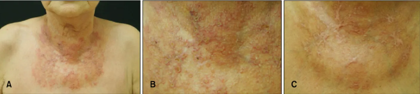
Fig. 1. (A) Several crusted papules on an erythematous telangiectactic patch on the anterior neck and upper chest. (B) Close-up view.
(C) Improved skin lesion with fibrotic scar after treatment with oral itraconazole 200 mg/day and topical sertaconazole application for 3 months.
http://dx.doi.org/10.5021/ad.2016.28.2.273
A Case of Cutaneous Protothecosis in an Immunocompetent Patient
Ju Yun Woo, Eun Ah Suhng, Ji Yeon Byun, Hae Young Choi, Sun Hee Sung
1, You Won Choi
Departments of Dermatology and 1Pathology, Ewha Womans University School of Medicine, Seoul, Korea
Dear Editor:
Protothecosis is a rare infection, caused by the genus Prototheca, achlorophyllous algae which are saprophytes in soil, and occasionally affect dogs and cats1. It is be- lieved that they may infect humans by traumatic in- oculation2.
A 74-year-old woman visited our clinic with prickling er- ythematous umbilicated papules on the anterior neck and upper chest which developed 2 months prior on top of an erythematous telangiectatic patch which had been present for 2 years (Fig. 1A, B). She had been treated at another clinic. with topical corticosteroids for 2 years for sus- pected eczema aggravated by rubbing. She had no history of trauma or other medical disease. She had lived with a pet dog for 18 years.
The patient’s complete blood count, renal and liver func- tion, and chest X-ray study were all normal. The HBs Ag,
HCV Ab, HIV Ab, and anti-nuclear Ab tests were negative.
The venereal disease research laboratory test (VDRL) was nonreactive. The histopathology revealed a granulomatous inflammation with necrosis in the dermis (Fig. 2A), contain- ing many non-budding spherical organisms. Multiple spor- angia containing endospores were observed with the mor- ula appearance that is the characteristic feature of the Prototheca species (Fig. 2B). The Periodic acid-Schiff stain- ing was positive and showed characteristic internal septa- tion and thick cell walls (Fig. 2C). Treatment with oral itra- conazole (200 mg/day) and topical sertaconazole for 12 weeks resulted in marked improvement of the skin lesions (Fig. 1C).
Cutaneous protothecosis mainly involves the extremities and its presentation is variable2. It can manifest as an er- ythematous plaque, or less commonly, in ulcerative or pu- rulent forms3-5. Our patient presented with erythematous
Letter to the Editor
274 Ann Dermatol
Fig. 2. (A) Granulomatous inflammation with necrosis in the dermis (H&E, ×12.5). (B) Multiple sporangia with in morula-like or cartwheel-like appearance (arrow) (H&E, ×400). (C) Multiple morula-like appearances highlighted by Periodic acid-Schiff stain (×400).
umbilicated papules on chest, which is a less common3,4. Prototheca have low virulence; hence, systemic or local predisposing factors are commonly found in affected pa- tients, such as steroid use, malignancy, diabetes mellitus, and organ transplantation3. Our patient had intact im- munity but was predisposed to local immunosuppression by long-term topical steroid application, skin damage by frequent rubbing, and the presence of a pet dog, which may have been the source of the algae. Protothecawick- erhamii and P.zopfii can mainly cause human proto- thecosis, and P.wickerhamii is the most commonly re- sponsible3,4. The sporangia of P.wickerhamii are round with symmetrical morula formation and a diameter of 7∼
13 μm, while the sporangia of P.zopfii are oval with rela- tively asymmetric random septation and a diameter of 14∼16 μm. P.wickerhamii is susceptible to antifungal agents and is easily treated, but P.zopfii is resistant to treatment.
In Korea, 11 cases of cutaneous protothecosis have been reported2. Most of the reported cases involved patients who were over the age of 60 years. The most commonly involved sites were the extremities, except one patient whose cheek was affected. Most patients had systemic predisposing factors and 2 patients had local predisposing factors (long-term topical steroid use). All of these cases
were confirmed with diagnostic tests; P.wickerhamii was identified in 10 cases and P. zopfii was identified in 1 case2.
We report a rare case of cutaneous protothecosis in an im- munocompetent patient who presented with erythematous umbilicated papules on the chest after long-term topical corticosteroid use, traumatic rubbing, and exposure to a pet dog.
REFERENCES
1. Mayorga J, Barba-Gómez JF, Verduzco-Martínez AP, Muñoz-Estrada VF, Welsh O. Protothecosis. Clin Dermatol 2012;30:432-436.
2. Seok JY, Lee Y, Lee H, Yi SY, Oh HE, Song JS. Human cutaneous protothecosis: report of a case and literature review. Korean J Pathol 2013;47:575-578.
3. Lass-Flörl C, Mayr A. Human protothecosis. Clin Microbiol Rev 2007;20:230-242.
4. Hillesheim PB, Bahrami S. Cutaneous protothecosis. Arch Pathol Lab Med 2011;135:941-944.
5. Todd JR, King JW, Oberle A, Matsumoto T, Odaka Y, Fowler M, et al. Protothecosis: report of a case with 20-year follow-up, and review of previously published cases. Med Mycol 2012;50:673-689.