Vol. 16, No. 3, September, 2004
고령의 대퇴골 전자간부 분쇄골절에서 시행한 무시멘트형 고관절 반치환술의 결과
황득수・곽상구・우세민
충남대학교 의과대학 정형외과학교실
목적: 고령의 환자에서 발생한 대퇴 전자간 불안정성 분쇄 골절에서의 무시멘트형 원추형 대퇴 주대(Conical stem)를 이용한 양극성 반치환술을 시행하여 단기 치료 결과를 보고하고자 한다.
대상 및 방법: 1998년 4월부터 2002년 5월까지 대퇴 전자간부 불안정성 분쇄골절로 진단받고 본원에서 양극성 고관절 반치환술을 시행 받은 51명의 환자 중 원추형 대퇴 주대(Conical stem)를 이용한 무시멘트형 양극성 고관절 반치환술 을 시행 받은 25명 중 1년 이상 추시 가능하였던 20명을 대상으로 하였다. 환자들의 평균 연령은 75.9세(범위, 61~91 세)이며, 성별은 남자가 2례이며 여자가 18례였다. 평균 추시 기간은 22개월(범위, 13~30개월)이였다. 단순 방사선상 골절 양상은 Evans 분류상 불안정성 골절, Kyle-Gustilo 분류 III 이상의 골절, AO 분류상 A2, Tronzo 분류에서는 III와 IV 골절에 해당되는 것을 대상으로 하였다. 수술 시간은 피부 절개 후 봉합까지 평균 40분(범위, 35~46분)이었 다. 수술 후 수직침강 정도, 내반 변형, 대퇴 주대 주위의 대퇴골 근위부 골절의 유합 정도 등을 측정하였다.
결과: 수술 소견상 골절 양상은 대부분 골다공증 때문에 술 전 방사선 사진보다는 분쇄 정도가 심하였다. 수술 후 보조기 (walker)를 이용한 보행 가능한 기간은 평균 6일(범위, 5~14일)정도였으며, 근위부 골절부위 유합은 17예에서 관찰되 었으며 유합되기까지의 기간은 평균 15.4주(범위,13.4~18.6주)였다. 대퇴 삽입물의 침강은 평균 4 mm(범위, 2~7 mm)로 나타났다. 수술 후와 이후의 경과관찰 사진에서 평가한 내반 변형의 정도는 평균 5.3도(범위, 0~12도)로 나타 났다. 수술 후 합병증으로는 수술 중 인공 삽입물 주위 골절이 1례, 그리고 수술 후 1례에서 탈구 소견을 보였다 결론: 고령의 불안정성 골절에서 대퇴골 불안정성 전자간 골절의 수술적 치료로 무시멘트형 대퇴 스템을 이용한 수술방법 이 임상적으로 비교적 우수한 결과를 보였다.
색인 단어: 대퇴골, 대퇴 전자간부 골절, 양극성 고관절 반치환술, 원추형 대퇴 주대
서 론
대퇴골 전자간부 분쇄 골절은 주로 노년층에서 호발하며 대부분의 환자에서 골다골증을 동반하고 골절의 불안정으 로 인해 치료의 어려움이 있으며, 골유합을 얻기 위한 여 러 가지 내고정물이 개발되어 사용되었지만 후유증이 많은 것으로 보고 되고 있다1 8 , 1 9 , 3 1 ). 기존의 대부분 대퇴 전자간 부 불안정성 골절 치료에 사용되어 왔던 역동적 기구들은 환자의 골이 약하고 골다공증이 있어 나사못을 견고히 삽
입할 수 없기 때문에 골이 유합되는 동안 체중 부하를 금 지시키고 고관절을 보호해야 하며, 유연성 골수강내 고정 술은 불안정성 전자간 골절의 치료에 있어서는 불량한 결 과 및 많은 합병증들이 보고 되어 왔다2 , 6 , 1 4 , 1 8 , 2 2 , 2 7 - 2 9 , 3 1 ). 골시 멘트를 사용하는 고관절 치환술에서는 안정된 고정과 조기 체중부하가 가능하다는 점 등 여러 가지 장점이 있어 많이 사용되어 왔으나, 수술 시간의 지연 및 시멘트 고정의 일 반적인 합병증, 장기간의 추시 관찰 상 골 용해가 나타났 고, 이것은 인공관절의 후기 이완과 밀접한 관련이 있음이 보고 되고 있다1 , 5 , 1 7 , 1 9 , 2 4 , 3 2 ). 이에 골시멘트를 사용하지 않는 대퇴 주대를 개발하게 되었고, 현재는 사용 빈도가 증가하 는 추세이다. 무시멘트형 대퇴 주대의 성공적인 고정은 초 기의 안정성과 후기의 생물학적 고정에 의해 의존된다9 -
1 3 , 1 9 ). 본원에서는 횡단면이 실린더형으로 고안된 원추형 대
퇴 주대(Conical stem, Protek AGⓇ, Berne, S w i t z e r l a n d )를 이용하여 치료하였던 고령의 대퇴골 전자 간부 분쇄 골절 중 최소한 1년 이상 추시 가능하였던 2 0명
※ 통신저자: 황 득 수
대전시중구대사동640
충남대학교의과대학정형외과학교실 Tel: 82-42-220-7349
Fax: 82-42-252-7098 E-mail: [email protected]
본 논문의요지는2003년 대한고관절학회추계학술대회에서발 표되었음.
에 대하여 수술 후 방사선학적 안정성를 비교하였고, 추시 중 임상적 양상과 합병증 그리고 방사선학적 변화를 평가 해 보고하고자 하였다.
연구 대상 및 방법
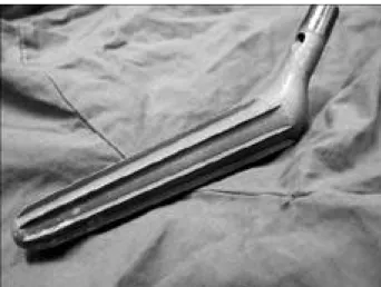
1 9 9 8년 4월부터 2 0 0 2년 5월까지 대퇴 전자간부 불안정 성 분쇄골절로 진단받고 본원에서 양극성 고관절 반치환술 을 시행 받은 5 1명의 환자 중 원추형 대퇴 주대를 이용한 무시멘트형 양극성 고관절 반치환술을 시행 받은 2 5명중 1 년 이상 추시 가능한 2 0명을 대상으로 하였다. 총 2 5명의 환자 중에서 수술 후 4주 이내 사망한 환자 2명과 추적 관 찰이 불가능한 3명은 대상에서 제외하였다. 환자들의 평균 연령은 7 5 . 9세(범위, 61~91세)이며, 평균 추시 기간은 2 2개월(범위, 13~30개월)이었다. 성별로 보면 여자가 1 8 례로 많았고, 좌측이 1 2례, 우측이 8례였다. 분류는 대퇴 골 전자간부 불안정성 분쇄 골절에서 AO 분류 A 2 , Kyle-Gustilo 분류2 6 )I I I와 IV , Tronzo 분류3 4 ) I I I와 I V 에 해당되는 환자였으며, 대퇴 내고정물로 사용된 원추형 대퇴 주대는 collarless, cone-shaped, coarse-blasted s u r f a c e로 처리된 Titanium 합금으로 원위부에 8개의 세로로 배열된 날(longitudinal ribs)을 갖고 있는 원추 형, 직선형 대퇴 삽입물이다(Fig. 1).
수술 술식은 측방 도달법이 4례, 후외방 도달법이 1 6례 였으며, 임상적으로는 Koval 분류2 5 )에 의한 수술전 보행 능력으로의 회복 정도를 분석하였으며, 수술 전과 최종 추 시 시기의 Harris 고관절 점수를 비교하였고, 대퇴부 동 통과 서혜부 동통 여부를 관찰하였다. 방사선학적으로는 수술 직후 및 최종 추시시 촬영한 전후면 및 측면 방사선 사진을 이용하였으며, 대퇴 삽입물의 안정성을 판단하는 소견인 주대의 수직 침강(subsidence) 정도는 술 후 방사 선 사진과 최종 추시 방사선 사진상의 인공 관절의 외상연 에서 대퇴골 대전자간의 근위첨부( t i p )까지 수직 거리를, 방사선상 확대율을 고려하여 형판을 이용하여 측정하였다.
또한 대퇴골 경간각 및 하지 단축을 측정하여 대퇴골 근위 부의 내반 변형이나 하지 단축 소견이 보이는지를 조사하 였고, 삽입물의 해리 및 골용해 여부를 관찰하였다.
결 과
수술 시간은 피부 절개에서 봉합까지 평균 4 0분(범위, 3 5 ~ 4 6분)이었으며, 평균 수술 중 출혈양은 600 cc(범위, 588~630 cc)이었다. 수술 후 평균 6일(범위, 5~14일)후 에 보행기를 이용한 부분적 체중부하를 시작하였다. 임상 적으로 K a v a l에 의한 보행 능력 정도는 수술 전 보행 능 력으로의 회복은 8례(40%), 수술 전보다 1단계이상 감소 한 경우가 1 2례( 6 0 % )였다(Table 1). Harris 고관절 점 수는 술전 평균 4 7 . 8점(범위, 28~65점)에서 수술 후 1 2 개월에 평균 8 6점(범위, 56~93점)으로 향상되었다. 대퇴 부 동통은 3례에서 있었으나 모두 1년이 지난 후 소실되었 다. 수술 중 대퇴골 근위부 골절에 대해 환상강선 고정을 시행하였으나, 추시상 이완이나 실패는 없었다(Fig. 2).
합병증으로 수술 중 대퇴 삽입물 주위 골절이 1례, 수술 후 탈구가 1례 있었다.
방사선학적으로 수직침강의 평균은 4 mm(범위, 2~7 m m )이었고, 의미 있는 수직 침강이 있었던 경우(5 mm 이상)는 2례(Fig. 3)에서 관찰되었으나 최종 추시시 사진 상 더 이상의 진행은 보이지 않았다. 방사선 사진상 내반 변형은 최종 추시 사진에서 건측과 비교하여 평균 5 . 3도 (범위, 0~12도)를 보였고, 최종 추시시 골내막 용해 소견 및 삽입물 해리 소견은 없었다. 3례에서 소전자 불유합 소
Fig. 1. Photograph showing a Conical stem(Protek AGⓇ, Berne, Switzerland) used in this study.
Table 1. Walking Ability by Koval25)
Walking ability Preoperative Postoperative
Independent community ambulators 9(45%) 5(25%)
Community ambulator with cane 6(30%) 6(30%)
Community ambulator with walker/crutches 3(15%) 5(25%)
Independent household ambulators 1(5%) 2(10%)
Household ambulator with cane 1(5%) 2(10%)
Household ambulator with walker/crutches - -
견이 보였으나 보행상의 문제점은 없었다.
고 찰
대퇴골 전자간 골절은 풍부한 해면골로 인해 불유합이나 대퇴골두의 무혈성 괴사 등의 합병증이 드물기 때문에 고 식적 치료 방법으로도 치료 될 수 있으나, 골절이 주로 고 령에서 빈발하므로 골절로 기인하는 합병증 이외에 고령에
Fig. 2. An unstable intertrochanteric comminuted fracture in 91 years old female( A) treated with primary bipolar hemiarthroplasty with Conical stem( B). In roentgenogram 22 months after operation shows no evidence of loosening(C).
Fig. 3. An unstable intertrochanteric comminuted fracture in 68 years old female( A) treated with primary bipolar hemiarthroplasty with Conical stem( B). In roentgenogram 22 months after operation, the shortening compared with normal side was 7.7mm(C).
A A
B B
C C
따르는 기존 질환의 악화 및 장기간의 침상 안정으로 인한 욕창, 폐렴, 폐색전증, 무기폐, 요로감염 등의 합병증으로 노년층 사망의 중요한 인자중의 하나로 지목되고 있다1 , 1 8 ). 최근에는 합병증과 사망률의 감소를 위해 수술적 정복과 금속 내고정 후, 조기 보행하는 것이 일반적인 치료법으로 간주되고 있다2 , 3 , 1 4 , 1 8 , 2 3 ). 금속 내고정물의 선택에 있어서 현 재 압박고 나사가 많이 이용되고 있으며 특히 일부 저자는 불안정 대퇴전자간 골절시 이것이 가장 적합한 수술 방법 이라 주장하였다1 8 , 2 0 ). 하지만 대퇴골 전자간 불안정 골절시 골절 정복 및 금속 내고정시 내고정 후 나사못의 이완, 내 반 변형, 고정물의 관절내 이동과 같은 고정실패 및 불유 합의 빈도의 우려성 때문에 조기 체중 부하를 주저하는 경 향이 있다2 , 7 , 8 , 1 4 , 1 8 , 2 8 , 2 9 ). 또한 고령의 환자들의 불안정성 대퇴 전자간 골절은 골다공증 때문에 견고한 내고정이 힘들고 또한 높은 유병률과 사망률을 보여주고 있다. 따라서 최근 에는 대퇴 전자간 불안정 골절에 있어서 인공 고관절 반치 환술을 실시하여 수술 후 합병증이 적고, 조기 보행을 시 킬 수 있으므로 술 후 보행 능력의 회복이 우수하다는 점 이 보고 되고 있다1 5 , 1 6 , 1 8 , 2 2 , 2 7 , 3 0 ). 본 연구에서는 대퇴골 전자 간 불안정성 골절에서 무시멘트형 원추형 대퇴 주대를 사 용하여 수술한 결과 수술 후 평균 6일에 조기 보행을 허용 할 수 있었으며 추시 방사선 사진에서 고정 실패나 내반 변형으로 인한 보행의 장애는 없었다.
Koval 등8 , 1 8 , 2 5 )은 보행 능력 회복을 예측할 수 있는 인자 로 연령, 골절 전의 보행 능력, 골절의 형태, 수술 전 위 험 요소 그리고 조기 체중 부하가 있다고 주장하였으며, Ceder 등7 , 8 , 1 8 )은 수술 후 2주 내에 조기 보행이나 개인위 생을 할 정도의 능력을 갖는 것이 예후에 있어서 중요성을 갖는다고 하였다. Koval 등에 의하면 제한된 체중 부하가 환자의 기능 회복을 지연시킬 수 있다고 주장하였으며, 고 령 환자에서의 조기 체중 부하는 환자 자신이 동통의 정도 및 자신의 생각에 따라 스스로 의식적인 제한을 하기 때문 에 불유합 및 수술적 정복 유지의 실패는 증가하지 않는다 고 하였다. 본 연구에서는 수술 후 약 5 ~ 7일후에 부분적 체중 부하를 시켰으며 조기 보행에 따른 문제점은 발생하 지 않았다. 원추형 주대는 5도의 원추형 형태를 가지는 주 대로서 Wagner 재치환 주대(revision stem)에서 원추형 고정의 개념으로 도입되어 고안된 것으로, 실린더 형의 디 자인은 주대 삽입 시 회전에 영향을 받지 않고 시술자가 원하는 대로 전염각을 조정할 수 있는 장점이 있다. 또한 횡단면상으로 8개의 세로로 배열된 날( r i b s )을 가지고 있 어, 이 비교적 예리한 날이 피질골에 단단히 밀착되어 회 전력에 대한 안정성을 얻을 수 있다1 9 , 3 5 ).
C a l l a g h a n등4 , 1 9 )은 대퇴부 동통은 수술 후 1년에 1 8 % , 2년에 1 6 %라고 보고하였으며 Engh 등1 2 , 1 9 )은 2년에서 7 . 8 %의 대퇴부 동통을 보고하였다. 본 례에서는 3례에서 수술 후 대퇴부 동통을 호소하였지만 1년 이내에 모두 소
실되었다.
합병증 중 Johansson 등1 9 , 2 1 )은 대퇴골 골절 치료 시 해 리, 불유합, 감염으로 불만족스러운 결과를 보고하였으며 수술결과에 나쁜 영향을 미친다고 보고하였다. 본 례에서 는 삽입물 해리 소견이나 감염은 없었으나, 3례에서 소전 자 불유합 소견이 보였다. 하지만 일상생활에 있어 보행에 문제는 없었다. 골용해 소견도 관찰되지 않았는데 이는 거 친 표면(blasted surface)의 주대가 금속 마모편의 형성 이 적고 대퇴 삽입물의 골 형성이 잘 일어나, 이미 형성된 폴리에틸렌 마모편이 주대를 따라 이동하는 것을 막고 있 기 때문으로 사료되었다.
결 론
횡단면이 실린더 형인 원추형 대퇴 주대에서 세로로 배 열된 날은 대퇴골의 피질골과 밀착시켜 초기의 안정화를 이루며, 골과 주대 사이에 미세운동을 최소화 시켜 생물학 적 고정에 좋은 결과를 얻을 수 있었다고 사료된다. 비록 짧은 추시 기간과 적은 증례이지만 고령의 대퇴골 불안정 성 전자간부 분쇄 골절에서 원추형 대퇴 주대를 사용하여 고관절 반치환술을 시행한 2 0례에서 금속 내고정술 시에 발생될 수 있는 합병증을 줄일 수 있었으며, 조기 보행을 시킬 수 있어서 비교적 양호한 결과를 보여 권유할 만한 수술 방법으로 판단되나, 더 적절한 평가를 위해서는 장기 적인 추시가 필요할 것으로 사료되었다.
REFERENCES
01) Beckenbaugh RD and Iliustrup DM : Total hip arthroplasty. A review of three hundred and thirty three cases with long follow up. J Bone Joint Surg, 60-A:306- 313, 1978.
02) Bickel WH and Jackson AE: Intertrochanteric fractures of the femur: An analysis of the end results of 126 fractures treated by various methods. Gynecol Obstet, 91:14, 1950.
03) Bridle SH, Patel AD, Bircher M and Calvert PT : Fixation of intertrochanteric fractures of the femur : A randomized prospective comparison of the Gamma nail and dynamic hip screw. J Bone Joint Surg, 73-A:330-334, 1991.
04) Callaghan JJ, Dysarts H and Savery CG : T h e uncemented porous coated anatomic total hip prosthesis.
Two year results of a prospective consecutive series. J Bone Joint Surg, 70-A:37-346, 1998.
05) Chandler HP, Reineck FT, Wixcon RL and McCathy JC: Total hip replacement in patients younger than thirty years old. A five year follow up study. J Bone Joint Surg, 63-A:1426-1434, 1981.
06) Cobelli NJ and Sadler AH : Ender-rod versus
compression screw fixation of hip fractures. Clin Orthop 201:123, 1985.
07) Ceder L Ekelund L and Inerot S: Rehabilitation after hip fracture in the elderly. Acta Orthop Scand, 50:60-68, 1979.
08) Ceder L, Thorngren KG and Wallden B: P r o g n o s t i c indicator and early home rehabilitation in elderly patients with hip fractures. Clin Orthop, 152:173-184, 1980.
09) Engh CA, Bobyn JD and Glassmann AH : P o r o u s coated hip replacement. The factors governing bone ingrowth. Stress shielding and clinical results. J Bone Joint Surg, 69-B: 45-55, 1987.
10) Engh CA and Bobyn JD: Principles, techniques, results and complications with a porous coated sintered metal stems. Instr Course Lect, 35:169-183, 1986.
11) Engh CA, Bobyn JD and Glassmann AH: S u r g i c a l principles in cementless total hip arthroplasty. Techniques in orthopaedics, 1:35-53, 1986.
12) Engh CA, Massin P and Suthers KE: Roentgenographic assessment of the biologic fixation of porous-surfaced femoral components. Clin Orthop, 257:107-128, 1990.
13) F e i g n J E , G o l d b e r g V M , D a v y D , P a r r J A a n d Stevenson S: The influence of surface-blasting on the incorporation of titanium-alloy implants in a rabbit intramedullary model. J Bone Joint Surg, 77-A:1380- 1394, 1995.
14) Fielding JW: Acontinuing end result study of displaced intracapsular fractures of the neck of the femur treated with the Pugh nail. J Bone joint Surg, 56-A:1464-1472, 1974.
15) Green S : Bipolar prosthetic replacement for the management of unstable intertrochanteric hip fracture in the elderly. Clin Orthop, 224:169-177, 1987.
16) Haentiens P, Casteleyn PP, De Boeck H, Handelberg F and Opdecam P: Treatment of unstable intertrochanteric and subtrochanteric fractures in elderly patients, J Bone Joint surg, 71-A:1214-1224, 1989.
17) Hungerford DS and Jones LC : The rationale for cementless total hip replacement, Orthop Clin North Am, 24(4):617-626, 1993.
18) Hwang DS, Lee KJ, and Choi JH: Recovery of Walking Ability after Treatment of Unstable Intertrochanteric Fractures in Elderly Patients-Comparison of compression hip screw to primary hemiarthroplasty. The Journal of Korean Fracture Society, 11(1):22-29, 1999.
19) Hwang DS, Yoon SH, Lee KJ, and Kim SB: Cementless Total hip arthroplasty using conical femoral stem in pediatric hip sequelae patients. J Korean Hip Society, 10(2):190-196, 1998.
20) Jensen JS, Sonne HS and Tondevold E : U n s t a b l e trochanteric fractures: A comparative analysis of four methods of internal fixation. Acta Orthop Scand, 51:949-
962, 1980.
21) Johansson JE, McBroom R, Barrington TW and Hunter GA : Fracture of the ipsilateral femur in patients with total hip replacement. J Bone Joint Surg, 63-A:143- 42, 1981.
22) Kang CN, Kim JO, Kim DW, Ko YD, Ko SH, and Lee K W: Comparison of compression hip screw to primary hemiarthroplasty of intertrochanteric fractures in elderly patients. J Korean Fracture Society 4:738-745, 1997.
23) Kenneth JK : Postoperative weight-bearing after a fracture of the femoral neck or an intertrochanteric fracture. J Bone Joint Surg, 80-A:352-356, 1998.
24) Kim YH and Suh JS : Low incidence of deep vein thrombosis after cementless total hip replacement. J Bone Joint Surg, 70-A:878-882, 1988.
25) Koval KJ, SKovron ML, Aharonoff GB, Meadows SE and Zuckerman JD : Ambulatory ability after hip fracture: A prospective study in geriatric patients. Clin Orthop, 310:150-159, 1995.
26) Kyle RF, Gustilo RB and Premer RF: Analysis of six hundred and twenty-two intertrochanteric hip fractures. J Bone Joint Surg, 61-A:216-221, 1979.
27) Lee JI, Son MH, Ho SK, Kwon YH, and Park JH:
Comparison of compression hip screw to hemiarthroplasty of unstable intertrochanteric fractures in elderly patients.
J Korean Fracture Society, 2:401-408, 1996.
28) Massie WK : Treatment of femoral neck fractures emphasizing long term follow-up observation on aseptic necrosis, Clin Orthop, 92:16-62, 1973.
29) Medoff, RJ and Maes K: A new devise for the fixation of unstable peritrochanteric fracture of the hip. J Bone Joint Surg, 73-A:1192-1199, 1991.
30) Moore MJ : Treatment of trochanteric fracture with special reference to complication. Am J Surg, 84:449-457, 1952.
31) Park MS and Choi SS: Bipolar hemiarthroplasty for the treatment of femoral neck and unstable intertrochanteric fracture in elderly patients. J Korean Fracture Society, 26(2):482-488, 1991.
32) Rorabeck CH, Borune RB and Nott L: Cemented vs noncemented total hips: A preliminary report. J Bone Joint Surg, 69-A:508, 1987.
33) Rothman RH and Cohn JC: Cemented versus cementless total hip arthroplasty. A critical review. Clin Orthop, 254:153-169, 1990.
34) Tronzo RG: Surgery of the hip joint. 1st ed, Philadelphia, Lea & Febiger: 559, 1973.
35) Wagner H: Conical stem fixation for cementless hip prosthesis for primary implantation and revisions. In:
Morscher EW ed. Endoprosthesics. Berlin, Springer: 258- 267, 1995.
Results of Cementless Hemiarthroplasty for Elderly Patients with Unstable Intertrochanteric Fractures
Deuk-Soo Hwang, M.D., Sang-Koo Kwak, M.D., Se-Min Woo, M.D.
Department of Orthopedic Surgery, Chungnam National University, Daejeon, Korea
Purpose: To evaluate the short term results of elderly patients with an unstable intertrochanteric fracture treated with bipolar hemiarthroplasty using a cementless conical stem.
Materials and Methods: From April 1998 to May 2002, Twenty-five patients underwent surgery using a cementless bipolar hemiarthroplasty with a conical femoral stem for an unstable intertrochanteric fracture at our hospital. The 20 patients who could be followed up were examined. The fracture types of the patients were an unstable fracture classified into grade III, and IV in the Kyle-Gustilo classification, type A2 in the AO classification and grade III, and Ⅳ fractures classified by the Tronzo classification. This study checked the degree of stem subsidence, the degree of varus deformity and stem osteolysis, loosening and the degree of union between the artificial joint and the proximal part of the fracture site.
Results: In most cases at the surgical fields comminution was was recognized as severe due to osteoporosis. These were a proximal fragment union in 17 cases with a union time of 15.4 weeks (range, 13.4~18.6 weeks). The average femoral stem subsidence was 4 mm(range, 2~7 mm). The average varus deformity was 5.3°(range, 0~12o). The postoperative complications encountered were one periprosthetic fracture during operation and one postoperative dislocation.
C o n c l u s i o n: Bipolar hemiarthroplasty with a noncement conical stem for unstable intertrochanteric fractures in elderly patients showed relative good clinical results.
Key Words: Femur, Intertrochanteric fracture, Bipolar hemiarthroplasty, Conical femoral stem ABSTRACT