활강 압박 고 나사를 이용한 불안정 대퇴골 전자간 골절 치료시 흔히 접하는 내 고정술의 실패 양상
김병순・조덕연・윤형구・신동은・한수홍・김재화・김동준
포천중문 의과대학교 분당 차병원 정형외과학교실
<국문초록>
목 적 : 활강 압박 고 나사를 이용한 불안정 대퇴골 전자간 골절 치료 시 흔히 접할 수 있는 내 고정 술 실패 양상과 관련된 위험 인자 및 대안을 알아보고자 한다.
대상 및 방법 : 1995년부터 2 0 0 1년까지 본원에 입원한 대퇴골 전자간 골절 1 1 3례 중 불안정 대퇴골 전 자간 골절로 활강 압박 고 나사를 이용하여 수술한 4 4례를 대상으로 하였다. 남자는 1 4례(32%), 여자가 3 0례(68%), 평균연령은 6 5세(22-90), 평균 추시 기간은 1 2개월( 8 - 2 2 )이였다.
골절의 분류는 Evans 분류를, 골 다공증에 대해서는 Singh index를 이용하였으며, 술 후 방사선 사진으 로 내 고정술 실패의 양상을 검사하였다.
결 과 : 불안정 대퇴골 전자간 골절 4 4례중 8례( 1 8 . 2 % )에서 내 고정술 실패를 보였으며, 이중3례( 6 . 8 % ) 에서 가압 나사못의 대퇴 골두 천공을 동반한 근위 골절편의 내반 변형, 4례( 9 . 1 % )에서 과도한 가압나 사못의 외하방 돌출을 동반한 근위 골절편의 내반 변형, 나머지 1례( 2 . 3 % )에서 금속판 고정 나사못의 고정 실패를 보였다.
결 론 : 활강 압박 고 나사 고정술을 이용한 불안정 대퇴골 전자부 골절 치료시 후내측 골편의 부적절 한 해부학적 고정 및 골 다공증의 정도가 내 고정술 실패의 가장 중요한 원인으로 사료되며, 불안정 골절 치료 시 후내측 골편의 정확한 해부학적 고정이 필요하며, 또한 내 고정시 가압 나사못을 고정시 킬 대퇴 근위부 외측면의 골절이 예상되는 경우에는 근위 대퇴부 골수강 내 고정술등을 고려하여야 할 것이다.
색인 단어 : 불안정 대퇴골 전자간 골절, 내 고정술 실패
※ 통신저자 : 김 병 순
경기도 성남시 분당구 야탑동 351 (우)463-712 포천중문의과대학교 분당 차병원 정형외과학교실 TEL: (031) 780-5270/5271 FAX : (031) 708-3578 E-mail: [email protected]
서 론
대퇴골전자간골절은 고령화사회의영향으로증 가 추세이며, 불안정 대퇴골 전자간 골절에서 활강 압박고나사를이용한내고정술이가장보편적으로 사용되고 있으나언제나성공적이지는않으며, 불안 정 대퇴골 전자간 골절시 활강 압박 고 나사를 이용 한 내 고정술실패율은 2 0 %1 )까지 보고 되고있다. 이 에 저자들은 활강 압박 고 나사를 이용한 불안정 대 퇴골전자간 골절치료 시접할수 있는내고정술실 패의흔한양상과일반적으로알려진내고정술실패 의위험 인자와의상관 관계및 이에대한 예방과대 안을알아보고자한다.
대상 및 방법
1 9 9 5년 5월부터 2 0 0 1년 3월까지 본원에 입원한 대 퇴골전자간골절 1 1 3례중불안정대퇴골 전자간골 절로본원에입원하여활강압박고나사를이용하여 수술한 4 4례를 대상으로 하였으며, 남자는 1 4례 (32%), 여자가 3 0례( 6 8 % )였고, 평균 연령은 술시 6 5 ( 2 2 - 9 0 )세였으며 평균 추시 기간은 1 2 ( 8 - 2 2 )개월이 었다. 골절 형태는 Evans 분류1 7 )상 전위되고 정복 후 골절편의밀착이없는군 2 9례, 분쇄골절 1 5례, 골절 선의 역전 0례였으며, 술 후 촬영한 고 관절 전후면 방사선사진으로건측과비교하여대퇴경간각의외 전혹은내전 ( > 10°) 여부와원위골절편의근위골 절 편에 대한 내측 혹은 외측 전위( > 5mm ) 여부 및 술 후 촬영한 측면 방사선 사진으로 원위 골절편의 근위골절편에대한전방혹은후방전위 ( > 5mm ) 여 부를결정하여정복의정확도2 )를평가하였다.
골 다공증은 수상 직후 촬영한 고 관절 전후면 방 사선사진에서건측 대퇴골근위부의 Singh index3 )를 이용하여 분류하였으며, gradeⅠ은 0례, gradeⅡ 4례, g r a d eⅢ 7례, gradeⅣ 1 5례, gradeⅤ 1 6례, gradeⅥ 2례였 다. 술후대퇴골두내가압나사못의위치는술후고 관절전후면방사선 사진상상・중・하로측면사진 상전・중・후로분류하여9개구역으로나누었다4 ). 각자료값에대한통계적유의성은 chi-square test와 F i s h e r’s exact test 방법을이용하였다.
술후정복의정확도 , 골다공증의정도 및가압나
사못의 대퇴 골두내 위치에 따라 각각의 내 고정술 실패양상을검사했으며, 그기준은가압나사못의대 퇴 골두 천공을 동반한 근위 골절편의 내반 변형, 과 도한가압나사못의외하방 돌출( > 20mm )을동반한 근위 골절편의 내반 변형 및 금속판 고정 나사못의 고정실패로정의하였다5 ).
결 과
1. 술후정복양상과내고정술실패
술 후대퇴 경간 각과 내고정술 실패율은 대퇴 경 간각이 내전된 례는 없었으며, 외전된 6례에서는 내 고정술실패를보이지않았으나, 해부학적범위에속 한 3 8례중 8례에서내고정술실패를보였다( Table 1 ).
따라서대퇴경간각과내고정술실패율은통계적으 로 유의한 상관관계를보이지 않았다 ( Fisher’s exact test , p > 0.5 ).
술후촬영한고관절전후면방사선사진상원위골 편의근위골편에 대한전위 여부와내고정술실패율 은 내측 전위된 8례중 6례, 해부학적 정복이 된 3 0례 중 1례, 외측 전위된 6례중1례에서 내고정술 실패가 발생하였다( Table 2 ). 따라서 원위골편의 근위골편 에 대한 내측전위가 있는 경우 내 고정술 실패가 유
‥
Table 1. Neck-shaft angle and fixation failure Reduction fixation failure
* %
Anatomical 8/38 21
Valgus > 10° 0/6 0
Valgus > 10° 0/0 0
(p > 0.5) (*=No. of fixation failure / No. of cases)
Table 2. Displacement of medial cortex of distal fragment in AP view and fixation failure Reduction fixation failure
* %
Anatomical 1/30 3.3%
Medialization(5mm) 6/8 75%
Lateralization(5mm) 1/6 16.6%
(p > 0.5) (*=No. of fixation failure / No. of cases)
의하게증가함을보여준다( F i s h e r’s exact test , p < 0.5 ).
술후촬영한고관절측면방사선사진상원위골편 의근위골편에대한전위여부와내고정술실패율은 후방 전위된 4례에서는 내 고정술 실패가 없었으나, 전방전위된 1 0례중 4례, 해부학적정복이된 3 0례중 4례에서 내 고정술 실패가 발생하였다( Table 3 ). 따 라서원위골편의근위골편에대한전위유무와내고 정술 실패율은 통계적으로 유의한 상관관계를 보이 지않았다. ( Fisher’s exact test , p > 0.5 ).
2. 골다공증
골 다공증과 내 고정술 실패율은 g r a d eⅡ 4례중 3 례, gradeⅢ 7례중 4례, gradeⅣ 1 5례중 1례에서 내 고 정술 실패를 보여( Table 4 ), chi-square test에 따라 골 다공증 정도가 심할수록내 고정술 실패율이 유의하 게증가함( p < .0001 )을보여준다.
3. 대퇴골두내가압나사못의위치
대퇴골두내가압나사못의 각위치별환자분포와 그에따른내고정술실패의 결과는그림 1과같으며
(Figure 1), 대퇴골두내가압나사못의위치와그에따
른내고정술실패는통계적으로유의한상관관계가 없었다( Fisher’s Exact Test , p > 0.5 ).
4. 내고정술의실패양상
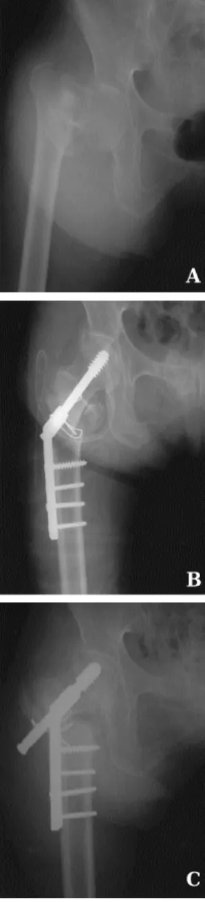
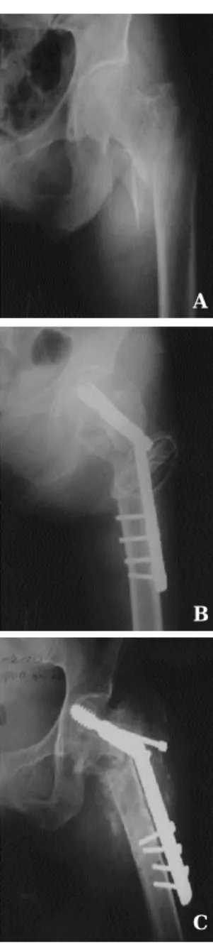
불안정 대퇴골 전자간 골절 4 4례중 8례( 1 8 % )에서 내고정술실패를보였다. 내고정술실패를보인 8례 중 3례( 6 . 8 % )에서가압나사못의대퇴골두천공을동 반한근위골절편의내반변형 ( Fig. 2), 4례( 9 . 1 % )에서 과도한가압나사못의외하방돌출을동반한근위골 절편의내반변형 (Fig. 3 ), 나머지1례( 2 . 3 % )에서는금 속판고정나사못의고정실패를나타내었다( Fig. 4).
고 찰
대퇴골전자간 골절에서 치료의원칙은수술을통 한조기재활및 기능회복에있으며, 현재대퇴골전 자간 골절 치료에 있어 가장 널리 사용되는 활강 압 박 고 나사는 나사의 끝이 무디며 외측 나사 지름이 크다는장점이있어, 이론적으로근위골절편의고정 의 향상과 triflanged 나사못에서의 예리한 모서리를 제거함으로압박고나사의대퇴골두관통의위험성 을감소시켜, 많은연구들에서좋은결과를보여주고 있다6 , 8 , 1 0 , 1 2 , 1 3 , 1 4 , 1 5 ).
‥
Table 3. Displacement of posterior cortex of distal fragment in axial view and fixation failure Reduction fixation failure
* %
Anatomical 4/30 13.3%
Anterior(5mm) 4/10 40%
Posterior(5mm) 0/4 0%
(p > 0.5) (*=No. of fixation failure / No. of cases)
Table 4. Displacement of posterior cortex of distal fragment in axial view and fixation failure Reduction fixation failure
* %
Ⅰ 0/0 0%
Ⅱ 3/4 75%
Ⅲ 4/7 57.1%
Ⅳ 1/15 6.6%
Ⅴ 0/16 0%
Ⅵ 0/2 0%
(p < .0001) (*=No. of fixation failure / No. of cases)
3 / 4
5 / 30 0 / 2 0 / 5
0 / 2 0 / 1
SUPERIOR
INFERIOR
ANTERIOR POSTERIOR
Figure 1. Location of the lag screw in femoral head and fixation failure
( P > 0.5 ) (No.of fixation failure / No.of cases)
그러나, 활강 압박 고 나사를 이용한 대퇴골 전자 간 골절의 내 고정술은 언제나 좋은 결과를 얻을 수 있는 간단한 술식은 아니며7) 불안정한 대퇴골 전자 간골절에서내고정술실패율은약 2 0 %까지보고된 바있다.
대퇴골 전자간 골절의 분류는 생역학적으로나 임 상적으로대부분안정성및불안정성골절로분류되 며, 1949년 E v a n s1 7 )는 골절 형태의 안정성 및 불안정 성 골절의 안정한 정복 전환 가능성에 근거한 분류
체계를통해대퇴골전자간골절에대한이해에많은 공헌을했으며정복시안정성에대한주요한요인이 후내측 피질골의 연속성 회복이라는 사실을 발견하 여후내측피질골의상태에따라대퇴골전자간골절 을안정성및불안정성골절로분류하였다.
L a s k i n등9 )에의하면대퇴골전자간골절의불안정
성은소전자 내측 b u t t r e s s의 분쇄골절이있을때발생 하며, 대퇴 전자간 골절의 안정성 혹은 불안정성은 수술전방사선사진에서소전자의분쇄와전위로예 Fig 2-A. Preoperative
roentgenogram of 76 years old female, Evans classification ( displaced not reduced ) and Singh index Ⅲ.
Fig2-B. Immediate postoperative roentgenogram shows
medialization ( >
5mm ) of medial cortex of the distal fragment.
Fig2-C. Postoperative 6 months follow up roentgenogram shows varus collapse of the proximal fragment with cutout of the lag screw.
A
B
C
Fig 3-A. Preoperative roentgenogram of 65 years old female, Evans classification ( comminuted ) and Singh index
Ⅱ.
Fig3-B. Immediate postoperative roentgenogram shows
medialization ( >
5mm ) of medial cortex of the distal fragment.
Fig3-C. Postoperative 3 months follow up roentgenogram shows varus collapse of the proximal fragment with excessive sliding of the lag screw.
A
B
C
상이가능하나불안정에대한특징적인소견은아니 며, 안정성에대한확실한 평가는 술 중근위대퇴골 의후내측부위의촉지에의해서가능하다하였다.
술후골절정복의정확도는근위대퇴골의후내측 피질골의정확한해부학적복구가이상적이며, Davis 등2 )은술후촬영한방사선전후면및측면사진상원 위 골편의 근위 골편에 대한 5mm 이상의 전위가 있 을 경우 내 고정술 실패가 증가한다 하였으나 원위 골편의전위방향에따른고정실패에대해선언급이
없었다. 이에저자는내고정술실패가일어난 8례에 서 술 후골절 정복 상태를분석하였으며 원위 골편 의근위 골편에 대한 내측 전위가 5mm 이상인 경우 에서유의하게내고정술실패가증가함을발견하였 다.
대퇴골전자간골절치료시근위부골편의견고한 고정은 해면 골 자체의 골밀도에 달려 있으므로 골 다공증은 치료 결과에 중요한 인자라고 생각되며, L a r o s등1 1 )은 골 다공증이 동반된 대퇴골 전자간 안 정 골절과 골 다공증이 없는 불안정 대퇴골 전자간 골절간의내고정술실패율을비슷하게발표함으로 써, 골 다공증의 중요성을 강조하였다. 본 논문에서 도 골 다공증의 정도가 심할수록 내 고정술 실패가 유의하게증가함을보였다.
C l e v e l a n d등4 )은 압박 고 나사의 대퇴 골두내 위치 가전상방 및상방또는전방위치에있을때대퇴골 두 천공의 발생 가능성이 많아 내 고정술 실패율이 높다고 하였으나, 본 논문에서는 대퇴 골두내 내 고 정물의위치와합병증발생과의관계에서유의한상 관관계를유추할수없었으며 , 가압나사못의이상 적 위치에 대해서는 아직도 논란의 대상이 되고 있 다1 6 ).
결 론
본연구에서나타난불안정대퇴골전자간골절에 대해시행한활강압박고나사고정술후내고정술 실패로는 가압 나사못의 대퇴 골두 천공을 동반한 근위 골절편의 내반 변형, 과도한 압박 고 나사못의 외하방 돌출을 동반한 근위 골절편의 내반 변형 및 금속판 고정 나사못의 고정 실패등이었으며, 활강 압박 고 나사 고정술을 이용한 불안정 대퇴골 전자 간골절치료시후내측골편의부적절한해부학적고 정 및 골 다공증의 정도가내 고정술 실패의가장 중 요한원인으로사료되며, 불안정골절치료시후내측 골편의 정확한 해부학적 고정이 필요하며, 또한 내 고정시 가압 나사못을 고정 시킬 대퇴골 근위부 외 측면의골절이 예상되는 경우에는근위대퇴부골수 강내고정술등을고려하여야할것이다.
Fig 4-A. Preoperative roentgenogram of 67 years old female, Evans classification ( displaced not reduced ) and Singh index Ⅲ.
Fig4-B. Immediate postoperative roentgenogram shows
medialization ( >
5mm ) of medial cortex of the distal fragment.
Fig4-C. Postoperative 1 month follow up roentgenogram shows loss of fixation of the plate-holding screws.
A
B
C
R E F E R E N C E
1. Baumgaertner MR, Chrostowski JH, Levy RN.:
Intertrochanteric hip fractures. In: Browner BD, Levine AM, Jupiter JB, et al., eds. Skeletal trauma, vol 2. Philadelphia: WB Saunders, 1992:1833-1881.
2. Davis, T.R.C., Sher, J.L., Horsmam, A., Simpson, M., Porter, B.B. and Chekett, R. G. : I n t e r t r o c h a n t e r i c femoral fractures : mechanical failure after internal fixation. J Bone Joint Surg, 72-B: 26-31,1990.
3. Singh M, Nagrath A and Maini P : Changes in Trabecular Pattern of the Upper End of the Femur as an Index of Osteoporosis. J Bone Joint Surg, 52A:
4 5 7 - 4 6 7 , 1 9 7 0
4. Cleveland, M., Bosworth, D.M., Thompson, F.R., Wilson, H.J. and Ishizuka, T. : A ten-year analysis of intertrochanteric fractures of the femur. . J Bone Joint Surg, 41-A: 1399-1408,1959.
5. Kim WY, Han CH, Park JI : Failure of intertrochanteric fracture fixation with a dynamic hip screw in relation to pre-operative fracture stability and osteoporosis. Int Orthop 2001;25(6):360-2
6. Kyle RF, Cabanela ME, Russel TA, et al. : Fractures of the proximal part of the femur. Instr Course Lect 1995;44:227-253.
7. Wolfgang, G.L., Bryant, M.H. and O’Neill, J.P. : Treatment of intertrochanteric fracture of the femur using sliding screw plate fixation : Clin. Orthop., 163:148-158, 1982
8. Zuckerman JD. : Comprehensive care of orthopaedic injuries in the elderly. Baltimore: Urban
and Schwarzenberg, 1990.
9. Laskin R, Gruber M and Zimmerman A : Intertrochanteric fractures of the hip in the elderly.
Clin. Orthop., 141:188-195, 1979
10. Zuckerman JD. Hip fracture. N Engl J Med 1 9 9 6 ; 3 3 4 : 1 5 1 9 - 1 5 2 5 .
11. Laros G and Moore J : Complication of fixation in intertrochanteric fractures. Clin. Orthop., 101:110- 119, 1974
12. Moon Myung Sang, Kim In , Chung Young Bok : A Clinical Study on Trochanteric Fractures of the Femur. J Korean Orthop Assoc. 12-2: 147-152, 1 9 7 7 .
13. Lee Han Koo, Chung Moon Sang Yang Young S i k : Failed Hip Nailing in Hip Fractures- A Radiological Analysis. J Korean Orthop Assoc. 11- 3: 531-541, 1976.
14. Bonarno, J.J., and Accettola, A.B. : Treatment of intertrochanteric fractures with a sliding nail-plate. J.
Trauma, 22-3 :205-215,1982
15. Jocobs, R.R., McClain, O., and Amstrong, H.J. : Internal fixation of intertrochanteric hip fractures : A clinical and biomechanical study, Clin. Orthop., 146:62-70, 1980
16. DeLee JC. : Fractures and dislocation of the hip. In:
Rockwood CJ,Jr. Green DP, eds. Fractures in adults.
2 nd ed. Vol. 2. Philadelphia, etc : JB Lippincott Co, 1984 : 1211-356
17. Evans E. : The treatment of trochanteric fractures of the femur. J Bone Joint Surg 1949;31B:190-203.
Common Modes of Fixation Failure with a Sliding Hip Screw encountered Unstable Intertrochanteric Fracture
Byung Soon Kim, M.D., Duck Yun Cho, M.D., Hyung Ku Yoon, M.D., Dong Eun Sin, M.D. Soo Hong Han, M.D., Jae Hwa Kim, M.D.,
Dong Jun Kim, M.D.
Department of Orthopedic Surgery,
Pundang CHA Hospital, College of Medicine, Pochon CHA University, Sungnam, Korea
The purpose of this study was to evaluate the common modes of fixation failure in unstable intertrochanteric fractures , related risk factors and the prevention of fixation failure.
Between 1995 and 2001, 44 patients who had sustained an unstable intertrochanteric fractures were assigned to be treated with a sliding hip screw. Men in 14 cases ( 32% ), women in 30 cases ( 68% ) , the average age at the operation was 65(22-90) years and the average duration of follow up was 12(8-22) months.
We classified the fracture patterns with Evans system and used Singh’s index for osteoporosis. And we examined the common modes of fixation failure with postoperative X-ray.
The fixation failure in unstable intertrochanteric fracture was 8 cases (18.2 % );
varus collapse of the proximal fragment with cutout of the lag screw was 3 cases (6.8%), varus collapse of the proximal fragment with excessive sliding of the lag screw was 4 cases (9.1%) and loss of fixation of the plate-holding screws was 1 case (2.3%).
The authors think that inadequate anatomical reduction of comminuted posteromedial fragment and severity of osteoporosis are main causes of fixation failure.
During operation for unstable intertrochanteric fractures, the most important point is accurate reduction of posteromedial fragment and the intramedullary hip screw like proximal femoral nail ( PFN ) may be considered to avoid fracture of lateral cortex that enter the lag screw, causing fixation failure.
Key Words : unstable intertrochanteric fractures, fixation failure Abstract
‥
‥