pISSN: 0378-6471⋅eISSN: 2092-9374
http://dx.doi.org/10.3341/jkos.2014.55.7.1099
Case Report
묻힌 시신경 유두 드루젠에서 동반된 시신경 유두 출혈 1예
A Case of Optic Disc Hemorrhage Associated with Buried Optic Nerve Head Drusen
최우곤⋅장지혜
Woo Gon Choi, MD, Ji Hye Jang, MD
대구파티마병원 안과
Department of Ophthalmology, Daegu Fatima Hospital, Daegu, Korea
Purpose: We report a case of optic disc hemorrhage associated with buried optic nerve head drusen in a pediatric patient.
Case summary: A 10-year-old female visited our clinic with a floating sensation in her left eye, 2 days in duration. Best corrected visual acuity was 1.0 in both eyes. Intraocular pressure, light reflex, relative afferent pupillary defect and color vision were normal. The patient showed a small optic disc with blurred, irregular margins in both eyes, and optic disc hemorrhage in the left eye on fundus examination. Visual field examination revealed an enlarged blind spot in the left eye. To achieve correct diagnosis, brain MRI was performed and revealed normal findings. On spectral-domain optical coherence tomography (OCT), hyper-re- flective and heterogeneous mass like lesions were found with buried optic nerve head drusen.
Conclusions: In general, optic nerve head drusen, has a good prognosis; however, visual field defects or hemorrhagic complica- tions can occur, therefore, correct diagnosis and regular follow-up are necessary.
J Korean Ophthalmol Soc 2014;55(7):1099-1105
Key Words: Buried optic nerve drusen, Optic disc hemorrhage, Pediatrics
■Received: 2014. 1. 25. ■ Revised: 2014. 2. 17.
■Accepted: 2014. 6. 9.
■Address reprint requests to Ji Hye Jang, MD
Department of Ophthalmology, Daegu Fatima Hospital,
#99 Ayang-ro, Dong-gu, Daegu 701-724, Korea Tel: 82-53-940-7545, Fax: 82-53-954-7417 E-mail: [email protected]
* This study was presented as an e-poster at the 111th Annual Meeting of the Korean Ophthalmological Society 2014.
ⓒ2014 The Korean Ophthalmological Society
This is an Open Access article distributed under the terms of the Creative Commons Attribution Non-Commercial License (http://creativecommons.org/licenses/by-nc/3.0/) which permits unrestricted non-commercial use, distribution, and reproduction in any medium, provided the original work is properly cited.
시신경 유두 드루젠은 좁은 공막관과 작은 크기의 시신 경 유두에 잘 발생하며, 축삭 대사 이상과 축삭 운반 변성 으로 생성된 점액 다당류 및 점액 단백질 덩어리들로 이루 어져 있다. 나이가 들면서 퇴행성으로 형성되는 황반 드루 젠과는 달리, 시신경 유두 드루젠은 소아에서도 발견이 되 며, 1,000명당 약 3-24명의 빈도로, 75% 이상에서 양측성으 로 관찰된다.1-3
성인의 경우는 칼슘 침착과 드루젠의 크기 증가로 인하 여 보이는 시신경 유두 드루젠(visible optic nerve head dru- sen)이 흔하며, 소아의 경우는 대부분이 묻힌 시신경 유두 드루젠(buried optic nerve head drusen)으로 안저검사에서 시신경 유두 부종과 감별하기가 어렵다.4,5
시신경 유두 드루젠은 증상없이 검사상 우연하게 발견되 는 경우가 대부분이며, 드물게 진행성의 시야 손상과 출혈 성 합병증이 동반되면서 중심시력저하가 발생하여 진단되 기도 한다.4 시신경 유두 드루젠의 약 2-10%에서 시신경 유 두 주변부로의 출혈이 동반되며, Sanders et al5은 동반되는 출혈 양상에 따라 시신경 유두하 출혈, 신경섬유층의 선상 출혈과 시신경 유두 부위에서 기인한 유리체 출혈로 크게 3가지로 구분하여 보고하였다.
국외 논문에는 시신경 유두 드루젠과 동반한 시신경 유 두 출혈에 관한 보고가 있지만 국내에서는 아직 보고된 바
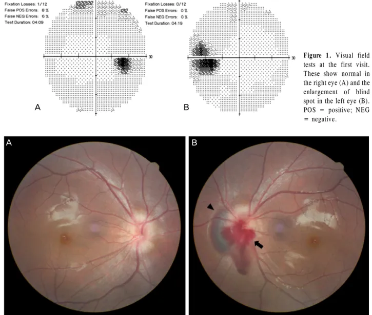
Figure 1. Visual field
tests at the first visit.These show normal in the right eye (A) and the enlargement of blind spot in the left eye (B).
POS = positive; NEG
= negative.
Figure 2. Fundus photographs at the first visit (A, B). These show bilateral small optic disc with blurred, irregular margin, sugges-
tive of optic nerve head drusen. The arrowhead indicates deep peripapillary hemorrhage. The arrow indicates splinter hemorrhage.가 없다. 이에 저자들은 10세 여아의 묻힌 시신경 유두 드 루젠과 동반된 시신경 유두 출혈 1예를 경험하였기에 이를 보고하고자 한다.
증례보고
10세 여아가 2일 전부터 시작된 좌안의 비문증을 주소로 본원 안과를 방문하였다. 본원 방문 당시, 교정시력은 우안 1.0, 좌안 1.0이었으며, 양안 동공크기, 안압, 동공반사, 구 심성동공운동 및 한천석 색각 검사는 정상이었다. 험프리 자동시야검사계(Humphrey Instruments, San Leandro, CA, USA)로 시행한 시야검사상 좌안의 맹점확대가 관찰되었다 (Fig. 1). 산동하여 시행한 안저검사에서는 양안 시신경 유 두 주변부로 경계가 명확하지 않은 흐림이 관찰되었고, 좌
안의 시신경 유두 및 주변부로 망막하 출혈, 선상 출혈 및 유리체 출혈이 관찰되었다(Fig. 2).
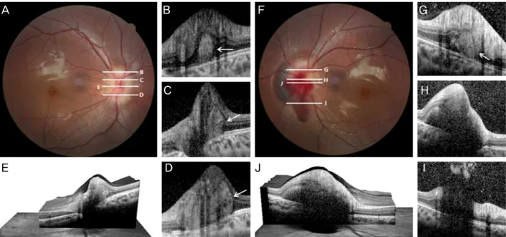
시신경 유두 경계부위의 흐림과 망막출혈의 원인을 감별 하기 위하여 뇌 조영증강 자기공명검사 및 스펙트럼영역 빛간섭단층촬영(Spectralis OCT®, Heidelberg Engineering, Heidelberg, Germany)을 추가로 시행하였다. 뇌 조영증강 자기공명검사는 정상소견이었으나, 스펙트럼영역 빛간섭단 층촬영에서는 좌안의 경우, 선상 출혈, 망막하 출혈 및 유 리체 출혈의 후방으로 그림자 효과에 의한 저반사 공간으 로 관찰되어 망막층의 구조적 변화를 정확히 알 수는 없었 으며, 우안의 경우, 외망막층과 망막색소상피층 사이에 위 치한 고반사의 비균질한 덩어리 형태의 묻힌 시신경 유두 드루젠과 함께 그 주변부로 울퉁불퉁한 망막층 내부의 윤 곽이 관찰되었고, 외망상층과 외과립층을 포함한 망막층의
A B
A B
Figure 3. Images showing multiple buried optic disc drusen within small optic disc of the right eye (A-E) and left eye (F-J). W hite
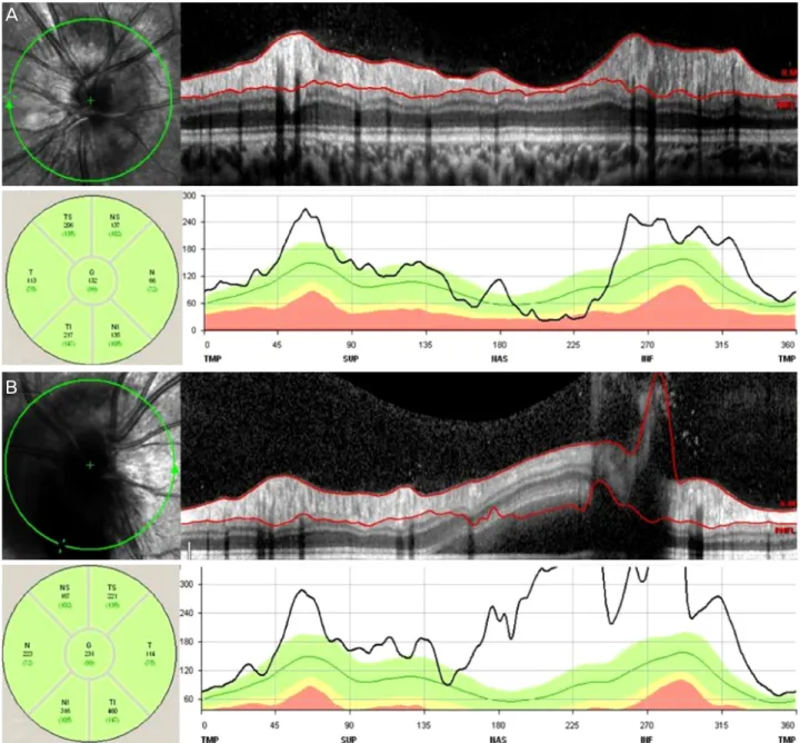
horizontal lines in fundus photographs (A, F) indicate the horizontal cross sections level of the optical coherence tomography (OCT) scan (B-E, G-J). E and J images show three-dimensional image-OCT on center of optic disc. These reveal buried optic nerve head drusen with a subretinal hyperreflective mass like lesion (B-D, G, white arrows) with optic disc elevation in both eyes. Deep peri- papillary hemorrhage and vitreous hemorrhage reflect hyporeflectivity on the subretinal and preretinal space in the left eye.전반적인 융기와 함께 드루젠 후방으로 감각신경망막층이 관찰되지 않는 등의 구조적 변화도 관찰되었다(Fig. 3). 또 한, 시신경 유두 부종과 감별하기 위하여 측정한 시신경 유 두 주변부 망막신경섬유층의 두께는 우안의 경우, 비측이 66 μm로 얇았으며, 이측은 113 μm로 정상보다 두껍게 관 찰되었고(Fig. 4A), 좌안에서는 시신경 유두 드루젠과 동반 된 망막 출혈로 인하여 정확한 망막신경섬유층의 두께를 측정할 수가 없었다(Fig. 4B).
한 달 뒤 본원 안과로 재방문하여 측정한 시력, 안압 등 의 기본적인 검사에서는 정상소견을 보였다. 좌안의 경우, 안저검사를 통하여 망막출혈의 감소를 확인하였고(Fig.
5A), 스펙트럼영역 빛간섭단층촬영으로 측정한 신경섬유층 의 비측 두께는 81.0 µm로 정상 두께와 비슷하거나 약간 얇게 관찰되었으며(Fig. 6), 시야검사상 좌안의 맹점확대가 한 달 전과 비교해서 큰 차이 없이 진행되지 않는 양상을 보여서 치료 없이 정기적으로 본원 안과 외래에서 추적 관 찰하였다(Fig. 5B).
고 찰
본 증례는 소아에서 양안에 묻힌 시신경 유두 드루젠과 동반하여 갑작스런 시신경 유두 출혈이 발생하였고 약물 치료 없이 경과가 호전된 환자로 국내에서는 보고된 적이 없다.
시신경 유두 드루젠은 황반 드루젠과는 달리 소아에서도 발견되는 드루젠이다. 시신경은 망막신경절세포로부터 나 온 축삭들로 이루어져 있으며, 시신경 유두에 모인 후 공막 관을 통해서 안구를 빠져나가게 되는데, 축삭들이 통과하 는 공막관의 크기가 작을수록, 축삭들이 밀집되어 압박을 받게 된다. 이러한 압박효과에 의해서 시신경이 붓게 되면 시신경 유두의 생리적 유두함몰이 없거나 작은 시신경 유 두를 가지게 되며, 축삭의 대사이상 및 축삭 운반의 변성으 로 인하여 발생한 점액 다당류 및 단백질 덩어리들이 시신 경 유두 주위로 축적되면서 시신경 유두 드루젠을 형성하 게 된다.4,6 이러한 드루젠들이 소아에서는 대부분 시신경 유두와 그 주변부의 심층부에 위치하여 묻힌 드루젠의 형 태로 관찰되는 반면에 성인에서는 나이가 들수록 드루젠 이 축적되고 그 범위가 넓어지면서 시신경 유두 경계부 밖 으로 침범하여서 눈에 보이는 드루젠의 형태로 관찰되는 경우가 많다.
시신경 유두 드루젠에 동반되는 대표적인 합병증으로는 시야결손, 시신경 유두 주변부 출혈 및 맥락막신생혈관의 생성이 있다.3-7 시야 결손을 일으키는 기전으로 제시되는 것은 시신경 유두 드루젠이 혈관들을 압박할 때 발생되는 허혈 현상으로 인한 기전과 시신경이 작은 공막관을 통과 할 때 받는 압박으로 발생한 축삭 운반 및 대사의 장애에 의한 기전이 있다.4 허혈 현상으로 인한 경우는 갑작스런 시야 상실이 발생할 수도 있는데, 대표적인 예로 앞허혈시
A B F G
E D J I
C H
Figure 4. Retinal nerve fiber layer (RNFL) measurement using spectralis optical coherence tomography (OCT) of both eyes. (A)
The RNFL thickness is increased in all sections except for nasal section by the underlying buried optic nerve head drusen in the right eye. (B) Because of deep peripapillary hemorrhage in the left eye, the RNFL thickness cannot be measured accurately. ILM = inner limiting membrane; TS = superotemporal; NS = superonasal; T = temporal; G = general; N = nasal; TI = inferotemporal; NI = inferonasal; TMP = temporal; SUP = superior; NAS = nasal; INF = inferior.신경병증과 망막중심동맥폐쇄 후에 발생되는 시야 결손이 있는데 반해서, 축삭의 운반 및 장애로 인한 경우는 서서히 진행하는 시야결손으로 나타나는 경우가 대부분이다. 또 다른 합병증으로 망막 출혈, 유리체 출혈과 같은 출혈성 합 병증이 있으며, 점차 커진 드루젠이 시신경 주변의 혈관들 을 직접 손상시켜 출혈을 일으키는 경우와 시신경 유두 주 위의 혈액순환 장애에 의한 허혈로 인하여 맥락막신생혈관 의 생성을 촉진시켜 망막하 출혈이 발생하는 경우가 있다.7 시신경 유두 주위에만 국한된 출혈성 합병증이 있는 경우
에서는 별다른 자각 증상이 없는 경우가 대부분이지만, 출 혈이 황반 중심부까지 영향을 미치는 경우에는 시력저하 등의 증상이 동반된다. 본 증례의 경우도 묻힌 시신경 유두 드루젠과 동반하여 시신경 유두 주변부로 다양한 형태의 망막출혈이 관찰되었지만, 황반 중심 부위까지 영향을 주 지 않아서 시력에는 영향이 없었으며, 비문증의 증상과 함 께 시야검사에서 좌안의 맹점확대가 확인되었다.
시신경 유두 드루젠의 진단하는 대표적인 방법으로는 안 저검사, B-scan 초음파검사, 컴퓨터 단층촬영, 형광안저촬
A
B
Figure 5. Fundus photograph and visual field test after 1 month. (A) Fundus photograph shows the disappearance of peripapillary
hemorrhage. (B) Visual field test shows the enlargement of blind spot, not much change comparing with visual field test of the first visit.POS = positive; NEG = negative.
Figure 6. Retinal nerve fiber layer (RNFL) measurement using spectralis optical coherence tomography (OCT) of the left eye after
the disappearance of peripapillary hemorrhage, after 1 month. The RNFL thickness is increased in all sections except for nasal sec- tion by the underlying buried optic nerve head drusen. ILM = inner limiting membrane; TS = superotemporal; NS = superonasal;T = temporal; G = general; N = nasal; TI = inferotemporal; NI = inferonasal; TMP = temporal; SUP = superior; NAS = nasal;
INF = inferior.
영 및 빛간섭단층촬영이 있다. 과거에 칼슘 침착물이 동반 된 시신경 유두 드루젠에 있어서 B-scan 초음파가 가장 정 확한 진단 방법이었지만, 최근에는 빛간섭단층촬영을 이용 함으로써, 비침습적인 방법으로도 짧은 시간에 시신경 유 두 후방까지도 정확한 관찰이 가능하다는 점에서 시신경 유두 드루젠의 진단 도구로 각광받고 있다.1,2,4,8 특히, 본 증
례와 같은 묻힌 시신경 유두 드루젠의 경우는 시신경 부종 과의 감별이 어렵다는 점에서, 시신경 주변부 망막신경섬 유층의 두께 값을 이용할 경우에 시신경 유두 부종과의 감별에 도움을 줄 수 있다는 장점이 있다.1,2 Lee et al2과 Johnson et al8에 의하면, 스펙트럼영역 빛간섭단층촬영으 로 측정한 망막신경섬유층의 비측 두께가 시신경 유두 드
A B
루젠과 시신경 유두 부종을 감별하는 데 있어서 가장 중 요한 요소라고 보고한 바 있다. Lee et al2은 비측 망막신 경섬유층의 두께가 78.0 µm 이하일 경우는 시신경 유두 드루젠을 시신경 유두 부종과 구분하는 데 있어서 80%의 민감도와 88.9%의 특이도를 가진다고 하였으며, Johnson et al8은 비측 망막신경섬유층의 두께를 86.0 µm를 기준으 로 하여 감별할 경우는 63%의 민감도를 가진다고 보고하 였다. 본 증례의 경우에서도 스펙트럼영역 빛간섭단층촬 영으로 측정한 망막신경섬유층의 두께가 우안의 경우 비 측만 66 µm로 얇게 관찰되었으며, 좌안도 첫 내원 당시에 는 망막출혈로 인한 그림자 효과로 망막신경섬유층의 정 확한 두께를 측정하지 못했으나, 한 달 뒤 시행한 추적 검 사에서 좌안의 비측 두께가 81.0 µm로 정상 두께와 비슷 하거나 약간 얇게 관찰되어서, 양안 모두 시신경 유두 부 종과 감별하여 시신경 유두 드루젠으로 쉽게 진단할 수 있 었다.
시신경 유두 드루젠의 치료에 대해서는 현재까지 알려 진 것이 없으나, 시신경 유두 드루젠의 크기와 수는 점점 증가하고, 이로 인해 시신경 손상이 서서히 진행하는 것으 로 알려졌다. 성인에 이르면 약 25-75%에서 시야결손이 관찰되며, 시신경 유두 드루젠의 수와 안압 상승 정도에 따라 시야 변화가 급격히 변할 수 있다.9 시신경 유두 모 양의 변화에 따른 시야손상이 관찰되는 경우에는 정기적 으로 시야 변화 및 망막신경섬유층 두께 변화를 관찰해야 한다. 또한 고안압이 존재할 시 안압하강제를 사용하여 시 신경 축삭과 시경절 세포의 변성을 최소화하여야 한다. 드 물게 시신경 유두 허혈로 인해 맥락막신생혈관이 발생할 수 있는데, 이 경우에는 광역학치료10나 항혈관내피세포성 장인자주입술11을 시행하여 신생혈관에 의한 시력 저하를 방지할 수 있다. 그러므로, 본 증례와 같이 시신경 유두 드 루젠에서 망막 출혈과 같은 출혈성 합병증이 동반되거나, 시야검사에서 드루젠에 의한 시야손상이 관찰된 경우는
추가적인 합병증 및 시야 결손의 진행을 예방하기 위해 정기적인 시력측정과 안압 측정 등의 기본적인 안과검사 는 물론이고 시야검사, 스펙트럼영역 빛간섭단층촬영 및 형광안저혈관조영술 등의 정기적인 추적검사가 도움이 될 것이다.
REFERENCES
1) Lee KM, Woo SJ, Hwang JM. Morphologic characteristics of optic nerve head drusen on spectral-domain optical coherence tomography.
Am J Ophthalmol 2013;155:1139-47.
2) Lee KM, Woo SJ, Hwang JM. Differentiation of optic nerve head drusen and optic disc edema with spectral-domain optical coher- ence tomography. Ophthalmology 2011;118:971-7.
3) Neffendorf JE, Mulholland C, Quinlan M, Lyons CJ. Disc drusen complicated by optic disc hemorrhage in childhood. Can J Ophthalmol 2010;45:537-8.
4) Auw-Haedrich C, Staubach F, Witschel H. Optic disk drusen. Surv Ophthalmol 2002;47:515-32.
5) Sanders TE, Gay AJ, Newman M. Hemorrhagic complications of drusen of the optic disk. Am J Ophthalmol 1971;71:204-17.
6) Mullie MA, Sanders MD. Scleral canal size and optic nerve head drusen. Am J Ophthalmol 1985;99:356-9.
7) Harris MJ, Fine SL, Owens SL. Hemorrhagic complications of op- tic nerve drusen. Am J Ophthmol 1981;92:70-6.
8) Johnson LN, Diehl ML, Hamm CW, et al. Differentiating optic disc edema from optic nerve head drusen on optical coherence tomography. Arch Ophthalmol 2009;127:45-9.
9) Grippo TM, Shihadeh WA, Schargus M, et al. Optic nerve head drusen and visual field loss in normotensive and hypertensive eyes.
J Glaucoma 2008;17:100-4.
10) Chaudhry NA, Lavaque AJ, Shah A, Liggett PE. Photodynamic therapy for choroidal neovascular memebrane secondary to optic nerve drusen. Ophthalmic Surg Lasers Imaging 2005;36:70-2.
11) Knape RM, Zavaleta EM, Clark CL 3rd, et al. Intravitreal bev- acizumab treatment of bilateral peripapillary choroidal neo- vascularization from optic nerve head drusen. J AAPOS 2011;
15:87-90.
= 국문초록 =
묻힌 시신경 유두 드루젠에서 동반된 시신경 유두 출혈 1예
목적: 소아에서 묻힌 시신경 유두 드루젠에 동반된 시신경 유두 출혈 1예를 경험하였기에 이를 보고하고자 한다.
증례요약: 10세 여아가 2일 전부터 시작된 좌안의 비문증을 주소로 내원하였다. 내원 당시 교정시력은 우안 1.0, 좌안 1.0이었고, 안압, 동공반사, 구심성동공운동 및 한천석 색각 검사는 정상이었다. 안저검사에서 양안의 시신경 유두 경계부위의 불규칙한 흐림과 좌안의 시신경 유두 출혈이 관찰되었고, 시야검사에서 좌안의 맹점확대가 관찰되었다. 시신경 유두 경계부위 흐림의 원인을 감별진단하기 위해 시행한 뇌 자기공명영상 결과 정상이었고, 스펙트럼영역 빛간섭단층촬영에서 고반사의 비균질한 덩어리의 형태로 묻힌 시신경 유두 드루젠이 관찰되었다.
결론: 시신경 유두 드루젠은 일반적으로 예후가 좋으나 진행하는 시야 장애나 출혈성 합병증을 일으킬 수 있으므로 정확한 진단과 추적관찰이 필요하다.
<대한안과학회지 2014;55(7):1099-1105>