218
Prognostic Significance of Immunohistochemical Expression of p53 Gene Product in O perable Breast Cancer
Hong Suk Song, M.D.
1, Young Rok Do, M.D.
1, Sun Hee Kang, M.D.
2, Ki Yong Jeong, M.D.
2and Yu Sa Kim, M.D.
2Departments of
1Internal Medicine and
2General Surgery, Dongsan Medical Center, Keimyung University School of Medicine, Daegu, Korea
Purpose: The aim of this study was to investigate the prognostic significance of the expression of p53 gene product in operable invasive breast cancer by performing immunohistochemical analysis.
M aterials and M ethods: Between January 1993 and December 2001, 440 operable invasive breast cancer patients underwent immunohistochemical staining for p53, and we retrospectively analyzed these results to- gether with the clinical outcomes.
Results: The overexpression of p53 was detected in 51.6% of the cases. The overexpression of p53 was inver- sely correlated with lymph node metastasis (p=0.005).
The tumor size, tumor histology, histologic grade, hor- monal receptor status and tumor stage were not related to the overexpression of p53. M ultivariate Cox regression analysis indicate that lymph node metastasis, tumor size and the p53 expression were the significant prognostic factors for overall survival; lymph node metastasis, the
estrogen receptor status and the p53 expression were the significant prognostic factors for relapse free survival. O n the subgroup analysis, the p53 non-expressors showed better 7-year overall survival (92.7% vs. 76.7% , respec- tively, p=0.011) and relapse free survival (74.9% vs. 57.8% , respectively, p=0.032) than did the p53 overexpressors for the patients with lymph node metastasis. However, for the patients without lymph node metastasis, the survival rates were not different for both the p53 non-expressors and the p53 overexpressors.
Conclusion: Immunohistochemical staining of the p53 gene product was an independent prognostic factor for predicting survival of the lymph node positive invasive breast cancer patients. (Cancer Res Treat. 2006;38:218- 223)
Key W ords: Breast neoplasms, Prognosis, p53, Immu- nohistochemistry
Correspondence: Hong Suk Song, Department of Internal Medicine, Dongsan Medical Center, Keimyung University School of Medi- cine, 194, Dongsan-dong, Jung-gu, Daegu 700-712, Korea. (Tel) 82-53-250-7436, (Fax) 82-53-425-6476, (E-mail) shs7436@
dsmc.or.kr
Received August 20, 2006, Accepted October 18, 2006
This study was supported by a grant of the Korean Health 21 R&D Project, Ministry of Heath & Welfare, Republic of Korea (0412-CR01- 0704-0001).
INTRODUCTION
Breast cancer has become the most common cancer for Kore- an females since 2002. A population-based cancer registry was established on January 1, 1997 to estimate the incidence of can- cer in Daegu, Korea. The age-standardized incidence rates (ASR) of breast cancer were 31.1 per 100,000 females as re- ported by the Daegu Cancer Registry in 2002. The median age of Korean breast cancer patients (early 40's) is younger than that in the Western developed countries, and the incidence of familiar breast cancer is relatively rare in Korea.
Great insight has recently been gained into the biological properties of tumor cells. The products of tumor suppressor ge- nes are of specific interest as they play important roles in a variety of critical and highly conserved cellular functions, in- cluding regulation of the cell cycle and apoptosis, differentia- tion, surveillance of genomic integrity and repair of DNA er- rors, signal transduction and cell adhesion. The p53 tumor suppressor gene normally regulates cell proliferation (1) and programmed cell death (2).
Abnormalities of the p53 tumor suppressor gene have been implicated in both tumorigenesis and tumor progression. The importance of the p53 expression has been extensively analyzed by immunohistochemical methods with regard to a plethora of human malignancies, including breast cancer, yet the role of p53 protein in breast cancer is incompletely understood. The prognostic roles of the p53 gene expression with regard to breast cancer remain controversial, and p53 mutation would be not a feasible prognostic marker in the routine diagnostic evaluation of breast cancer.
The purpose of this study was to determine the prognostic significance of immunohistochemical staining the p53 expre- ssion in operable breast adenocarcinoma.
MATERIALS AND METHODS
1) Patients and setting
From January 1993 to December 2001, we performed immu- nohistochemical analyses of p53 gene product in 440 of the 813 operable invasive breast cancers at our hospital. We reviewed the clinicopathologic parameters, the size of the tumor, the pathologic type, histologic grade, nuclear grade, hormone re- ceptor status and the TNM stage. Staging evaluation was done by the guidelines of the American Joint Committee on Cancer, 5th edition.
2) Immunohistochemical staining
Immunohistochemical staining was performed using the avidin- biotin-peroxidase complex with monoclonal antibodies created against p53 (NCL-p53-DO7, Novocastra Laboratories, Newcas- tle, UK). Representative paraffin blocks containing the tumors isolated from each case were sectioned into 5μm slices and these were affixed to slides; they were then dried for 1 hour at 60oC. The sections were deparaffinized in xylene and next rehydrated with a descending series of alcohol solutions. The endogenous peroxidase activity was blocked by 3% hydrogen peroxidase for 15 minutes, and this was followed by washing with phosphate buffered saline (PBS), at a pH of 7.2. The sec- tions were then subjected to a heat antigen retrieval process by autoclaving them with 1% zinc sulfate solution for 5 minutes.
After cooling for 20 minutes at room temperature, the sections were incubated with 10% normal horse serum (Vectastatin Elite kit) for 30 minutes. After decanting away the excess serum, the sections were incubated with primary antibody for 2 hours at 37.0oC. For the p53 study, DO7 monoclonal antibody was used at a 1:100 dilution (Novocastra, Newcastle, UK). The sections were subsequently incubated with prediluted biotinylated anti- mouse immunoglobulin (Vectastain Elite kit) for 30 minutes at 37oC. After washing with PBS, the sections were reacted with peroxidase-conjugated streptoavidin (Dako) at a dilution of 1:
500 for 30 minutes at 37oC. After washing with PBS, the per- oxidase activity was evaluated with 3,3'-deaminobenzidine te- trahydrochloride (DAB), and the sections were counterstained with Meyer's hematoxylin.
Two pathologists, who were kept “blinded” to the clinical outcomes and features of the patients, independently evaluated all the immunohistochemical slides. The tumors with positive nuclear staining in more than 1% of tumor cells were inter- preted as having a positive expression.
3) Statistical analysis
Statistical analysis was performed using chi square tests to compare percentages in the cross tabulations, and independent sample T tests were used to compare the means. Survival curves were generated by the Kaplan-Meier method, and they were compared by using the log-rank test. To determine the significant prognostic factors related to overall survival and relapse-free survival (including all of the loco-regional recur- rences and distant metastasis), multivariate analysis was per- formed with the Cox proportional hazards regression model. All
the significance levels refer to two-sided tests. p values less than 0.05 were considered significant.
Statistical analyses were performed using SPSS for Windows 12.0.
RESULTS
1) Patients characteristics
All of the patients were female and the median age of the subjects was 47.8 years. The mean diameter of the tumors was 3.1 cm (range: 0.0~15.0 cm). Histologically, infiltrating ductal cancer was most common (388 patients, 88.2%), and lobular carcinoma was noted in 19 patients (4.3%), medullary carcino- ma was noted in 15 patients (3.4%), mucinous carcinoma was noted in 12 patients (2.7%), papillary carcinoma was noted in 7 patients (1.6%), and tubular carcinoma was noted in 12 (2.7%) patients. Mastectomy was performed for 367 patients (83.4%) and breast conserving surgery with radiotherapy was performed for 73 patients (16.6%). Adjuvant hormonal therapy was performed in 298 patients (67.7%) and adjuvamt chemo- therapy was performed in 366 patients (83.2%) (Table 1). Adju- vant chemotherapy was performed with the cyclophosphamide +methotrexate+5-FU (CMF) regimen for 239 patients (65.3%), with adriamycin+cyclophosphamide (AC) for 79 patients (21.6%), with 5-FU+epirubicin+cyclophosphamide (FEC) for 40 patients (10.9%), and with other combination chemothera- pies for 8 patients.
2) Correlation with clinicopathologic parameters The staging was as follows; 168 patients were the T1 stage (40.4%), 214 were T2 (51.4%), 32 were T3 (7.7%), and 2 were T4 (0.5%), 214 patients were N0 (48.6%), 226 patients were N1 (51.4%), and stage I was noted in 107 patients (24.3%), stage IIA was noted in 171 patients (38.9%), stage IIB was noted in 138 patients (31.4%), and stage IIIA was noted in 24 (4.4%) patients. There were 290 patients (66.1%) in the estro- gen receptor (ER) positive or progesterone receptor (PR) posi- tive group (66.1%) and 149 patients (33.9%) were in the ER- PR-group.
Positive staining for p53 were found in 227 of the 440 examined tumor samples (51.6%). Lymph node metastasis was more common in the p53 non-expressors (58.2%) than in the p53 overexpressors (44.9%) (p=0.005) (Table 1). No significant correlation were observed with regard to the overexpression of p53 and the tumor size, histologic grade, nuclear grade, vascular invasion, tumor stage or hormonal status.
3) Survival analysis
The 7-year overall survival rate and 7-year relapse-free sur- vival rate of the total patient population were 90.7%, and 78.2%, respectively. According to the stage, the 7-year overall survival rate of stage I was 97.9%, it was 94.8% for stage IIA, 82.4% for stage IIB and 75.6% for stage III (p=0.000). The 7-year overall survival rate of the PR positive patients were better than that of the ER negative patients (93.4% vs. 86.7%, respectively, p=0.019), and the 7-year relapse-free survival rate of the ER positive patients was better than that for the ER
Table 1. Clinical characteristics of the operable breast cancer patients according to the p53 expression
p53 positive (n=227) p53 negative (n=213) p value
Age (mean±SD) 46.8±10.6 49.0±11.0 0.034
Tumor size (cm) 3.2±2.0 3.0±2.0 0.281
Histology Infiltrating ductal (NOS) 204 (89.9%) 184 (86.4%) 0.143
Invasive lobular 7 (3.0%) 12 (5.6%)
Medullary 8 (3.5%) 7 (3.3%)
Mucinous 3 (1.3%) 9 (4.2%)
Papillary 4 (1.8%) 3 (1.4%)
Tubular 4 (1.8%) 8 (3.8%)
Histologic grade 1 4 (3.2%) 9 (6.6%) 0.231
2 41 (32.8%) 52 (38.2%)
3 80 (64.0%) 75 (55.1%)
Nuclear grade 1 7 (5.4%) 17 (12.1%) 0.085
2 46 (35.4%) 54 (38.3%)
3 68 (52.3%) 56 (39.7%)
4 9 (6.9%) 14 (9.9%)
T 1 92 (43.2%) 76 (37.4%) 0.635
2 103 (48.4%) 111 (54.7%)
3 17 (8.0%) 15 (7.4%)
4 1 (0.5%) 1 (0.5%)
N 0 125 (55.1%) 89 (41.8%) 0.005
1 102 (44.9%) 124 (58.2%)
Stage I 67 (29.5%) 40 (18.8%) 0.060
IIA 83 (36.6%) 88 (41.3%)
IIB 64 (28.2%) 74 (34.7%)
IIIA 13 (5.7%) 11 (5.2%)
ER+ or PR+ 140 (61.9%) 150 (70.4%) 0.276
ER-PR- 86 (38.1%) 63 (29.6%)
Operation Mastectomy 184 (81.4%) 182 (85.4%) 0.139
Breast conserving surgery 42 (18.6%) 31 (14.6%)
Hormonal therapy 156 (73.2%) 142 (62.6%) 0.017
Adjuvant chemotherapy 192 (84.6%) 174 (81.7%) 0.418
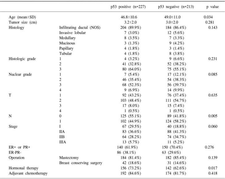
Fig. 1. Kaplan-Meier overall survival curve in operable breast can- cer patients according to p53 expression.
negative patients (81.3% vs. 74.6%, respectively, p=0.035). The 7-year overall survival rate of patients with p53 overexpression was worse than that of patients with p53 non-expression (87.1%
vs. 94.3%, respectively, p=0.041, Fig. 1), but there was no significant differences in terms of the relapse-free survival rate (p=0.072) between the patients with p53 overexpression and the patients with p53 non-expression.
According to multivariate Cox regression analysis, lymph node metastasis, tumor size and p53 expression (hazard ratio=3.1, 95% CI: 1.02-9.44, p=0.046) were the important prognostic factors for the overall survival rates, and lymph node metas- tasis, ER status and p53 expression (hazard ratio=1.8, 95% CI:
1.15-2.82, p=0.011) were the important prognostic factors for the relapse-free survival rates (Table 2, 3).
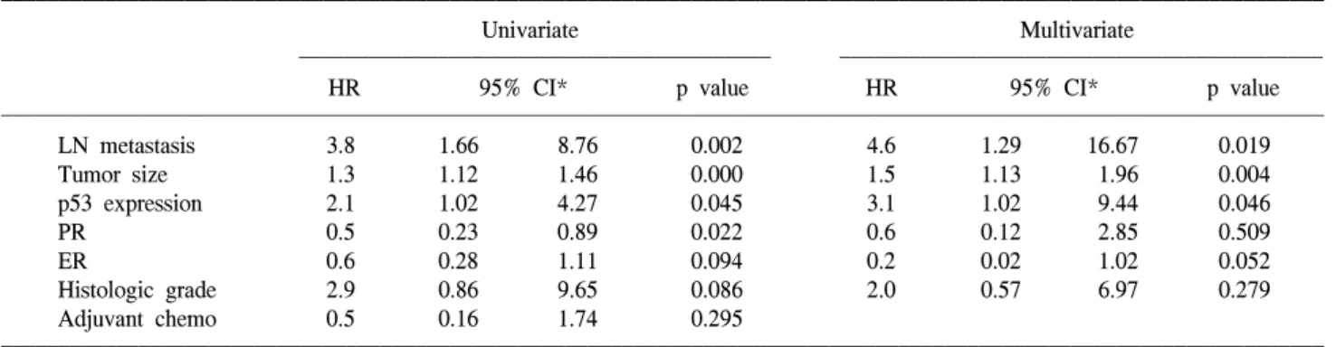
On the subgroup analysis, the p53 non-expressors had better 7-year overall survival and relapse free survival rates than did the p53 overexpressors for the patients with lymph node metas- tasis (7-year OS: 92.7% vs. 76.7%, respectively, p=0.011, 7- year RFS: 74.9% vs. 57.8%, respectively, p=0.032), but there were no significant differences of the overall survival and
Table 2. Cox regression analysis of overall survival for the operable breast cancer patients
Univariate Multivariate
HR 95% CI* p value HR 95% CI* p value
LN metastasis 3.8 1.66 8.76 0.002 4.6 1.29 16.67 0.019
Tumor size 1.3 1.12 1.46 0.000 1.5 1.13 1.96 0.004
p53 expression 2.1 1.02 4.27 0.045 3.1 1.02 9.44 0.046
PR 0.5 0.23 0.89 0.022 0.6 0.12 2.85 0.509
ER 0.6 0.28 1.11 0.094 0.2 0.02 1.02 0.052
Histologic grade 2.9 0.86 9.65 0.086 2.0 0.57 6.97 0.279
Adjuvant chemo 0.5 0.16 1.74 0.295
*confidence interval.
Table 3. Cox regression analysis of relapse free survival for the operable breast cancer patients
Univariate Multivariate
HR 95% CI* p value HR 95% CI* p value
Lmph node meta 3.5 2.07 5.79 0.000 3.6 2.11 6.31 0.000
Tumor size 1.2 1.05 1.28 0.005 1.1 0.99 1.24 0.063
Adjuvant chemo 0.4 0.18 0.96 0.040 0.9 0.37 2.14 0.790
p53 1.5 0.96 2.32 0.074 1.8 1.15 2.82 0.011
ER 0.6 0.40 0.97 0.037 0.6 0.40 0.97 0.036
PR 0.8 0.51 1.21 0.276
Histologic grade 1.2 0.72 2.13 0.434
*confidence interval.
Fig. 2. Kaplan-Meier overall survival curve in operable lymph node-negative and lymph node-positive breast cancer patients according to p53 expression.
relapse free survival rates for the patients without lymph node metastasis (Fig. 2).
DISCUSSION
The p53 tumor suppressor gene is located in the short arm
of human chromosome 17 (17p13.1), and it encodes a 53-kd multifunctional transcription factor that plays a pivotal role in multiple cellular processes such as cell cycle control, apoptosis, DNA repair and angiogenesis, and it plays an important role for controlling of tumors by regulating the expression of vas- cular endothelial growth factor. Mutation of the p53 gene is the most common genetic alterations in human cancer. The p53
gene is composed of 11 exons; the region encompassing exons 5 through 9 of the p53 gene contains 80~90% of the known p53 gene mutations (3). Mutation in the p53 gene leads to loss of its usual negative growth regulation and more rapid cell proliferation (4). The levels of wild-type p53 protein are nor- mally extremely low and due to its short half-life, it is un- detectable by standard immunohistochemical staining in nor- mal cells and tissues. Conversely, the mutated p53 protein ac- cumulates in the nucleus either by binding with other oncogene proteins or by its half-life being prolonged (5,6). A previous study has shown that the nuclear accumulation of p53 was consistent with the mutation rates, and the nuclear accumulation of p53 exhibited the same subtype specificity; simple immu- nohistological methods can provide strong evidence of such mutations (7).
The expression of the p53 gene can be easily detected by immunohistochemical methods in a wide variety of human malignancies, including breast cancer, lung cancer (4), colon cancer (8) and ovarian cancer (9). The College of American Pathologists Consensus Statement in 1999 revealed that p53 had a category ll role as a prognostic factor in breast cancer and this remains to be validated via statistically robust studies (10). In previous studies, p53 overexpression was found in 14~
58% of breast cancer cases (5,11~15), and it was found in 51.6% (227/440) of our breast cancer cases. The majority of tumors exhibited p53 located in the nucleus. This is consistent with previous studies and it appears to occur via the regulation of transcriptional factors.
Many studies showed a significant relationship between p53 overexpression and high tumor grade (2,10,16), and inverse cor- relation was noted with the ER status (2,10,16,17). Davidoff et al (5) found significant associations between p53 overexpres- sion and late stage, metastatic spread and a low concentration of progesterone receptors. Lipponen et al (12) found that p53 overexpression was associated with the ductal type tumor, high- grade tumors, dense inflammatory cell infiltrate, a high S-phase fraction, a high mitotic frequency and high values for the nu- clear factors. These data suggest that p53 overexpression may be a marker of more aggressive carcinoma. In our study, p53 overexpression was found to be significantly lower in the pa- tients with lymph node matastasis; p53 overexpression was de- tected in 58.2% (124/213) of the patients without lymph node metastasis, but it was detected in only 44.9% (102/227) of the patients with lymph node metastasis (p=0.005). This is contrast to the results reported by Overgaard et al (18). No significant correlations were detected with regard to the overexpression of p53 and tumor size, vascular invasion, histologic grade, tumor stage, or hormonal status in our study of operable breast cancer patients. Our study data suggest that p53 overexpression may not be a marker of tumor aggressiveness.
Many studies have investigated the association between p53 gene overexpression and the clinical outcome of breast cancer, and most investigators have reported poorer overall and dis- ease-free survival for the breast cancer patients with p53 mutation (11,16,18~20). However, different studies have shown that the prognostic power of p53 overexpression is likely to be weak and it is probably not of clinical value. Barnes et al (11) found that patients who expressed p53 had a worse prognosis for disease-free survival and overall survival, and this
was seen for patients with a 10-year median follow-up, and the effect was most apparent for the patients who suffered with infiltrating lobular and grade II infiltrating ductal carcinomas.
Meta-analysis of eleven studies revealed that the relative esti- mated hazard ratio of overall survival was 2.0 (confidence in- terval: 1.7~2.5) (21). In our study, the seven-year overall sur- vival rate of all the patients was 90.7% and the relapse-free survival rate was 78.2%. The 7-year overall survival rate of the patients without a p53 expression was better than that of the patients with a p53 overexpression (94.3% vs. 87.1%, respec- tively, p=0.041), but the relapse-free survival rate was not statistically different between the p53 expressors and the p53 non-expressors (82.1% vs. 74.3%, respectively, p=0.072). Ac- cording to multivariate Cox regression analyses, lymph node metastasis, tumor size and p53 expression were the important prognostic factors for the overall survival rates; lymph node metastasis, the ER status and p53 expression were the important prognostic factors for the relapse-free survival rates in our study.
Assessment of the prognostic markers that are independent of the axillary lymph node status for breast cancer is a major concern for the application of adjuvant treatment regimens. In previous studies, the p53 expression was a useful marker for lymph node negative patients (22,23). Rosen et al (19) and Ferrero et al (24) conducted p53 studies on 440 and 297 node- negative breast cancer patients, respectively, with a median fol- low-up of 10 years. They found that p53 was not a reliable prognostic indicator for the node-negative breast cancer pati- ents. And, Levesque et al (17) and Gohring et al (13) have both identified p53 overexpression as a significant predictor of outcome appear to be limited to the node-positive patients. In our study, for the patients with lymph node metastasis, the p53 non-expressors showed better 7-year overall survival and rel- apse free survival rates than did the p53 overexpressors (OS:
92.7% vs. 76.7%, respectively, p=0.011; RFS 57.8% vs. 74.9%, respectively, p=0.032), but there were no significant differences of the overall survival and relapse free survival rates for the patients without lymph node metastasis, according to the p53 expression.
CONCLUSIONS
In our retrospective analysis, immunohistochemical staining for p53 protein proved to be a clinically useful prognostic indicator for the cases of operable breast cancer, but this was limited to patients with lymph node metastasis. Further pro- spective studies are warranted in order to clarify the rela- tionship between tumor suppressor proteins, the breast cancer prognosis and the cell cycle regulation associated with the ac- tion of these proteins.
REFERENCES
1. Adair FE, Berg J, Joubert L, Robbins GF. Long-term follow- up of breast cancer patients: the 30-year report. Cancer. 1974;
33:1145-50.
2. Friedrichs K, Gluba S, Eidtmann H, Jonat W. Overexpression of p53 and prognosis in breast cancer. Cancer. 1993;72:3641-7.
3. Iggo R, Gatter K, Bartek J, Lane DP, Harris AL. Increased
expression of mutant form of p53 oncogene in primary lung cancer. Lancet. 1990;335:675-9.
4. Finlay CA, Hinds PW, Levine AJ. The p53 proto-oncogene can act as a suppressor of transformation. Cell. 1989;57:1083-93.
5. Davidoff AM, Herndon JE, Glover NS, Kerns BJM, Pence JC, Iglehart JD, et al. Relation between p53 overexpression and established prognostic factors in breast cancer. Surgery. 1991;
110:259-64.
6. Finlay CA, Hinds PW, Tan TH, Eliyahu D, Oren M, Levine AJ. Activating mutations for transformation by p53 produce a gene product that forms an hsc70-p53 complex with an altered half-life. Mol Cell Biol. 1988;8:531-9.
7. Campo E, de la Calle-Martin O, Miquel R, Palacin A, Romero M, Fabregat V, et al. Loss of heterozygosity of the p53 gene and p53 protein expression in human colorectal carcinomas.
Cancer Res. 1991;51:4436-42.
8. Marks JR, Davidoff AM, Kerns BJ, Humphrey PA, Pence JC, Dodge RK, et al. Overexpression and mutation of p53 in epithelial ovarian cancer. Cancer Res. 1991;51:2979-84.
9. Fitzibbons PL, Page DL, Weaver D, Thor AD, Allred DC, Clark GM, et al. Prognostic factors in breast cancer. College of American Pathologists Consensus Statement 1999. Arch Pathol. 2000;124:966-78.
10. Isola F, Visakorpi T, Holli K, Kallioniemi OP. Association of overexpression of tumor suppressor protein p53 with rapid cell proliferation and poor prognosis in node-negative breast cancer patients. J Natl Cancer Inst. 1992;84:1109-14.
11. Barnes DM, Dublin EA, Fisher CJ, Levison DA, Milles RR.
Immunohistochemical detection of p53 protein in mammary carcinoma: an important new independent indicator of pro- gnosis? Hum Pathol. 1993;24:469-76.
12. Lipponen P, Ji H, Aaltomaa S, Syrjanen S, Syrjanen K. P53 protein expression in breast cancer as related to histopatholo- gical characteristics and prognosis. Int J Cancer. 1993;55:51-6.
13. Gohring UJ, Scharl A, Heckel C, Ahr A, Crombach G. P53 protein in 204 patients with primary breast carcinoma: immu- nohistochemical detection and clinical value as a prognostic factor. Arch Gynecol Obstet. 1995;256:139-46.
14. Sirotkovic-Skerlev M, Krizanae S, Kapitanovic S, Husnjak K, Unusic J, Pavelic K. Expression of c-myc, erbB-2, p53 and nm23-H1 gene product in benign and malignant breast lesions coexpression and correlation with clinicopathologic parame
ters. Exp Mol Pathol. 2005;79:42-50.
15. Rahko E, Blanco G, Bloigu R, Soini Y, Talvensaari-Mattila A, Jukkola A. Adverse outcome and resistance to adjuvant antiestrogen therapy in node-positive postmenopausal breast cancer patients-The role of p53. Breast. 2006;15:69-75.
16. Beck T, Weller EE, Weikel W, Brumm C, Wilkens C, Knaps- tein PG. Usefulness of immunohistochemical staining for p53 in the prognosis of breast carcinomas: correlations with esta- blished prognosis parameters and with the proliferation marker, MIB-1. Gynecol Oncol. 1995;57:96-104.
17. Levesque MA, Katsaros D, Yu H, Giai M, Genta F, Roagna R, et al. Immunofluorometrically determined p53 accumulation as a prognostic indicator in Italian breast cancer patients. Int J Cancer. 1998;79:147-52.
18. Overgaard J, Yilmaz M, Guldberg P, Hansen LL, Alsner J.
TP53 mutation is an independent prognostic marker for poor outcome in both node-negative and node-positive breast can- cer. Acta Oncol. 2000;39:327-33.
19. Rosen PP, Lesser ML, Arroyo CD, Cranor M, Borgen P, Norton L. p53 in node-negative breast carcinoma. an immu- nohistochemical study of epidemiological risk factors, histolo- gic features, and prognosis. J Clin Oncol. 1995;13;821-30.
20. Lai H, Ma F, Trapido E, Meng L, Lai S. Spectrum of p53 tumor suppressor gene mutations and breast cancer survival.
Breast Cancer Res Treat. 2004;83:57-66.
21. Pharoah PD, Day NE, Caldas C. Somatic mutations in the p53 gene and prognosis in breast cancer: a meta analysis. Br J Cancer. 1999;80:1968-73.
22. Jansson T, Inganas M, Sjogren S, Norberg T, Lindgren A, Holmberg L, et al. P53 status predicts survival in breast cancer patients treated with or without postoperative radiotherapy: a novel hypothesis based on clinical findings. J Clin Oncol.
1995;13:2745-51.
23. Elledge RM, Fuqua SA, Clark GM, Pujol P, Allred DC, McGuire WL. Prognostic significance of p53 gene alterations in node-negative breast cancer. Breast Cancer Res Treat.
1993;26:225-35.
24. Ferrero JM, Ramaioli A, Formento JL, Francoual M, Etienne MC, Peyrottes I, et al. P53 determination alongside classical prognostic factors in node-negative breast cancer: an evaluation at more than 10-year follow-up. Ann Oncol. 2000;11:393-7.