INTRODUCTION
Most studies examining the clinical significance of p53 al- terations have demonstrated that accumulation of mutant p53 in breast tumors generally correlates with aggressive clinico- pathological factors: estrogen receptor (ER) and progesterone receptor (PR) negativity, high proliferation rate, high histolog- ical grade, aneuploidy, and poor survival outcome [1-5]. De- spite many previous studies demonstrating a poor outcome in
cases of p53 overexpression, some studies have shown no sta- tistically significant association with or only a trend towards a poor outcome [6-9]. Recently, a few studies, based on distinct subgroups determined by ER, PR, and human epidermal growth factor receptor 2 (HER2) status, suggested that p53 overexpression plays different prognostic roles depending on ER/PR and HER2 status. Because of the increased prevalence of p53 overexpression in hormone receptor (HR)-negative breast cancers, p53 has been validated as a prognostic marker in HR-negative breast cancers. In addition, the prognostic power of p53 was partially demonstrated in the triple-negative subtype (ER-/PR-/HER2-), which has an aggressive clinical course and a lack of molecular markers [10-12]. Meanwhile, attempts to more accurately predict survival outcomes in ER- positive breast cancers have also been made [13,14]. There- fore, these contradictory results need to be addressed by fo- cusing separately on the prognostic and predictive effects of
Effect Modification of Hormonal Therapy by p53 Status in Invasive Breast Cancer
Sei Hyun Ahn*, Hwa Jung Kim1,*, Wonshik Han2, Jihyoung Cho3, Gyungyub Gong4, Kyung Hae Jung5, Sung-Bae Kim5, Byung Ho Son, Jong Won Lee
Departments of Surgery and 1Biostatistics and Clinical Epidemiology, Asan Medical Center, University of Ulsan College of Medicine, Seoul; 2Department of Surgery, Seoul National University College of Medicine, Seoul; 3Department of Surgery, Keimyung University School of Medicine, Daegu; Departments of
4Pathology and 5Oncology, Asan Medical Center, University of Ulsan College of Medicine, Seoul, Korea ORIGINAL ARTICLE
Purpose: We aimed to confirm the prognostic and predictive val- ue of p53 expression, particularly in invasive breast cancer pa- tients, according to immunohistochemical hormone receptor (HR) and human epidermal growth factor receptor 2 (HER2) status.
Methods: Immunohistochemical data for p53, estrogen receptor, progesterone receptor, and HER2 expression from a total of 15,598 patients were retrospectively retrieved from the web- based database of the Korean Breast Cancer Society. Overall survival (OS) and breast cancer-specific survival (BCSS) were calculated and compared using the Kaplan-Meier method and log-rank test, respectively. Multivariate analyses were performed using a stratified Cox proportional hazard regression model. A model evaluating interactions between p53 expression and both hormonal therapy and chemotherapy was used to determine the treatment benefit from both modalities. Results: The prognostic value of p53 for OS and BCSS was most significant in the HR+/
HER2- subgroup, with hazard ratios of 1.44 (95% confidence in- terval [CI], 1.08-1.93) and 1.47 (95% CI, 1.09-1.99), respectively.
The p53 overexpression hazard ratios were of borderline signifi- cance for the HR+/HER2+ subgroup and were not significant for the HR-/HER2+ and HR-/HER2- subgroups. The model with in- teraction terms revealed that hormonal therapy significantly inter- acts with p53 status (p=0.002 and p=0.007 for OS and BCSS, respectively), suggesting an insignificant prognostic value for p53 status (p=0.268 and p=0.296 for OS and BCSS, respectively).
An interaction between chemotherapy and p53 status was not found in this model. Conclusion: p53 overexpression has inde- pendent prognostic value, particularly in cases of HR+/HER2- in- vasive breast cancer, which may be due to effect modification of hormonal therapy dependent on p53 status.
Key Words: Breast neoplasms, Drug resistance, Tumor suppressor protein p53
Correspondence to: Jong Won Lee
Department of Surgery, Asan Medical Center, University of Ulsan College of Medicine, 88 Olympic-ro 43-gil, Songpa-gu, Seoul 138-736, Korea Tel: +82-2-3010-5603, Fax: +82-2-474-9027
E-mail: [email protected]
*These authors contributed equally to this work.
Received: August 27, 2013 Accepted: December 13, 2013
Cancer
http://dx.doi.org/10.4048/jbc.2013.16.4.386 http://ejbc.kr
p53 for each distinct subtype.
A previous study of node-negative breast cancer patients who were not treated with adjuvant systemic therapy demon- strated that there is a correlation between p53 and HER2 over- expression, and their independent poor prognostic effects on long-term survival is commonly due to increased cell prolifer- ation [15]. In terms of an interaction between p53 overexpres- sion and the proliferation rate, a study on p53 accumulation as a variable for prognosis demonstrated that the p53 hazard ra- tio for distant metastasis paradoxically decreased for higher cell proliferation indexes. This result suggested that p53 accu- mulation provided prognostic information for a subset of pa- tients with slowly proliferating tumors but not for those with rapidly proliferating tumors [1]. Concomitantly, p53 overex- pression has been reported to predict a reduced response to adjuvant treatment [16,17]. However, a retrospective analysis of a series of 1,716 breast cancer patients from the Danish Breast Cancer Cooperative Group 77c study, failed to demon- strate a distinct association between p53 positivity and tamox- ifen treatment [18]. Because of the small number of patients in almost all previous studies, investigation of the prognostic and predictive relevance of p53 accumulation with respect to breast cancer subtypes has been inconclusive.
In the present study, we evaluated the prognostic value of p53 overexpression using a nationwide dataset registered by the Korean Breast Cancer Society (KBCS). In subgroup analy- sis, we also sought to determine whether the prognostic im- plications of p53 overexpression differ according to the breast cancer subtype and whether the prognostic power of p53 overexpression results from differences in the adjuvant treat- ment response, as indicated by previous data [16,17].
METHODS
The KBCS has collected breast cancer data since 1996, as described in previous reports [19-21]. Briefly, the Korean Breast Cancer Registry System (KBCRS) is a web-based data- base prospectively maintained by the KBCS. Breast surgeons in 102 teaching hospitals throughout Korea participate in this program. Essential information includes sex; age; surgical methods; cancer stage based on the American Joint Commit- tee on Cancer classification; immunohistochemical (IHC) staining results for ER, PR, HER2, and p53; adjuvant treat- ment modalities; and patient survival data. The KBCRS data do not include the type or date of tumor recurrence, only the cause or date of death.
For this study, retrospective IHC data on p53, ER, PR, and HER2 status from 60 hospitals were retrieved from the KBCRS.
A total of 15,598 patients diagnosed between 1999 and 2006
were enrolled in this study. Patients who received preoperative systemic therapy or had metastatic breast cancer were exclud- ed. Individual institutions assessed the levels of ER, PR, HER2, and mutant p53 according to their own IHC assay methods.
The cutoff value for p53 expression was 10% at all involved in- stitutions, and IHC results for p53 were dichotomized as nega- tive or positive. HR status was determined as positive in cases of ER positivity and/or PR positivity and as negative in cases of both ER and PR negativity. Therefore, all invasive breast can- cers were stratified into four subgroups on the basis of IHC HR and HER2 status.
The chi-square test was used to identify differences in vari- ables between groups according to p53 status (Table 1). The prognostic role of p53 for overall survival (OS) and breast cancer-specific survival (BCSS) was estimated by univariate analysis using the Kaplan-Meier method and log-rank test.
Multivariate analyses of the prognostic role of p53 in each subtype were assessed using a stratified Cox proportional haz- ard regression model, and a likelihood test for homogeneity was performed to determine whether the overall hazard ratio should be summarized across all subtypes. In analyses of treat- ment benefits, first-order interaction terms between p53 over- expression and adjuvant treatments such as hormonal therapy and chemotherapy were entered into a Cox proportional haz- ard regression model to evaluate their prognostic effect on OS and BCSS. The threshold of statistical significance was 0.05.
All statistical analyses were performed using SPSS version 12.0 (SPSS Inc., Chicago, USA) and SAS version 9.2 (SAS Inc., Cary, USA).
RESULTS
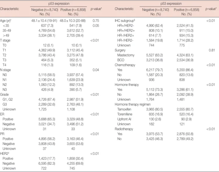
p53 overexpression was statistically associated with ad- vanced pathological stage, high tumor grade, ER negativity, PR negativity, and HER2 positivity (Table 1).
The median follow-up duration was 53 months (range, 0-125 months). The 5-year OS rate was 88.0% for p53-positive patients and 91.3% for p53-negative patients (p<0.01). The 5-year BCSS rate was 88.5% for p53-positive patients and 91.8% for p53-negative patients (p<0.01) (Figure 1A). Figure 1 shows the results of subgroup analyses. p53 accumulation was of prognostic value in terms of OS and BCSS, irrespective of tumor size, nodal status, and age. Relatively weak statistical significance for p53 status as a prognostic factor was observed for the group with negative nodal status (p=0.02 and p=0.05 for BCSS and OS, respectively), compared to the group with positive nodal status (p<0.01 for BCSS and OS). With re- gard to tumor grade, the prognostic significance of p53 accu- mulation disappeared in the grade 3 group (p=0.21 and p=
0.25 for BCSS and OS, respectively). The significant prognos- tic role of p53 in the HR+/HER2- (p<0.01 for BCSS and OS) and the HR+/HER2+ (p=0.02 for BCSS and OS) IHC subgroups completely disappeared when the HR-/HER2- (p=
0.98 and p=0.81 for BCSS and OS, respectively) and HR-/
HER2+ (p=0.17 and p=0.10 for BCSS and OS, respectively) IHC subgroups were examined.
Six factors, including tumor size, nodal status, tumor grade, age <35 years, chemotherapy received, and p53 overexpres- sion, were entered into a stratified Cox regression model to evaluate their prognostic effects on survival. The adjusted haz- ard ratios for all of these factors were significant. The hazard ratio for p53 overexpression was statistically significant only for
patients with HR+/HER2- tumors; the hazard ratio of death was significantly higher for patients with p53-positive tumors than for those with p53-negative tumors (OS: hazard ratio, 1.44; 95% confidence interval [CI], 1.08-1.93 and BCSS: hazard ratio, 1.47; 95% CI, 1.09-1.99). Such a difference was not ob- served in patients with HR-/HER2+ or HR-/HER2- tumors.
The hazard ratios of p53 overexpression for OS and BCSS for the HR-/HER2+ and HR-/HER2- subgroups were 1.25 (95%
CI, 0.96-1.60) and 1.21 (95% CI, 0.94-1.57), and 0.94 (95% CI, 0.73-1.20) and 0.92 (95% CI, 0.71-1.18), respectively. For the HR+/HER2+ subgroup, the association of p53 overexpression with poor prognosis had borderline significance (OS: hazard ratio, 1.27; 95% CI, 0.98-1.66 and BCSS: hazard ratio, 1.26; 95%
Table 1. Clinicopathological characteristics of 15,598 patients according to expression of p53 determined by immunohistochemistry Characteristic
p53 expression
p-value* Characteristic
p53 expression
p-value*
Negative (n=8,740)
No. (%) Positive (n=6,858)
No. (%) Negative (n=8,740)
No. (%) Positive (n=6,858) No. (%)
Age (yr)† 48.1±10.4 (19-91) 48.0±10.3 (20-88) 0.75 IHC subgroup§ <0.01
<35 637 (7.3) 541 (7.9) 0.05 HR+/HER2- 4,990 (62.4) 2,524 (41.5)
35-49 4,769 (54.6) 3,612 (52.7) HR+/HER2+ 808 (10.1) 911 (15.0)
>49 3,334 (38.1) 2,705 (39.4) HR-/HER2+ 614 (7.7) 934 (15.3)
T stage <0.01 HR-/HER2- 1,584 (19.8) 1,714 (28.2)
T0 12 (0.1) 10 (0.1) Unknown 744 775
T1 4,362 (49.9) 3,112 (45.4) Surgery 0.81
T2 3,786 (43.4) 3,275 (47.8) Mastectomy 5,527 (63.2) 4,324 (63.1)
T3 464 (5.3) 352 (5.1) BCO 3,213 (36.8) 2,534 (36.9)
T4 116 (1.3) 109 (1.6) Chemotherapy <0.01
N stage 0.04 Yes 6,217 (79.7) 5,200 (86.4)
N0 5,115 (58.5) 3,937 (57.4) No 1,587 (20.3) 820 (13.6)
N1 2,136 (24.4) 1,639 (23.9) Unknown 936 838
N2 1,063 (12.2) 892 (13.0) Hormone therapy <0.01
N3 426 (4.9) 390 (5.7) Yes 5,112 (73.3) 3,286 (61.1)
Grade <0.01 No 1,864 (26.7) 2,092 (38.9)
G1, G2 4,726 (67.4) 2,987 (51.9) Unknown 1,764 1,481
G3 2,289 (32.6) 2,763 (48.1) Hormone therapy regimen
Unknown 1,725 1,108 Tamoxifen 3,965 (80.5) 2,555 (80.7)
ER <0.01 Toremifene 835 (16.9) 520 (16.4)
Positive 5,688 (65.3) 3,329 (48.8) Upfront AI 130 (2.6) 90 (2.9)
Negative 3,021 (34.7) 3,496 (51.2) Unknown 182 121
Unknown 31 33 Radiotherapy <0.01
PR <0.01 Yes 3,975 (53.7) 2,876 (50.8)
Positive 4,895 (56.2) 3,163 (46.4) No 3,425 (46.3) 2,789 (49.2)
Negative 3,808 (43.8) 3,655 (53.6)
Unknown 37 40
HER2‡ <0.01
Positive 1,423 (17.7) 1,858 (30.4) Negative 6,595 (82.3) 4,255 (69.6)
Unknown 722 745
ER=estrogen receptor; PR=progesterone receptor; HER2=human epidermal growth factor receptor 2; IHC=immunohistochemical; HR=hormone receptor;
BCO=breast-conserving operation; AI=aromatase inhibitor.
*The chi-square test was used to identify differences in variables between groups according to the p53 status; †Mean±SD (range); ‡The results of immunohisto- chemical HER2 expression were scored as 0 to 3+ and dichotomized into negative (0, 1+, or 2+) or positive (3+); §IHC subgroup determined based on ER, PR, and HER2 status. HR+ means ER positive and/or PR positive.
http://dx.doi.org/10.4048/jbc.2013.16.4.386 http://ejbc.kr Figure 1. (A) Breast cancer-specific survival (BCSS) (left) and overall survival (OS) (right) according to p53 status in the overall series. Subgroup analy- ses (B) by tumor size: ≤2 cm vs. >2 cm (left, BCSS; right, OS); (C) by lymph node status: negative vs. positive (left, BCSS; right, OS). (A-E) p53 is significantly prognostic in all subgroups except in the subgroup with grade 3.
(Continued to the next page)
Survival probability
0 20 40 60 80 100 120 Time (mo)
1.0
0.9
0.8
0.7
0.6
0.5
p<0.01
A BCSS
p53 negative p53 negative-censored p53 positive p53 positive-censored
Survival probability
0 20 40 60 80 100 120 Time (mo)
1.0
0.9
0.8
0.7
0.6
0.5
p<0.01
OS p53 negative p53 negative-censored p53 positive p53 positive-censored
Time (mo)
Survival probability
0 12 24 36 48 60 72 84 96 108 120 1.0
0.9
0.8
0.7
0.6
0.5
Tumor size >2 cm p<0.01
Tumor size ≤2 cm p<0.01
OS
≤2 cm p53 negative
>2 cm p53 negative
≤2 cm p53 negative-censored
>2 cm p53 negative-censored
≤2 cm p53 positive
>2 cm p53 positive
≤2 cm p53 positive-censored
>2 cm p53 positive-censored BCSS
≤2 cm p53 negative
>2 cm p53 negative
≤2 cm p53 negative-censored
>2 cm p53 negative-censored
≤2 cm p53 positive
>2 cm p53 positive
≤2 cm p53 positive-censored
>2 cm p53 positive-censored
B
Survival probability
0 12 24 36 48 60 72 84 96 108 120 Time (mo)
1.0
0.9
0.8
0.7
0.6
0.5
Tumor size >2 cm p<0.01
Tumor size ≤2 cm p<0.01
OS BCSS
Node (-) p53 negative Node (-) p53 negative
Node (+) p53 negative Node (+) p53 negative
Node (-) p53
negative-censored Node (-) p53
negative-censored
Node (+) p53
negative-censored Node (+) p53
negative-censored
Node (-) p53 positive Node (-) p53 positive
Node (+) p53 positive Node (+) p53 positive
Node (-) p53
positive-censored Node (-) p53
positive-censored
Node (+) p53
positive-censored Node (+) p53
positive-censored
C
Survival probability Survival probability
0 12 24 36 48 60 72 84 96 108 120 0 12 24 36 48 60 72 84 96 108 120
Time (mo) Time (mo)
1.0
0.9
0.8
0.7
0.6
0.5
1.0
0.9
0.8
0.7
0.6
0.5
Node (-) p=0.02 Node (-) p=0.05
Node (+) p<0.01 Node (+) p<0.01
http://ejbc.kr http://dx.doi.org/10.4048/jbc.2013.16.4.386 Figure 1. (Continued from the previous page) (D) by grade: grade1 or 2 vs. grade 3 (left, BCSS; right, OS); and (E) by age: <35 years vs. 35 to 49 years vs. >50 years old (left, BCSS; right, OS). (A-E) p53 is significantly prognostic in all subgroups except in the subgroup with grade 3. (F) OS and BCSS according to p53 status by possible intrinsic subtypes only by three immunohistochemical markers showed that the prognostic role of p53 persists only for the luminal A (hormone receptor (HR) positive/human epidermal growth factor receptor 2 (HER2) negative; blue line) and luminal B (HR positive/HER2 positive; green line) subtype and not for the HER2 (HR negative/HER2 positive; gray line) or TN (HR negative/HER2 negative; red line) subtype.
LA=luminal A; LB=luminal B; TN=triple negative.
Survival probability
0 12 24 36 48 60 72 84 96 108 120 Time (mo)
1.0
0.9
0.8
0.7
0.6
E
Survival probability
0 12 24 36 48 60 72 84 96 108 120 Time (mo)
1.0
0.9
0.8
0.7
0.6
Survival probability
0 12 24 36 48 60 72 84 96 108 120 Time (mo)
1.0
0.9
0.8
0.7
0.6
F
Survival probability
0 12 24 36 48 60 72 84 96 108 120 Time (mo)
1.0
0.9
0.8
0.7
0.6
OS
Grade 1/2 p53 negative Grade 3 p53 negative Grade 1/2 p53 negative-censored
Grade 3 p53 negative-censored Grade 1/2 p53 positive Grade 3 p53 positive
Grade 1/2 p53 positive-censored
Grade 3 p53 positive-censored
OS
OS
Survival probability
0 12 24 36 48 60 72 84 96 108 120 Time (mo)
1.0
0.9
0.8
0.7
0.6
0.5
Grade 1/2 p<0.01
Grade 3 p=0.21
D
Survival probability
0 12 24 36 48 60 72 84 96 108 120 Time (mo)
1.0
0.9
0.8
0.7
0.6
0.5
Grade 1/2 p<0.01
Grade 3 p=0.25 BCSS
Grade 1/2 p53 negative Grade 3 p53 negative Grade 1/2 p53 negative-censored
Grade 3 p53 negative-censored Grade 1/2 p53 positive Grade 3 p53 positive
Grade 1/2 p53 positive-censored
Grade 3 p53 positive-censored
BCSS
BCSS p53 (-)
p53 (-)
p53 (-)
p53 (-) p53 (-)
p53 (-)
p53 (-)
p53 (-) p53 (-)
p53 (-)
p53 (-) p53 (-)
p53 (-)
p53 (-) Age 35-49, p<0.01
LA, p<0.01
Age 35-49, p<0.01
LA, p<0.01 Age ≥50, p<0.01
LB, p=0.02
Age ≥50, p<0.01
LB, p=0.02 Age <35, p<0.01
TN, p=0.98
HER2, p=0.17 HER2, p=0.10
Age <35, p<0.01
TN, p=0.81 p53 (+)
p53 (+)
p53 (+)
p53 (+) p53 (+)
p53 (+)
p53 (+)
p53 (+) p53 (+)
p53 (+)
p53 (+) p53 (+)
p53 (+)
p53 (+)
http://dx.doi.org/10.4048/jbc.2013.16.4.386 http://ejbc.kr Table 3. Cox proportional hazards regression model for breast cancer-specific and overall survival considering interactions between p53 and adjuvant treatments
Variable BCSS OS
Hazard ratio (95% CI) p-value Hazard ratio (95% CI) p-value
Model without interaction terms
p53 overexpression 1.16 (1.00-1.34) 0.04 1.18 (1.02-1.35) 0.02
Age <35 yr 1.38 (1.10-1.71) <0.01 1.33 (1.07-1.66) 0.01
Tumor size >2 cm 1.98 (1.67-2.36) <0.01 1.92 (1.62-2.26) <0.01
Lymph node positivity 3.70 (3.12-4.39) <0.01 3.57 (3.03-4.21) <0.01
Grade 3 1.50 (1.29-1.74) <0.01 1.48 (1.28-1.72) <0.01
Chemotherapy 0.71 (0.54-0.93) 0.01 0.63 (0.49-0.82) <0.01
IHC subgroup
HR+/HER2- 1 (reference) 1 (reference)
HR+/HER2+ 1.95 (1.57-2.43) <0.01 1.88 (1.52-2.33) <0.01
HR-/HER2+ 3.03 (2.45-3.75) <0.01 2.93 (2.38-3.61) <0.01
HR-/HER2- 2.60 (2.15-3.14) <0.01 2.51 (2.09-3.01) <0.01
Model with interaction terms
Interaction term, p53 by hormonal therapy 1.46 (1.11-1.91) 0.01 1.51 (1.16-1.97) <0.01
Interaction term, p53 by chemotherapy 0.84 (0.53-1.31) 0.43 0.86 (0.56-1.31) 0.47
p53 overexpression 1.21 (0.76-1.94) 0.43 1.16 (0.75-1.81) 0.51
Age <35 yr 1.39 (1.14-1.71) <0.01 1.36 (1.11-1.66) <0.01
Tumor size >2 cm 2.10 (1.79-2.48) <0.01 2.04 (1.74-2.39) <0.01
Lymph node positivity 3.52 (2.99-4.13) <0.01 3.40 (2.91-3.97) <0.01
Grade 3 1.63 (1.41-1.87) <0.01 1.61 (1.40-1.84) <0.01
Chemotherapy 0.77 (0.56-1.07) 0.12 0.71 (0.52-0.97) 0.03
Model with reclassification
Age <35 yr 1.39 (1.14-1.71) <0.01 1.36 (1.11-1.66) <0.01
Tumor size >2 cm 2.11 (1.79-2.48) <0.01 2.04 (1.74-2.39) <0.01
Lymph node positivity 3.51 (2.99-4.13) <0.01 3.40 (2.91-3.97) <0.01
Grade 3 1.63 (1.41-1.87) <0.01 1.61 (1.40-1.84) <0.01
Chemotherapy 0.71 (0.56-0.90) <0.01 0.66 (0.53-0.83) <0.01
Hormonal therapy, no & p53 negative 1 (reference) 1 (reference)
Hormonal therapy, yes & p53 negative 0.49 (0.40-0.60) <0.01 0.49 (0.40-0.60) <0.01
Hormonal therapy, no & p53 overexpression 1.02 (0.84-1.25) 0.84 1.00 (0.82-1.22) 0.97
Hormonal therapy, yes & p53 overexpression 0.73 (0.60-0.89) <0.01 0.76 (0.63-0.92) <0.01
BCSS=breast cancer-specific survival; OS=overall survival; CI=confidence interval; IHC=immunohistochemical; HR=hormone receptor; HER2=human epider- mal growth factor receptor 2.
Table 2. Adjusted hazard ratios for the immunohistochemical subtypes for breast cancer-specific and overall survival from a stratified Cox proportional hazard model
Characteristic
Hazard ratio* (95% CI) in each IHC subgroup Likelihood test for homogeneity,
p value
Adjusted hazard ratio† (95% CI) in all IHC
subgroups
HR+/HER2- HR+/HER2+ HR-/HER2+ HR-/HER2-
BCSS OS BCSS OS BCSS OS BCSS OS BCSS OS BCSS OS
p53 overexpression 1.45
(1.13-1.86)‡ 1.46
(1.15-1.85)‡ 1.17
(0.83-1.63) 1.15
(0.82-1.59) 1.16
(0.85-1.57) 1.18
(0.87-1.60) 1.02
(0.82-1.28) 1.05
(0.84-1.30) 0.23 0.28 1.16
(1.00-1.34)‡ 1.18 (1.02-1.35)‡
Age <35 yr 1.94
(1.34-2.81)‡ 1.84
(1.27-2.66)‡ 1.70
(1.03-2.80)‡ 1.63
(0.99-2.67) 1.20
(0.71-2.04) 1.15
(0.68-1.95) 1.22
(0.88-1.69) 1.20
(0.87-1.66) 0.23 0.25 1.38
(1.10-1.71)‡ 1.33 (1.07-1.66)‡ Tumor size >2 cm 2.38
(1.75-3.22)‡ 2.10
(1.58-2.79)‡ 1.55
(1.03-2.34)‡ 1.53
(1.02-2.28)‡ 1.95
(1.34-2.83)‡ 2.07
(1.43-3.00)‡ 1.79
(1.38-2.33)‡ 1.80
(1.39-2.32)‡ 0.36 0.58 1.98
(1.67-2.36)‡ 1.92 (1.62-2.26)‡ Lymph node
positivity 3.06
(2.25-4.17)‡ 2.94
(2.19-3.95)‡ 4.88
(3.02-7.88)‡ 4.65
(2.92-7.40)‡ 4.99
(3.39-7.36)‡ 5.21
(3.54-7.65)‡ 3.29
(2.58-4.21)‡ 3.12
(2.46-3.96)‡ 0.12 0.05 3.70 (3.12-4.39)‡ 3.57
(3.03-4.21)‡
Grade 3 1.85
(1.44-2.38)‡ 1.80
(1.41-2.30)‡ 1.40
(1.00-1.96)‡ 1.39
(1.00-1.93)‡ 1.18
(0.86-1.62) 1.20
(0.88-1.64) 1.32
(1.03-1.69)‡ 1.30
(1.02-1.65)‡ 0.12 0.15 1.50
(1.29-1.74)‡ 1.48 (1.28-1.72)‡ Chemotherapy
performed 0.92
(0.59-1.44) 0.77
(0.51-1.14) 0.63
(0.35-1.13) 0.64
(0.36-1.12) 0.81
(0.35-1.88) 0.53
(0.27-1.07) 0.44
(0.27-0.73)‡ 0.47
(0.28-0.77)‡ 0.19 0.48 0.71
(0.54-0.93)‡ 0.63 (0.49-0.82)‡ CI=confidence interval; IHC=immunohistochemistry; HR=hormone receptor; HER2=human epidermal growth factor receptor 2; BCSS=breast cancer-specific survival; OS=overall survival.
*Hazard ratios in each subgroup from Cox proportional hazard model with adjustment for six characteristics, including tumor size, nodal status, tumor grade, age less than 35 years, chemotherapy received, and p53 overexpression; †Adjusted hazard ratios across all IHC subgroups through likelihood test for homogeneity;
‡p<0.05.
CI, 0.96-1.65). By adjustment in a stratified Cox regression model, p53 overexpression was found to be an independent predictor for 5-year OS and BCSS along with conventional prognostic factors such as tumor size, nodal status, tumor grade, age <35 years, and chemotherapy received (Table 2).
Possible interactions between p53 and adjuvant treatment were explored. A significant interaction was observed between p53 expression and hormonal therapy (p=0.002 and p=0.007 for OS and BCSS, respectively) (Table 3), suggesting that the effects of hormonal therapy may differ according to p53 sta- tus. Such an interaction was not observed for chemotherapy (p=0.233 and p=0.300 for OS and BCSS, respectively) (Table 3). Although IHC subgroup, p53 overexpression, tumor size, nodal status, tumor grade, age <35 years, and chemotherapy received were significant independent factors affecting OS and BCSS in a model without interaction terms, p53 overexpres- sion alone was not significant in a second model with interac- tion terms (p=0.268 and p=0.296 for OS and BCSS, respec- tively) (Table 3). The response to hormonal therapy was great- er in p53-negative patients (hazard ratio, 0.49 for OS and BCSS; p<0.01) than in p53-positive patients (hazard ratio, 0.76 and 0.73 for OS and BCSS, respectively; p<0.01), indicat- ing an effect modification of hormonal therapy according to p53 status. Data for 10,073 patients were analyzed in the sec- ond model because of missing information on adjuvant treat- ments.
DISCUSSION
The present study confirmed that nuclear accumulation of the mutant p53 protein correlates with a poor prognosis, as previously observed in several studies. Patients with p53 over- expression had a worse OS than those without p53 accumula- tion regardless of tumor size, nodal metastasis, and age at diag- nosis. The most noteworthy finding in this study was that the prognostic power of p53 varied according to IHC subtypes representing tumor biology: these variations were likely caused by the effect modification of hormonal treatment by p53 status.
Our study demonstrated that the differences in OS and BCSS between patients with and without p53 accumulation reached significance in the HR+/HER2- subgroup, whereas these differences were of borderline significance in the HR+/
HER2+ subgroup and of no significance in the HR-/HER2+
and HR-/HER2- subgroups (Table 2). In addition, Kaplan- Meier analysis revealed that p53 accumulation was a signifi- cant prognostic factor only for patients with grade 1 and 2 his- tology, compared with patients with grade 3 histology. Silves- trini et al. [22] suggested that p53 accumulation had no prog- nostic value for contralateral breast failure or locoregional re-
lapse after radical or conservative surgery plus radiotherapy.
However, it was a significant predictor for distant metastasis, which resembled the predictive pattern observed for hormone receptors [1,22]. This result indicates the possibility of interac- tions between p53 accumulation and hormone receptor sta- tus. Therefore, in our study, patients were stratified according to IHC subtypes to evaluate any correlation with survival out- come. The results supported the prognostic value of p53 only for patients with HR+/HER2- tumors. The nonsignificant prognostic value of p53 overexpression for aggressive sub- groups such as the HR-/HER2+, HR-/HER2-, and high-grade subgroups can be explained as follows: p53 overexpression is consistently correlated with high malignant potential, but prognostic value may be weakened by the much more promi- nent effects of strong prognosticators in aggressive breast can- cer subtypes. Silvestrini et al. [1] examined a relatively large series of 1,400 patients to validate p53 accumulation as a con- tinuous variable associated with prognosis and showed that the hazard ratio for distant metastasis increased with increas- ing p53 accumulation up to a value of 12% positive cells and paradoxically decreased thereafter, possibly because of the sig- nificant interaction between p53 expression and the cell prolif- eration index. p53 accumulation is reported to provide prog- nostic information in a subset of patients with slowly prolifer- ating tumors but not in those with rapidly proliferating tumors [1]. This finding led us to consider the possibility of strength- ening the prognostic power of p53 by evaluating p53 in con- junction with ER, PR, and HER2 to elucidate a subtype for which p53 has strong prognostic power. This subgroup was demonstrated to be HR+/HER2- breast cancer in this study.
Experimental approaches to determine a plausible mecha- nism for the interaction of ER and p53 have suggested the possibility of cross talk between pathways mediated by ER and p53. When deregulated, ERα becomes abnormally prolifera- tive and greatly contributes to the onset and progression of breast cancer [23]. Similar to ERα, the tumor suppressor p53 plays a central role in many cellular processes, such as cell cy- cle regulation, apoptosis, senescence, and differentiation [24].
Both ERα and p53 play a pivotal role in normal mammary de- velopment and in breast cancer oncogenesis. Sayeed et al. [25]
proposed a novel mechanism by which ERα opposes p53-me- diated apoptosis in breast cancer cells that involves direct binding of ERα to the promoters of p53 target genes, such as survivin and multidrug resistance gene 1. They also showed, in a small-sized retrospective study that analyzed the response to tamoxifen therapy in a subset of 35 patients with ER-positive breast cancer expressing either wild-type or mutant p53, that the presence of wild-type p53 is an important determinant of a positive therapeutic response [26]. Further investigation of
http://dx.doi.org/10.4048/jbc.2013.16.4.386 http://ejbc.kr
the predictive value of p53 for adjuvant endocrine therapy in this study (Table 3) showed a significant association, and the treatment benefit in patients without mutant p53 overexpres- sion was much greater than that in patients with mutant p53 overexpression. The results of our large-scale analysis support the finding by Konduri et al. [26] that the presence of wild- type p53 is an important determinant of a positive therapeutic response in ER-positive breast cancers.
The findings of this study can be applied to several clinical circumstances. First, our findings refine previous luminal IHC biomarker signatures. Millar et al. [13] suggested that predict- ing the outcome of ER-positive breast cancer is improved us- ing a marker panel comprised of HER2, Ki-67, and p53. They modified the conventional working definition for classifying ER-positive breast cancers, previously based on HER2 overex- pression alone, into two distinct subtypes, by analyzing HER2, Ki-67, and/or p53, the five IHC biomarker classifier [13]. In addition, the 2011 St. Gallen International Conference Expert Panel strongly supported the addition of Ki-67 to ER, PR, and HER2, the four IHC biomarker classifier for defining luminal A or B subtypes with a cutoff point of 14% for the Ki-67 label- ing index [27]. The present study suggests that p53 overex- pression can be incorporated into a more refined luminal IHC biomarker signature and supports the validity of Millar’s five IHC biomarker classifier [13]. Second, mutant p53 protein status can be included in the process of determining whether to extend letrozole treatment after 5 years of tamoxifen treat- ment. This is particularly relevant for patients with HR-posi- tive breast cancer as most instances of relapse and breast can- cer mortality occur after 5 years [28]. Although an effective benefit with extended adjuvant letrozole treatment was con- firmed irrespective of patient age and nodal status in terms of disease-free survival (DFS) and distant DFS [29,30], the OS advantage was demonstrated only in node-positive breast can- cer, casting doubt on the safety of extended adjuvant letrozole therapy. Therefore, when deciding whether to use extended adjuvant letrozole therapy, clinicians and patients should con- sider the benefit of adjuvant endocrine treatment in addition to the residual risk of relapse, comorbidities, and individual preferences.
One major limitation of this study is that, despite the general consensus on IHC assay methods and the cutoff criteria for re- ceptor positivity in the KBCRS, the IHC results have not been centrally validated. However, it may be justifiable to interpret the clinical role of mutant p53 accumulation as the analyses in this study were performed after dichotomization according to a cutoff value of 10%. An additional limitation of this study is the lack of consideration for the effect of Ki-67 on survival out- comes. The results of this study, therefore, should be interpret-
ed with caution because it is possible that the unique prognos- tic value of p53 could be confounded by the Ki-67 index given the importance of the proliferation index in HR-positive breast cancer. Finally, an inherent limitation of this study is that data on adjuvant therapy are not mandatory in KBCRS. As men- tioned in the methods section, we could only analyze data for 10,073 out of 15,598 patients in the second model with inter- action terms. However, importantly, the results of this large, multi-institution analysis further suggests that HR-positive breast cancer patients with IHC p53 accumulation have worse OS and BCSS than those without p53 accumulation. Further- more, clinicians should note that this finding could result from the association between p53 overexpression and hormonal therapy response and may help when making clinical deci- sions based on the possible effect modification by p53 status, particularly in patients with HR-positive breast cancer.
CONFLICT OF INTEREST
The authors declare that they have no competing interests.
REFERENCES
1. Silvestrini R, Daidone MG, Benini E, Faranda A, Tomasic G, Boracchi P, et al. Validation of p53 accumulation as a predictor of distant metastasis at 10 years of follow-up in 1400 node-negative breast cancers. Clin Cancer Res 1996;2:2007-13.
2. Krajewski S, Krajewska M, Turner BC, Pratt C, Howard B, Zapata JM, et al. Prognostic significance of apoptosis regulators in breast cancer.
Endocr Relat Cancer 1999;6:29-40.
3. Malamou-Mitsi V, Gogas H, Dafni U, Bourli A, Fillipidis T, Sotiropou- lou M, et al. Evaluation of the prognostic and predictive value of p53 and Bcl-2 in breast cancer patients participating in a randomized study with dose-dense sequential adjuvant chemotherapy. Ann Oncol 2006;
17:1504-11.
4. Marks JR, Humphrey PA, Wu K, Berry D, Bandarenko N, Kerns BJ, et al. Overexpression of p53 and HER-2/neu proteins as prognostic mark- ers in early stage breast cancer. Ann Surg 1994;219:332-41.
5. Han JS, Cao D, Molberg KH, Sarode VR, Rao R, Sutton LM, et al. Hor- mone receptor status rather than HER2 status is significantly associated with increased Ki-67 and p53 expression in triple-negative breast carci- nomas, and high expression of Ki-67 but not p53 is significantly associat- ed with axillary nodal metastasis in triple-negative and high-grade non- triple-negative breast carcinomas. Am J Clin Pathol 2011;135:230-7.
6. Haerslev T, Jacobsen GK. An immunohistochemical study of p53 with correlations to histopathological parameters, c-erbB-2, proliferating cell nuclear antigen, and prognosis. Hum Pathol 1995;26:295-301.
7. Barbareschi M, Caffo O, Veronese S, Leek RD, Fina P, Fox S, et al. Bcl-2 and p53 expression in node-negative breast carcinoma: a study with long-term follow-up. Hum Pathol 1996;27:1149-55.
8. Lê MG, Mathieu MC, Douc-Rasy S, Le Bihan ML, Adb El All H, Spiel- mann M, et al. c-myc, p53 and bcl-2, apoptosis-related genes in infiltrat-
ing breast carcinomas: evidence of a link between bcl-2 protein over- expression and a lower risk of metastasis and death in operable patients.
Int J Cancer 1999;84:562-7.
9. Reed W, Hannisdal E, Boehler PJ, Gundersen S, Host H, Marthin J. The prognostic value of p53 and c-erb B-2 immunostaining is overrated for patients with lymph node negative breast carcinoma: a multivariate analysis of prognostic factors in 613 patients with a follow-up of 14-30 years. Cancer 2000;88:804-13.
10. Chae BJ, Bae JS, Lee A, Park WC, Seo YJ, Song BJ, et al. p53 as a specific prognostic factor in triple-negative breast cancer. Jpn J Clin Oncol 2009;
39:217-24.
11. Jung SY, Jeong J, Shin SH, Kwon Y, Kim EA, Ko KL, et al. Accumulation of p53 determined by immunohistochemistry as a prognostic marker in node negative breast cancer: analysis according to St Gallen consen- sus and intrinsic subtypes. J Surg Oncol 2011;103:207-11.
12. Bidard FC, Matthieu MC, Chollet P, Raoefils I, Abrial C, Dômont J, et al. p53 status and efficacy of primary anthracyclines/alkylating agent- based regimen according to breast cancer molecular classes. Ann Oncol 2008;19:1261-5.
13. Millar EK, Graham PH, McNeil CM, Browne L, O’Toole SA, Boulg- hourjian A, et al. Prediction of outcome of early ER+ breast cancer is improved using a biomarker panel, which includes Ki-67 and p53. Br J Cancer 2011;105:272-80.
14. Mauri FA, Maisonneuve P, Caffo O, Veronese S, Aldovini D, Ferrero S, et al. Prognostic value of estrogen receptor status can be improved by combined evaluation of p53, Bcl2 and PgR expression: an immunohis- tochemical study on breast carcinoma with long-term follow-up. Int J Oncol 1999;15:1137-47.
15. Isola J, Visakorpi T, Holli K, Kallioniemi OP. Association of overexpres- sion of tumor suppressor protein p53 with rapid cell proliferation and poor prognosis in node-negative breast cancer patients. J Natl Cancer Inst 1992;84:1109-14.
16. Elledge RM, Gray R, Mansour E, Yu Y, Clark GM, Ravdin P, et al. Accu- mulation of p53 protein as a possible predictor of response to adjuvant combination chemotherapy with cyclophosphamide, methotrexate, fluorouracil, and prednisone for breast cancer. J Natl Cancer Inst 1995;
87:1254-6.
17. Bergh J, Norberg T, Sjögren S, Lindgren A, Holmberg L. Complete se- quencing of the p53 gene provides prognostic information in breast cancer patients, particularly in relation to adjuvant systemic therapy and radiotherapy. Nat Med 1995;1:1029-34.
18. Knoop AS, Bentzen SM, Nielsen MM, Rasmussen BB, Rose C. Value of epidermal growth factor receptor, HER2, p53, and steroid receptors in predicting the efficacy of tamoxifen in high-risk postmenopausal breast cancer patients. J Clin Oncol 2001;19:3376-84.
19. Ahn SH, Son BH, Kim SW, Kim SI, Jeong J, Ko SS, et al. Poor outcome of hormone receptor-positive breast cancer at very young age is due to tamoxifen resistance: nationwide survival data in Korea: a report from the Korean Breast Cancer Society. J Clin Oncol 2007;25:2360-8.
20. Han W, Kang SY; Korean Breast Cancer Society. Relationship between age at diagnosis and outcome of premenopausal breast cancer: age less than 35 years is a reasonable cut-off for defining young age-onset breast cancer. Breast Cancer Res Treat 2010;119:193-200.
21. Moon HG, Han W, Noh DY. Comparable survival between pN0 breast cancer patients undergoing sentinel node biopsy and extensive axillary dissection: a report from the Korean Breast Cancer Society. J Clin Oncol 2010;28:1692-9.
22. Silvestrini R, Daidone MG, Luisi A, Boracchi P, Mezzetti M, Di Fronzo G, et al. Biologic and clinicopathologic factors as indicators of specific relapse types in node-negative breast cancer. J Clin Oncol 1995;13:697- 704.
23. Deroo BJ, Korach KS. Estrogen receptors and human disease. J Clin In- vest 2006;116:561-70.
24. Riley T, Sontag E, Chen P, Levine A. Transcriptional control of human p53-regulated genes. Nat Rev Mol Cell Biol 2008;9:402-12.
25. Sayeed A, Konduri SD, Liu W, Bansal S, Li F, Das GM. Estrogen recep- tor alpha inhibits p53-mediated transcriptional repression: implications for the regulation of apoptosis. Cancer Res 2007;67:7746-55.
26. Konduri SD, Medisetty R, Liu W, Kaipparettu BA, Srivastava P, Brauch H, et al. Mechanisms of estrogen receptor antagonism toward p53 and its implications in breast cancer therapeutic response and stem cell reg- ulation. Proc Natl Acad Sci U S A 2010;107:15081-6.
27. Goldhirsch A, Wood WC, Coates AS, Gelber RD, Thürlimann B, Senn HJ, et al. Strategies for subtypes: dealing with the diversity of breast can- cer: highlights of the St. Gallen International Expert Consensus on the Primary Therapy of Early Breast Cancer 2011. Ann Oncol 2011;22:
1736-47.
28. Early Breast Cancer Trialists’ Collaborative Group (EBCTCG). Effects of chemotherapy and hormonal therapy for early breast cancer on re- currence and 15-year survival: an overview of the randomised trials.
Lancet 2005;365:1687-717.
29. Goss PE, Ingle JN, Martino S, Robert NJ, Muss HB, Piccart MJ, et al.
Randomized trial of letrozole following tamoxifen as extended adjuvant therapy in receptor-positive breast cancer: updated findings from NCIC CTG MA.17. J Natl Cancer Inst 2005;97:1262-71.
30. Muss HB, Tu D, Ingle JN, Martino S, Robert NJ, Pater JL, et al. Efficacy, toxicity, and quality of life in older women with early-stage breast cancer treated with letrozole or placebo after 5 years of tamoxifen: NCIC CTG intergroup trial MA.17. J Clin Oncol 2008;26:1956-64.