Prognostic Significance of Immunohistochemical Expression of p53 and Retinoblastoma Gene Protein
(pRB) in Curatively Resected Gastric Cancer
Hong Suk Song, M.D., In Ho Kim, M.D.
2, Soo Sang Sohn, M.D.
2, Kun Young Kwon, M.D.
3and Won Sik Lee, M.D.
4Departments of Internal Medicine, General Surgery
2and Pathology
3, Keimyung University School of Medicine, Daegu, Korea; Department of Internal Medicine, Fatima Hospital
4, Daegu, Korea
Background : The aim of this study was to determine the prognostic significance of the expression of p53 and retinoblastoma (Rb) gene products in cases of curatively resected gastric adenocarcinoma, by immunohistochemical analysis.
Methods : Between January 1996 and December 2001, 736 curatively resected gastric cancer patients underwent immunohistochemical staining for p53 or Rb proteins (pRb), and we retrospectively analyzed the correlation of our results with the clinical outcomes of these cases.
Results : High levels of expression of p53 (>25% p53-positive cells) and Rb (>50% Rb-positive cells) proteins were detected in 40.1% and 43.7% of cases, respectively. Tubular type was found to frequently exhibit higher levels of p53 expression (high expression in 44.2%) than signet ring cell type (high expression in 26.0%) (p=0.042). The incidence of vascular invasion was lower in the high pRb expressors (43.2%) than in the pRb low expressors (56.8%), but this was not a statistically significant discrepancy (p=0.063). Preoperative CEA levels were found to be low in high pRb expressors: initial CEA level in the high pRb expressors was 2.31±3.30 ng/mL, and was 5.18±24.80 ng/mL in the low pRb expressors (p=0.033). Tumor depth and node metastasis were both independent of the levels of expression of p53 and Rb proteins. The seven-year overall survival rate and relapse-free survival rates of patients were 87.2% and 75.7%, respectively. Multivariate Cox regression analysis indicated that tumor stage, tumor size, patient age and pRb expression were the significant prognostic factors with regard to overall survival, and tumor stage and age were both significant factors with regard to relapse-free survival.
Conclusion : Immunohistochemical staining of retinoblastoma gene products was an independent prognostic factor for the prediction of overall survival in curatively resected gastric cancer patients.
Key Words : Gastric cancer, Prognosis, p53, Rb gene, Immunohistochemistry
∙Received : July 27, 2004
∙Accepted : September 22, 2004
∙Correspondence to : Hong Suk Song, M.D., Department of Hematooncology, Dongsan Medical Center, Keimyung University School of Medicine, 194 Dongsan-dong, Choong-Koo, Daegu, 700-712, Korea Tel : 82-53-250-7436, Fax : 82-53-250-7434
E-mail : [email protected]
INTRODUCTION
Gastric cancer is one of the most common cancers, repre- senting the second leading cause of cancer deaths in Korea. A population-based cancer registry was established on January 1, 1997 to estimate the incidence of cancer in Daegu. The
age-standardized incidence rates (ASR) of gastric cancer were
73.5 per 100,000 males and 28.9 per 100,000 females, as
reported by the Daegu Cancer Registry in 2002. Recently, the
proportion of gastric cancer cases among all malignancies is
declining: annual reported cases of gastric cancer constituted
24.1% of all cancers in 1990, and 20.8% of all cancers in 2000,
Number of patients Male 736
Sex Male
Female
494 (67.1%) 242 (32.9%)
Age, years Mean
Range
57.1 19-80
Operation method Total gastrectomy
Subtotal gastrectomy D1, D1 + α D2, D2 + α
114 (15.5%) 622 (84.5%) 232 (31.5%) 504 (68.5%)
Stage T1/T2/T3/T4
N0/N1/N2 IA IB II IIIA IIIB
378/138/217/3 467/173/96 338 (45.9%) 104 (14.1%) 115 (15.6%) 106 (14.4%) 73 (9.9%)
Follow-up period, months Median
Range
37.8 0.8-97.3 Table 1. Clinical characteristics of curatively resected gastric cancer patients
as reported by the Korea Cancer Registry Program. This decline appears to be due principally to changes in diet and food preparation, as well as an increased incidence of early diagnosis of gastric cancer.
Greater insight has recently been gained into the biological properties of tumor cells. Tumor suppressor gene products are of specific interest, as they play important roles in cell cycle regulation. The p53 tumor suppressor gene normally regulates cell proliferation
1, 2)and programmed cell death
3, 4). Abnormalities of the p53 tumor suppressor gene have been implicated in both tumorigenesis and tumor progression. The retinoblastoma gene is a prototype of the tumor suppressor gene which controls the cell cycle at the G1 phase
5, 6), and the Rb gene product (pRb) functions as a signal transducer, connecting the cell cycle with the transcriptional machinery. The Rb gene product, pp110Rb, is a nuclear phosphoprotein which exhibits DNA binding pro- perties
6), and it is cyclically phosphorylated and dephosphorylated during the cell cycle, playing a significant role in regulation
5-7). Loss of pRB function deprives the cell of an important mechanism for halting proliferation
8). The importance of p53 expression has been extensively analyzed with regard to a plethora of human malignancies, including gastric cancer, by immunohistochemical methods, while the role of retinoblastoma gene protein expression has been studied, but certainly not to so significant a degree
9). The prognostic roles of the above gene expressions with regard to gastric cancer remain contro- versial. The purpose of this study was to determine the prognostic significance of p53 and Rb protein expression in curatively resected gastric adenocarcinoma.
MATERIALS AND METHODS
Patients and setting
From January 1996 to December 2001, 2,104 pathologically confirmed gastric cancer patients were registered in our hospital. Of these, 1,158 were curatively resected patients. In 736 of these patients, immunohistochemical analyses of p53 or Rb proteins were performed. We reviewed the clinicopathological parameters of TNM stage, the World Health Organization classifications, histological grades, Lauren classifications, Ming classifications, vascular invasion rates, and nerve invasion rates in all 736 patients. Staging evaluation was done according to the 5th edition guidelines published by the American Joint Committee of Cancer.
Curative resection was defined as the removal of all gross
tumors, and the demonstration of tumor-negativity by microscopic
examination, in both proximal and distal surgical margins. Total
gastrectomy was performed in 114 patients (15.5%), subtotal
gastrectomy in 622 patients (84.5%), D1 and D1+α resection in
232 patients (31.5%), and D2 and D2+α resection in 504
patients (68.5%). 494 (67.1%) of these patients were male, and
242 (32.9%) were female. The mean age of the subjects was
57.1 years (range: 19-80 years). Staging was as follows: IA in
338 (45.9%) patients, IB in 104 patients (14.1%), II in 115
patients (15.6%), IIIA in 106 patients (14.4%), and IIIB in 73
patients (9.9%). The median follow-up duration was 37.8
months (range: 0.8 to 97.3 months) (Table 1).
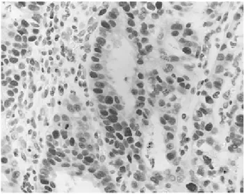
Figure 1. Immunohistochemical staining for Rb evidences high expression in the nuclei of tumor cells (×400).
Immunohistochemical staining
Immunohistochemical staining was performed using the avidin-biotin-peroxidase complex with monoclonal antibodies against p53 (NCL-p53-D07, Novocastra Laboratories, Newcastle, United Kingdom), and Rb (14001A, Pharmingen, USA). Repre- sentative paraffin blocks containing tumors isolated from each case subject were sectioned into 5 μm slices and affixed to slides, then dried for 1 hour at 60℃. The sections were deparaffinized in xylene, and rehydrated with a descending series of alcohol concentrations. Endogenous peroxidase activity was blocked by 3% hydrogen peroxidase for 15 minutes, followed by washing with phosphate buffered saline (PBS), at a pH of 7.2. The sections were then subjected to a heat antigen retrieval process, by autoclaving with 1% zinc sulfate solution for 5 minutes. After 20 minutes of cooling at room temperature, the sections were incubated with 10% normal horse serum (Vectastain Elite kit) for 30 minutes. After decanting away the excess serum, sections were incubated with primary antibody for 2 hours at 37℃. In the p53 study, DO7 monoclonal antibody was used at a 1:100 dilution (Novocastra, Newcastle, UK), and in the Rb study, the monoclonal antibody NCL-Rb1 was used, at a 1:500 dilution (Pharmingen, USA). The sections were subsequently incubated with prediluted biotinylated anti-mouse immunoglobulin (Vectastain Elite kit) for 30 minutes at 37℃.
After washing with PBS, the sections were allowed to react with peroxidase-conjugated streptoavidin (Dako, USA) at a dilution of 1:500 for 30 minutes at 37℃. After washing with PBS, peroxidase activity was evaluated with 3,3'-diaminobenzidine tetrahydrochloride (DAB), and the sections were counterstained with Mayer's hematoxylin.
All sections were examined by 2 pathologists, who were blind to the clinical outcomes and features of the patients. The sections were scored according to the percentage of positive cells using the following categories: 0=negative, 1=1~10%, 2=11~25%, 3=26~50%, 4=51~75% and 5=76~100%. A breast cancer biopsy specimen showing intense uniform positivity for pRb was used as a positive control, as was a squamous cell lung cancer biopsy specimen exhibiting uniform positivity for p53.
In the statistical analysis of p53 and Rb expression, the tmors were further categorized, into three and two groups respectively. Tumors expressing p53 protein in over 25% of cells with strong staining were designated as high p53 expressors, and tumors expressing p53 protein in 1~25% of cells or weak staining were designated as low p53 expressors.
Tumors exhibiting no p53 expression were designated negative p53 expressors. With regard to pRb expression, tumors expressing pRb in over 50% of cells with strong staining were designated high pRb expressors, and tumors expressing pRb in less than 50% of cells or weak staining were designated as low
pRb expressors (Figure 1). These classifications were predicated on the observations of previous studies
10).
Statistics
Statistical analysis was performed using χ-2 tests, in order to compare percentages in cross tabulations, and independent sample t-tests were used to compare the means. Survival curves were generated by the Kaplan-Meier method, and compared using the log-rank test. To determine the significant prognostic factors in terms of survival and relapse-free survival, multivariate analysis was performed with the Cox proportional hazards regression model. All significance levels listed refer to two-sided tests. p values of <0.05 were considered to be significant.
Statistical analyses were performed using SPSS for Windows 11.0 (Chicago, Il, USA).
RESULTS
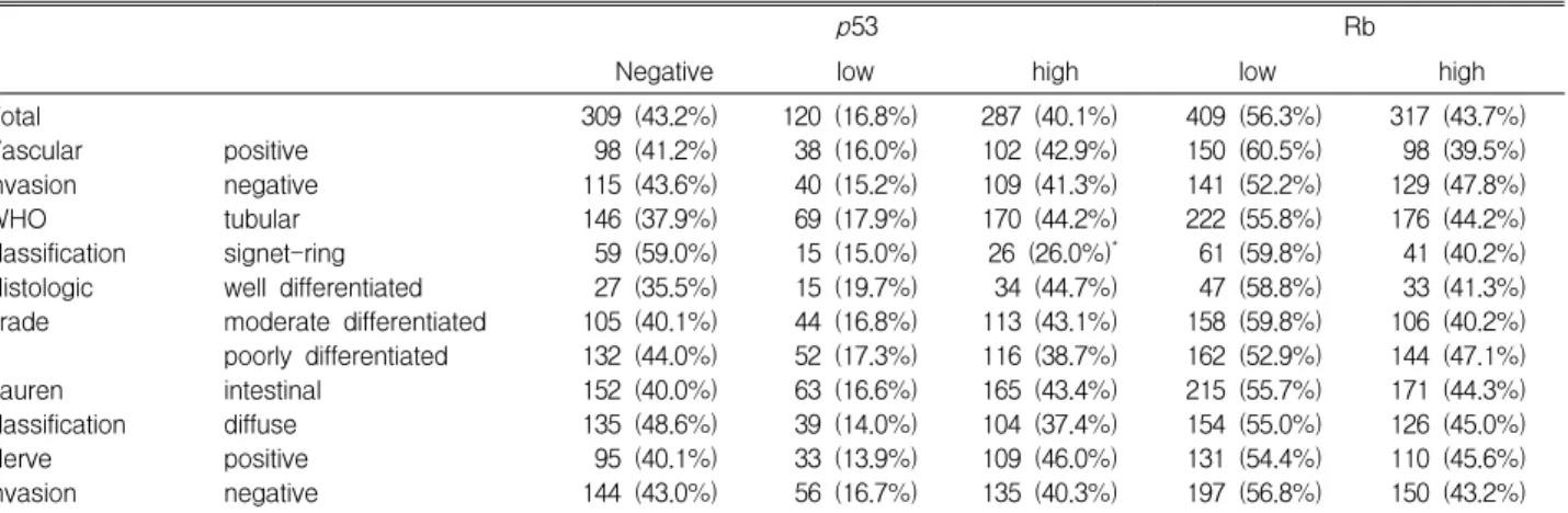
Tumor samples with high, low and no p53 protein expression were found in 287 (40.1%), 120 (16.8%) and 309 (43.2%) of the 716 samples. High and low pRb expressors were found in 317 (43.7%) and 409 (56.3%) of the 726 samples examined, respectively (Table 2).
Correlation with clinicopathologic parameters
According to the classifications provided by the World Health
Organization, tubular-type adenocarcinoma (low and high p53
expressors in 17.9% and 44.2%) was frequently more p53
positive than signet ring cell type (low and high p53 expressors
in 15.0% and 26.0%) (p=0.042). The incidence of vascular
p53 Rb
Negative low high low high
Total Vascular invasion WHO classification Histologic grade
Lauren classification Nerve invasion
positive negative tubular signet-ring well differentiated moderate differentiated poorly differentiated intestinal
diffuse positive negative
309 (43.2%) 98 (41.2%) 115 (43.6%) 146 (37.9%) 59 (59.0%) 27 (35.5%) 105 (40.1%) 132 (44.0%) 152 (40.0%) 135 (48.6%) 95 (40.1%) 144 (43.0%)
120 (16.8%) 38 (16.0%) 40 (15.2%) 69 (17.9%) 15 (15.0%) 15 (19.7%) 44 (16.8%) 52 (17.3%) 63 (16.6%) 39 (14.0%) 33 (13.9%) 56 (16.7%)
287 (40.1%) 102 (42.9%) 109 (41.3%) 170 (44.2%) 26 (26.0%)
*34 (44.7%) 113 (43.1%) 116 (38.7%) 165 (43.4%) 104 (37.4%) 109 (46.0%) 135 (40.3%)
409 (56.3%) 150 (60.5%) 141 (52.2%) 222 (55.8%) 61 (59.8%) 47 (58.8%) 158 (59.8%) 162 (52.9%) 215 (55.7%) 154 (55.0%) 131 (54.4%) 197 (56.8%)
317 (43.7%) 98 (39.5%) 129 (47.8%) 176 (44.2%) 41 (40.2%) 33 (41.3%) 106 (40.2%) 144 (47.1%) 171 (44.3%) 126 (45.0%) 110 (45.6%) 150 (43.2%)
*