Vol. 67, No. 5, November, 2004
349
서 론
유방암은 세계적으로 증가 추세에 있는 여성 암으로(1) 우리나라에서도 최근 급격히 증가하여 여성암 중 첫 번째 로 많이 발생하는 암이다.(2) 지금까지 알려진 예후 인자로 종양의 크기, 림프절 전이 상태, 환자의 연령, 에스트로겐 수용체 유무, 조직학적 분화도, 병기 등이 있고(3) 이들 중 액와부 림프절 전이 유무가 가장 중요한 예후인자로 알려 져 있지만,(4) 림프절 전이가 없는 경우에서도 10년 재발률 이 20%까지 보고되고 있다.(5) 최근 림프절 전이가 없는 유 방암에서, 예후 예측인자로 종양의 크기, 조직학적 분화도, 에스트로겐 수용체 유무 등이 알려져 있으며, 그 외 p53 유 전자, DNA배수성, Ki-67, nm23, 표피성장인자(Epidermal Growth Factor, EGF), ErbB2 등이 연구되고 있다. 이 같은 연구 결과로, 최근 ErbB2 과발현이 유방암 환자에서 특정 항암 치료제인 Herceptin (trastuzumab)과 같은 항체를 이용 한 단일 항체 면역 치료의 치료 표지자로 널리 이용되고 있어, ErbB2에 대한 관심이 증가하고 있다.(6)
c-erbB2 유전자의 산물인 ErbB2 단백(ErbB2)은 타이로신
액와부 림프절 전이가 없는 유방암에서 Akt 발현의 의의
고려대학교 의과대학 외과학교실, 1병리학교실
한형준․유영동․이재복․손길수․배정원․김애리1․김인선1․구범환
Significance of Akt Expression in Node-nega- tive Breast Cancer
Hyung Joon Han, M.D., Young Dong You, M.D., Jae Bok Lee, M.D., Gil Soo Son M.D., Jung Won Bae, M.D., Ae Ree Kim, M.D.1, In Sun Kim M.D.1 and Bum Hwan Koo, M.D.
Background: Patients with lymph node-negative breast cancer show a 10-year recurrence rate of approximately 20%. In node-negative breast cancer, the prognostic factors are age, menopause, tumor size, hormone receptors, p53, DNA ploidy, Ki-67 index (Ki-67) and c-erbB2. Of these, ErbB2 (the protein of the c-erbB2 gene) is a member of the receptor tyrosine kinase family. Overexpression of ErbB2 is known to regulate cell proliferation, differentiation, growth and apoptosis via the ErbB2/Phosphoinositol 3-Kinase (PI 3-K)/Akt signaling pathway. Therefore, it is important to identify high- risk patients that would benefit from adjuvant therapies related with ErB2. For this purpose, the prognostic relevance of the ErbB2/PI 3-K/Akt pathway was examined in node-negative breast cancer.
Methods: A retrospective analysis was performed on the hospital records of all 72 patients diagnosed with breast can- cer, and who underwent surgical treatment between January 1996 and December 2003. Clinicopathological data were compared with the results of immunohistochemical staining using the phospho-specific antibody for the expression of Akt.
Results: The mean age of the patient's was 48.6 years.
Phospho-Akt (pAkt) was expressed in 24 cases (33.3%), but there was no statistical relationship between pAkt expression and the known prognostic factors of breast cancer. There was no statistical significance in the survival rates between the pAkt positive and negative expression groups (P=0.123).
In the ErbB2 positive patients, the expression of pAkt was associated with a shorter disease-free survival (P=0.045),
and the disease-free survival was shorter in patients whose tumors expressed pAkt and had a high level of Ki-67 (P=0.040).
Conclusion: The co-expression of ErbB2 and pAkt positivity implied a poor prognosis in node-negative breast cancer patients, and the co-expression of high Ki-67 and pAkt positivity also revealed a poor prognosis in these patients.
These results show that the expression of pAkt could be considered a prognostic marker of node-negative breast can- cer with ErbB2 positive expression and high levels of Ki-67.
(J Korean Surg Soc 2004;67:349-355)
Key Words: Akt, ErbB2, Node-negative breast cancer, Prog- nosis
중심 단어: Akt, ErbB2, 액와부 림프절 음성 유방암, 예후
ꠏꠏꠏꠏꠏꠏꠏꠏꠏꠏꠏꠏꠏꠏꠏꠏꠏꠏꠏꠏꠏꠏꠏꠏꠏꠏꠏꠏꠏꠏꠏꠏꠏꠏꠏꠏꠏꠏꠏꠏꠏꠏꠏꠏꠏꠏꠏꠏꠏꠏ Departments of Surgery and 1Pathology, College of Medi- cine, Korea University
책임저자:구범환, 서울시 구로구 구로동 80번지 ꂕ 152-703, 고려대학교 구로병원 외과학교실 Tel: 02-818-6672, Fax: 02-859-5941
E-mail: [email protected]
접수일:2004년 6월 21일, 게재승인일:2004년 10월 15일
ꠏꠏꠏꠏꠏꠏꠏꠏꠏꠏꠏꠏꠏꠏꠏꠏꠏꠏꠏꠏꠏꠏꠏꠏꠏꠏꠏꠏꠏꠏꠏꠏꠏꠏꠏꠏꠏꠏꠏꠏꠏꠏꠏꠏꠏꠏꠏꠏꠏꠏꠏꠏꠏꠏꠏꠏꠏꠏꠏꠏꠏꠏꠏꠏꠏꠏꠏꠏꠏꠏꠏꠏꠏꠏꠏꠏꠏꠏꠏꠏꠏꠏꠏꠏꠏꠏꠏꠏꠏꠏꠏꠏꠏꠏꠏꠏꠏꠏꠏꠏꠏꠏꠏꠏꠏꠏꠏꠏꠏꠏꠏꠏꠏꠏꠏ 활성화 효소 수용체(Receptor tyrosine kinases)에 속한다. 표
피성장인자 관련 성장인자 군의 펩티드와 ErbB2 수용체의 세포 바깥쪽 도메인(domain)과 결합하여 homodimer나 he- terodimer를 형성하고, 리간드(ligand)가 결합하여 세포 내 타이로신 활성화 효소 수용체를 활성화시키면, auto- phosphorylation을 일으켜 세포 내 결합 부위 변화로 리간드 가 결합할 수 있는 도메인이 제공되면서, 세포 내 신호전달 에 자극을 준다.(7)
ErbB2는 유선(mammary glands) 발달의 모든 단계에서 발 현되며 소엽의 분화와 수유에 관여한다. ErbB2가 비정상적 으로 과발현하거나 증폭되면 정상적인 세포의 생물학적 조 절이 붕괴되고 유선조직에 종양이 발생하는데, ErbB2가 homo- 혹은 heterodimerization되어 활성화되면 하부 신호전 달 물질을 인산화(phosphorylation)시키고 차례로 신호전달 cascades를 활성화하는, SOS-Ras-Raf-MEK-Mitogen-activated protein kinase(MAPK) 신호전달체계와 Phosphatidylinositol 3-kinase (PI-3K)/Akt 신호전달체계가 ErbB2의 과발현에 의 한 암 증식에 관여하는 대표적인 기전이다.(7,8) 암세포 생 존의 신호 전달 체계 중 Akt에 의한 전달 체계가 중요한 역 할을 하고 있으며, PI 3-K/Akt 신호 전달 체계를 억제하는 암 억제 유전자가 많은 종류의 암에서 발견되고 있어, Akt의 과발현이 암 발생과 진행에 연관이 있음을 알 수 있다.(9) 본 연구는 림프절 전이가 없는 유방암 환자에서 ErbB2, pAkt를 면역조직화학 염색방법으로 확인하고, 유방암의 예후 인자와 ErbB2/PI 3-K/Akt 신호전달체계와의 상관 관계를 조 사하여, Akt 양성발현이 림프절 전이가 없는 유방암 환자에 서 고위험군 분류 지표로서의 유용성을 확인하고자 하였다.
방 법
1) 연구 대상
l996년 1월부터 2003년 l2월까지 고려대학교 의과대학 외 과학교실에서 유방의 원발성 침윤성 관암종으로 진단을 받 고, 수술 후 추적 관찰이 가능하였던 497명 중, 액와부 림프 절 전이가 없으면서, 조직 보존 상태가 양호한 72명을 대상 으로 하였다. 액와부 림프절 전이 유무는 액와부 림프절 곽 청술 후 조직 병리적 검사로 진단하였다. 평균 추적 관찰 기간은 2.47 (±1.71)년이었으며, 각 환자의 의무기록과 병 리보고서를 후향적으로 분석하였다(Table 1).
2) 연구방법
(1) Akt와 ErbB2의 면역조직화학 염색: 항 ErbB2 항체는 Phospho-HER2/ErbB2 (Tyr877) (Cell Signaling, MA, USA)를 사용하였고, 항 Akt 항체는 Phospho-Akt (Ser473) (Cell Sig- naling, MA, USA)를 사용하였다. 면역조직화학 염색은 조직 을 4μm 두께로 박절하여 유리 슬라이드에 부착시키고, xylene에 5분간 3회 세척하여 incubation 후 파라핀 제거와
함수과정을 거쳤다. 증류수로 5분간 2회 세척하고, Phos- phate Buffered Saline (PBS, 0.58M Na2HPO4, 0.17M NaH2PO4, 0.68M NaCl)에 5분간 세척하고, antigen un- masking을 위해 10 mM sodium citrate buffer에 넣어 전자레 인지에서 10분간 강열로 중탕한 후 실온에서 20분간 방치 하였다. 이후 증류수에 5분간 3회 세척하고 1% hydrogen Peroxide 에 10분간 incubation한 후, 다시 5분간 3회 증류수 로 세척하고 PBS에 5분간 세척하였다. 상온에서 100∼400 ul의 blocking solution (PBS에 5% horse serum)으로 각 section을 처리한 후에 blocking solution을 제거하고, 각
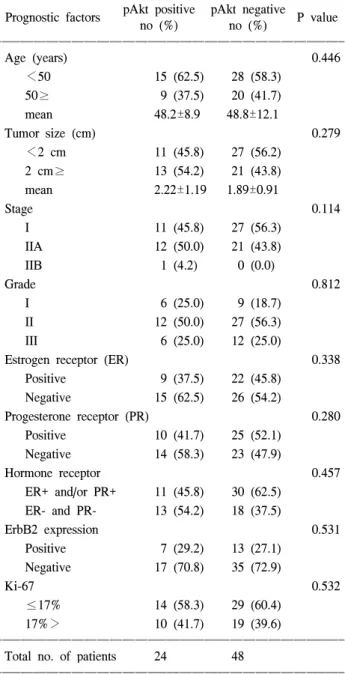
Table 1. Correlation between pAkt (phospho-Akt) expression and clinicopathologic data in 72 node negative breast cancers ꠚꠚꠚꠚꠚꠚꠚꠚꠚꠚꠚꠚꠚꠚꠚꠚꠚꠚꠚꠚꠚꠚꠚꠚꠚꠚꠚꠚꠚꠚꠚꠚꠚꠚꠚꠚꠚꠚꠚꠚꠚꠚꠚꠚꠚꠚꠚꠚꠚꠚꠚꠚꠚꠚꠚ
pAkt positive pAkt negative
Prognostic factors P value
no (%) no (%)
ꠏꠏꠏꠏꠏꠏꠏꠏꠏꠏꠏꠏꠏꠏꠏꠏꠏꠏꠏꠏꠏꠏꠏꠏꠏꠏꠏꠏꠏꠏꠏꠏꠏꠏꠏꠏꠏꠏꠏꠏꠏꠏꠏꠏꠏꠏꠏꠏꠏꠏꠏꠏꠏꠏꠏ
Age (years) 0.446
<50 15 (62.5) 28 (58.3)
50≥ 9 (37.5) 20 (41.7)
mean 48.2±8.9 48.8±12.1
Tumor size (cm) 0.279
<2 cm 11 (45.8) 27 (56.2)
2 cm≥ 13 (54.2) 21 (43.8)
mean 2.22±1.19 1.89±0.91
Stage 0.114
I 11 (45.8) 27 (56.3)
IIA 12 (50.0) 21 (43.8)
IIB 1 (4.2) 0 (0.0)
Grade 0.812
I 6 (25.0) 9 (18.7)
II 12 (50.0) 27 (56.3)
III 6 (25.0) 12 (25.0)
Estrogen receptor (ER) 0.338
Positive 9 (37.5) 22 (45.8)
Negative 15 (62.5) 26 (54.2)
Progesterone receptor (PR) 0.280
Positive 10 (41.7) 25 (52.1)
Negative 14 (58.3) 23 (47.9)
Hormone receptor 0.457
ER+ and/or PR+ 11 (45.8) 30 (62.5) ER- and PR- 13 (54.2) 18 (37.5)
ErbB2 expression 0.531
Positive 7 (29.2) 13 (27.1)
Negative 17 (70.8) 35 (72.9)
Ki-67 0.532
≤17% 14 (58.3) 29 (60.4)
17%> 10 (41.7) 19 (39.6)
ꠏꠏꠏꠏꠏꠏꠏꠏꠏꠏꠏꠏꠏꠏꠏꠏꠏꠏꠏꠏꠏꠏꠏꠏꠏꠏꠏꠏꠏꠏꠏꠏꠏꠏꠏꠏꠏꠏꠏꠏꠏꠏꠏꠏꠏꠏꠏꠏꠏꠏꠏꠏꠏꠏꠏ Total no. of patients 24 48
ꠏꠏꠏꠏꠏꠏꠏꠏꠏꠏꠏꠏꠏꠏꠏꠏꠏꠏꠏꠏꠏꠏꠏꠏꠏꠏꠏꠏꠏꠏꠏꠏꠏꠏꠏꠏꠏꠏꠏꠏꠏꠏꠏꠏꠏꠏꠏꠏꠏꠏꠏꠏꠏꠏꠏ
ꠏꠏꠏꠏꠏꠏꠏꠏꠏꠏꠏꠏꠏꠏꠏꠏꠏꠏꠏꠏꠏꠏꠏꠏꠏꠏꠏꠏꠏꠏꠏꠏꠏꠏꠏꠏꠏꠏꠏꠏꠏꠏꠏꠏꠏꠏꠏꠏꠏꠏꠏꠏꠏꠏꠏꠏꠏꠏꠏꠏꠏꠏꠏꠏꠏꠏꠏꠏꠏꠏꠏꠏꠏꠏꠏꠏꠏꠏꠏꠏꠏꠏꠏꠏꠏꠏꠏꠏꠏꠏꠏꠏꠏꠏꠏꠏꠏꠏꠏꠏꠏꠏꠏꠏꠏꠏꠏꠏꠏꠏꠏꠏꠏꠏꠏ
section에 blocking solution으로 희석한 100∼400 ul의 일차 항체를 가하고 4oC에서 overnight incubation 시켰다. 여분의 antibody solution을 제거하고 PBS에 5분간 세 번 세척한 후 2차 항체를 투여하고 상온에서 30분간 incubation하였다.
ABC reagent (Vectastatin ABC kit, Vector Laboratories, Inc., Burlingame, CA)로 상온에서 30분간 incubation한 후, 2차 항 체 solution을 제거하고 PBS에 5분간 3번 세척하였다. 각 section에 100∼400 ul ABC reagent를 다시 넣고, 상온에서 30분간 incubation 시킨 후, ABC reagent를 제거하고 PBS에 5분간 세 번 세척한 다음 1 mg/ml DAB (diaminobenzidine tetra-hydrochloride 10 ml)로 발색한 후 Hematoxylin 염색으 로 대조 염색을 하였다.
(2) 판정: ErbB2의 면역조직화학 염색의 판정은, 종양세 포의 세포막에 강하게 갈색으로 염색된 것을 양성으로 판 정하였다(Fig. 3). Akt의 면역조직화학 염색 판정은, 세포질 과 세포막에 갈색으로 염색된 종양세포가 전체 유방 종양 조직의 10% 이상이면 pAkt 양성으로 판정하였다(Fig. 4).
(3) 통계 검증: 면역조직화학 염색에 의한 pAkt 양성발현 여부를 기존에 알려진 임상병리학적 예후 인자인 연령, 폐 경 유무, 종양의 크기, 병기, 조직학적 분화도, 여성 호르몬 수용체 유무, Ki-67지수, ErbB2 과발현과 비교하고, 생존율 을 분석하였다. 통계분석은 SPSS 10.0 통계 프로그램을 이 용하였으며, 유방암 예후인자와 pAkt 발현과의 관계는 카 이 제곱 검증으로 분석하였고, 생존율과 무병생존율의 비 교는 Kaplan-Meier방법을 통한 log-rank test를 실시하여 P값 이 0.05 이하일 때를 통계적인 유의한 차이로 판단하였다.
결 과
1) 임상 소견
연령 분포는 28∼71세였으며, 평균 연령은 48.6 (±11.1)
세였다. pAkt 양성발현(양성)을 보인 환자의 평균 연령은 48.2 (±8.9)세, pAkt 음성발현(음성)은 48.8 (±12.1)세로, pAkt 양성과 음성군 간에 평균 연령은 비슷하였고, 두 군간 에 통계적 유의성은 없었다(Table 1).
2) 종양의 크기
평균 종양 크기는 2.0 (±1.02) cm였고, 2.0 cm 이상이 34명 (47.2%), 2.0 cm 미만이 38명(52.8%)이었고, pAkt 양성군에 서 종양 크기의 평균은 2.22 (±1.19) cm, pAkt 음성군은 1.89 (±0.91) cm였다. 종양의 크기가 2 cm 이상에서 2 cm 미만보다 pAkt 양성군이 높았으나 통계적 유의성은 없었다 (Table 1).
3) 조직학적 분화도
Nottingham(10) 조직학적 분화도에 따른 분류에서, 등급 Table 2. Statistical comparison of disease free survivals (DFS)
between phosporylated Akt (pAkt) expression and vari- ous prognostic factors of node-negative breast cancer ꠚꠚꠚꠚꠚꠚꠚꠚꠚꠚꠚꠚꠚꠚꠚꠚꠚꠚꠚꠚꠚꠚꠚꠚꠚꠚꠚꠚꠚꠚꠚꠚꠚꠚꠚꠚꠚꠚꠚꠚꠚꠚꠚꠚꠚꠚꠚꠚꠚꠚꠚꠚꠚꠚꠚ
Log-rank test for DFS Prognostic factors
P value
ꠏꠏꠏꠏꠏꠏꠏꠏꠏꠏꠏꠏꠏꠏꠏꠏꠏꠏꠏꠏꠏꠏꠏꠏꠏꠏꠏꠏꠏꠏꠏꠏꠏꠏꠏꠏꠏꠏꠏꠏꠏꠏꠏꠏꠏꠏꠏꠏꠏꠏꠏꠏꠏꠏꠏ
Age<50 0.212
Tumor≥2 cm 0.216
Grade 0.293
ER- 0.727
PR- 0.254
ErbB2 (+) 0.045*
Ki-67>17 0.040*
pAkt (+) 0.728
ꠏꠏꠏꠏꠏꠏꠏꠏꠏꠏꠏꠏꠏꠏꠏꠏꠏꠏꠏꠏꠏꠏꠏꠏꠏꠏꠏꠏꠏꠏꠏꠏꠏꠏꠏꠏꠏꠏꠏꠏꠏꠏꠏꠏꠏꠏꠏꠏꠏꠏꠏꠏꠏꠏꠏ
*Statistically significant.
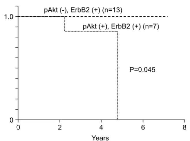
Fig. 1. Kaplan-Meier survival plots of patients with ErbB2 positive node-negative breast cancer according to pAkt expression.
Log-rank test: P=0.045.
Fig. 2. Kaplan-Meier survival plots of patients with higher Ki-67 index node-negative breast cancer in relation to pAkt expression. Log-rank test: P=0.040.
ꠏꠏꠏꠏꠏꠏꠏꠏꠏꠏꠏꠏꠏꠏꠏꠏꠏꠏꠏꠏꠏꠏꠏꠏꠏꠏꠏꠏꠏꠏꠏꠏꠏꠏꠏꠏꠏꠏꠏꠏꠏꠏꠏꠏꠏꠏꠏꠏꠏꠏꠏꠏꠏꠏꠏꠏꠏꠏꠏꠏꠏꠏꠏꠏꠏꠏꠏꠏꠏꠏꠏꠏꠏꠏꠏꠏꠏꠏꠏꠏꠏꠏꠏꠏꠏꠏꠏꠏꠏꠏꠏꠏꠏꠏꠏꠏꠏꠏꠏꠏꠏꠏꠏꠏꠏꠏꠏꠏꠏꠏꠏꠏꠏꠏꠏ
1, 2, 3은 각각 15명(20.8%), 39명(54.2%), 18명(25.0%)였고, pAkt 발현양성군은 등급 1, 2, 3에서 각각 6명(25.0%), 12명 (50.0%), 6명(25.0%)를 보였고, pAkt음성군과 통계적 유의성 은 없었다(P=0.812, Table 1).
4) 호르몬 수용체
에스트로겐 수용체(ER)는 대상 환자 72명 중 31명에서 양성이었고(ER+ 43.1%), 프로게스테론 수용체(PR)는 24명 에서 양성(PR+ 56.9%)이었다. pAkt 발현양성군은 ER+
37.5%, PR+ 41.7%였고, ER+혹은 PR+ 45.8%였다. pAkt 발현이 없던 환자에서는 ER+ 45.8%, PR+ 52.1%, ER+ 혹 은 PR+ 62.5%를 보였다. pAkt 발현 양성 유무와 호르몬 수용체 유무와는 통계적으로 유의한 차이가 없었다(ER P=0.338, PR P=0.280, ER/PR P=0.457, Table 1).
5) ErbB2
ErbB2는 발현양성 20예(27.8%), 음성 52예(72.2%)였고, ErbB2양성인 경우, pAkt양성군이 7예(29.2%)를 보였으며, pAkt양성군과 음성군 간 통계적 유의성은 없었다(Table 1).
6) Ki-67 지수
Ki-67 지수는 전체 평균 값은 17.6 (±13.2)였고, pAkt양성 군인 경우 평균 Ki-67 지수는 20.5, pAkt음성군은 16.2를 보 였다. 전체에서 Ki-67 지수가 평균보다 높을 경우가 43명 (59.7%)이었는데, pAkt양성군에서 Ki-67 지수가 평균보다 높은 예는 10명(41.7%)으로 pAkt음성군의 Ki-67 지수가 평 균보다 높은 19명(39.6%) 보다 높았으나, pAkt양성군과 음 성군 간에 통계적 유의성은 없었다(Table 1).
7) pAkt 발현과 생존율과의 관계분석(Table 2, Fig. 1, 2) 총 72명 중 5명에서 재발하였고, 그 중 2명에서 pAkt발현 양성을 보였고, Ki-67 지수는 1명에서 평균보다 높았다. 사 망한 2명은 모두 pAkt발현양성이었고, Ki-67 지수도 모두 평 균보다 높았다. 전체 환자의 5년 생존율(OS)은 86.6%였고 5년 무병생존율(DFS)은 78.4%였는데, pAkt 발현양성군은 5 년 생존율 80.0%, 5년 무병생존율 51.0%로 낮았으나 통계적 으로 유의한 차이는 없었다(DFS P=0.077, OS P=0.076).
pAkt발현과 유방암 예후인자에 따른 생존율과 무병생존율 의 비교에서는, ErbB2양성발현과 높은 Ki-67를 나타내는 환 자에서만 통계적으로 유의한 무병생존율의 차이를 보였다 (Table 2). ErbB2양성발현인 경우, 5년 무병생존율은 pAkt 발 현양성군 79.6%로 pAkt음성군 100%보다 통계적으로 유의 하게 낮았다(P=0.045, Fig. 1). 또한 Ki-67 지수가 평균보다 높은 예에서 5년 무병생존율은 pAkt 발현양성군 62.2%로 pAkt음성군 100%보다 통계적으로 유의하게 낮았다(P=
0.040, Fig. 2).
고 찰
Akt 신호 전달 체계는 종양형성에 중요한 역할을 한 다.(11) Akt는 serine/threonine kinase로, PI 3-K의 신호 전달 체계 중 Akt는 타이로신 활성화 효소 수용체에 연관되어, PI 3-K의 인산화에 의하여 활성화된다. Akt는 Bad, forkhead transcription factor, caspase 9와 같은 세포예정사 전구단백질 을 인산화하고 미토콘드리아에서 cytochrome c의 배출을 막 고 세포예정사를 억제하여 세포 생존을 증가시키며,(12) cyclin-dependent kinase inhibitor의 조절로 세포 증식에 관여 한다.(13) Akt는 표피 성장 인자(EGF),(14) 인슐린 유사 성장 Fig. 3. Invasive ductal carcinoma showing positive immunohisto-
chemical staining for phosphorylated Akt (pAkt). Fig. 4. Invasive ductal carcinoma showing positive immunohisto- chemical staining of ErbB2.
ꠏꠏꠏꠏꠏꠏꠏꠏꠏꠏꠏꠏꠏꠏꠏꠏꠏꠏꠏꠏꠏꠏꠏꠏꠏꠏꠏꠏꠏꠏꠏꠏꠏꠏꠏꠏꠏꠏꠏꠏꠏꠏꠏꠏꠏꠏꠏꠏꠏꠏꠏꠏꠏꠏꠏꠏꠏꠏꠏꠏꠏꠏꠏꠏꠏꠏꠏꠏꠏꠏꠏꠏꠏꠏꠏꠏꠏꠏꠏꠏꠏꠏꠏꠏꠏꠏꠏꠏꠏꠏꠏꠏꠏꠏꠏꠏꠏꠏꠏꠏꠏꠏꠏꠏꠏꠏꠏꠏꠏꠏꠏꠏꠏꠏꠏ 인자-I (Insulin-like growth factor-I),(14) heregulin(15) 등의 성
장 인자의 신호 전달 체계 하부(downstream)에 위치하며, 이 성장 인자들은 PI 3-K/Akt 신호 전달 체계를 통해, 에스트로 겐 수용체-α 활성화와 호르몬 비의존 성장을 유도한다. 암 세포 생존의 신호 전달 체계 중 Akt에 의한 전달 체계가 중 요한 역할을 하고 있으며, PI 3-K/Akt 신호 전달 체계를 억 제하는 암 억제 유전자가 많은 종류의 암에서 발견되고 있 어, Akt의 과발현 혹은 활성화가 암 발생과 진행에 연관이 있음을 알 수 있다.(9) 최근 연구에서, 액와부 림프절 음성 인 유방암 환자에서 Akt 활성화가 암의 공격성을 반영하여, 생존율에 독립적인 예후 인자로 알려지고 있다.(16) 본 연 구에서는 림프절 음성인 유방암 환자에서 Akt 발현 유무만 으로는 독립적인 예후 인자로서 통계적 유의성이 없었다.
이런 차이점은 유방암 발암 과정에서 Akt 신호 전달 체계뿐 만 아니라 다른 신호 전달 체계도 유방암 발생에 관여하는 것으로 생각되며, ErbB2/MAPK, Prostaglandin, AP-1 신호전 달체계 등이 알려져 있다.(17)
ErbB2의 과발현은 일반적으로 유전자 증폭과 관련 있고 이때 인산화된 형태를 보이며 이는 유방암 환자의 25∼30%
에서 발견되고, 무병 생존율과 생존율을 낮추는 것으로 알 려져 있다.(18) ErbB3는 ErbB2의 과발현과 함께 유방암 환 자에서 과발현이 나타나고, ErbB2의 변이가 있는 경우 선택 적으로 ErbB3의 발현이 항진된다. ErbB2의 비활성화는 ErbB3의 타이로신 인산화를 감소시키며, ErbB3는 타이로신 활성화 효소가 없기 때문에 인산화되고 신호 전달을 위해 서 dimerization이 필요하다.(19) ErbB3는 PI 3-K의 p85 부분 에 대한 결합 부위를 보유하고, 신호전달 체계에 관여하며, ErbB2가 과발현되었을 때 PI 3-kinase/protein kinase B의 활 성화가 감소되어 있어,(20) 활성화된 ErbB2의 신호전달에 서 ErbB3가 신호전달 체계를 자극하는 역할을 맡고 있음을 알 수 있다. ErbB2의 과활성에 의한 암 발생에서 ErbB3가 타이로신 인산화되어 발견되는 것으로, ErbB3의 활성이 중 요한 역할을 하는 것을 알 수 있다.(21) ErbB2와 ErbB3의 heterodimer가 homodimer에 비하여 분열을 최대로 촉진하 여,(22) 세포 활성, 성장과 형질 전환을 일으킨다. 표피 성장 인자 수용체와 ErbB2는 유방암을 포함한 여러 종류의 암에 서 과발현되어 있으며, 나쁜 예후와도 연관성을 보인다.(23) 세포 활성은 암 발생의 전구 단계로 받아들여지면서 특 정 신생물의 생물학적 형질을 예측할 수 있는 세포 증식능 에 관심이 모아졌다. Ki-67을 발현하는 세포의 숫자가 종양 의 조직학적 등급과 관련이 있고 이는 임상적 예후를 예측 할 수 있다고 보고되고 있다.(24) Ki-67은 원발성 유방암에 서, 치료 표지자로서는 유용하지 않다는 보고가 있는 반면, 액와부 림프절 음성인 유방암의 재발에 Ki-67이 종양의 크 기와 S기 분획과 함께 통계적 유의성을 가진 독립적인 예후 인자가 될 수 있다는 의견도 있다.(25) 다른 연구에서도 Ki-67과 무배수성(Anueploidy)이 관련이 있으며 종양의 악
성도와도 관계가 있다고 보고하고 있다.(26) 또한, 표피성장 인자 수용체와 ErbB2, Ki-67가 동시에 과발현하였을 때, 유 방암 환자에서 특히 다른 임상 병리적 변수와 같이 예후에 중요한 영향을 미치는 인자라고 평가하여 보고하고 있 다.(27) 본 연구에서는 Ki-67 지수는 단일 변수로는 재발과 생존에 통계적 유의성을 보이지는 않았지만, pAkt 양성군 에서 Ki-67 지수가 평균보다 높을 경우, 생존율에 대한 예후 인자로 통계적 유의성을 보였다.
ErbB2양성발현은 에스트로겐 수용체 및 프로게스테론 수용체와 역 상관 관계에 있으며, 액와부 림프절 음성인 환 자보다 양성인 환자에서 자주 나타난다.(28) 에스트로겐 수 용체 신호 전달 체계와 ErbB 타이로신 활성화 효소 수용체 간 교차 결합(cross coupling)이 유방암 세포에서 관찰되 며,(29) 에스트로겐 수용체-α도 ErbB2발현을 조절할 수 있 다. 또한, 유방암 세포에서 heregulin에 의한 ErbB2발현은 에 스트로겐 수용체-α의 직접적이고 빠른 인산화를 유도하고, 세포 성장과 에스트로겐 수용체-α 조절을 이루게 한다.(29) 에스트로겐 수용체는 세포 성장 조절에 중요한 역할을 하 는데, 에스트로겐 수용체 발현은 호르몬 치료의 표지자로 사용되고 있다. 세포 특이적 항에스트로겐 효과를 보이는 타목시펜(Tamoxifen)은 경쟁적으로 에스트로겐 수용체-α 에 결합하여, 유방 상피 세포의 에스트로겐 활성화로 인한 성장을 억제한다. 대부분은 에스트로겐 수용체 양성인 경 우 타목시펜 치료에 반응하지만, 타목시펜 치료에 저항성 이 있는 경우가 있다.(30) 이는 활성화 효소의 발현으로 에 스트로겐 수용체-α 인산화로 성장 인자들에 유도된 에스트 로겐 수용체-α의 활동성이 원인으로 생각된다.(30) 최근 연 구들에서 호르몬 치료에 저항성을 보이는 경우를 에스트로 겐 수용체-α의 작용으로 보고 있다. 앞으로 에스트로겐, ErbB2, 에스트로겐 수용체-α와 호르몬 치료에 대해 연구가 더 필요할 것으로 보인다.
본 연구에서는 액와부 림프절 음성인 유방암 환자에서 pAkt발현과 예후인자로 연령, 폐경, 종양의 크기, 병기, 조 직학적 등급, 에스트로겐 수용체 유무, Ki-67 지수, ErbB2의 상관성을 알아보았다. ErbB2과발현과 pAkt발현과 환자의 무병생존율이 ErbB2과발현과 pAkt음성군의 무병생존율보 다 낮았고, 평균보다 높은 Ki-67 지수와 pAkt발현군의 무병 생존율은 평균보다 낮은 Ki-67 지수와 pAkt음성군의 무병 생존율보다 낮았다.
결 론
원발성 침윤성 유방암 중 액와부 림프절 전이가 없는 72 예를 대상으로 면역조직화학 염색으로 ErbB2와 pAkt발현 유무를 통해 Akt가 유방암의 예후 인자와의 관련성 및 예후 에 미치는 영향을 알아본 결과, Ki-67 지수와 ErbB2발현은 pAkt발현 사이에 유의성을 보여, 무병생존율 비교에서 높은
ꠏꠏꠏꠏꠏꠏꠏꠏꠏꠏꠏꠏꠏꠏꠏꠏꠏꠏꠏꠏꠏꠏꠏꠏꠏꠏꠏꠏꠏꠏꠏꠏꠏꠏꠏꠏꠏꠏꠏꠏꠏꠏꠏꠏꠏꠏꠏꠏꠏꠏꠏꠏꠏꠏꠏꠏꠏꠏꠏꠏꠏꠏꠏꠏꠏꠏꠏꠏꠏꠏꠏꠏꠏꠏꠏꠏꠏꠏꠏꠏꠏꠏꠏꠏꠏꠏꠏꠏꠏꠏꠏꠏꠏꠏꠏꠏꠏꠏꠏꠏꠏꠏꠏꠏꠏꠏꠏꠏꠏꠏꠏꠏꠏꠏꠏ Ki-67 지수와 ErbB2발현은 각각 pAkt발현군에서 통계적으
로 유의한 낮은 무병생존율을 보였다. 이상의 결과로, 유방 암 조직에서 Akt발현은 액와부 림프절 전이가 없는 유방암 환자에서 ErbB2발현이나 높은 Ki-67를 보이는 경우 유방암 의 불량한 예후인자로 고려할 수 있을 것으로 판단된다.
REFERENCES
1) Seymour I, Schwartz G, Tom Shires, Frank C Spencer, John M Daly, Josef E Fischer, et al. Breast. In: Bland Kirby I, Michael P Vezeridis, Edward M Copeland III. Principles of Surgery. 7th ed. New York: McGraw-Hill; 1999. p.533-99.
2) Health and Welfare Ministry. Report of the central cancer registry in 2001, Republic of Korea 2003.
3) Page DL, Jensen RA, Simpson JF. Routinely available indica- tors of prognosis in breast cancer. Breast Cancer Res Treat 1998;51:195-208.
4) Fisher B, Slack NH. Number of lymph nodes examined and prognosis of breast carcinoma. Surg Gynecol Obstet 1970;
131:79-88.
5) Valagussa P, Bonadonna G, Veronesi U. Patterns of relapse and survival in operable breast carcinoma with positive and negative axillary nodes. Tumori 1978;64:241-58.
6) Hortobagyi GN. Opportunities and challenges in the devel- opment of targeted therapies. Semina Oncol 2004;31(1 Suppl 3):21-7.
7) Olayioye MA, Neve RM, Lane HA, Hynes NE. The ErbB signaling network: receptor heterodimerization in development and cancer. EMBO J 2000;19:3159-67.
8) Robinson MJ, Cobb MH. Mitogen-activated protein kinase pathways. Curr Opin Cell Biol 1997;9:180-6.
9) Cantley LC, Neel BG. New insights into tumor suppression:
PTEN suppresses tumor formation by restraining the phos- phoinositide 3-kinase/AKT pathway. Proc Natl Acad Sci USA 1999;96:4240-5.
10) Elston CW, Ellis IO. Pathologic prognostic factors in breast cancer. The value of histologic grade in breast cancer: ex- perience from a large study with long-term follow-up. Histo- pathology 1991;19:403-10.
11) Testa JR, Bellacosa A. AKT plays a central role in tumori- genesis. Proc Natl Acad Sci USA 2001;98:10983-5.
12) Datta SR, Dudek H, Tao X, Masters S, Fu H, Gotoh Y, et al. Akt phosphorylation of BAD couples survival signals to the cell-intrinsic death machinery. Cell 1997;91:231-41.
13) Zhou BP, Liao Y, Xia W, Spohn B, Lee MH, Hung MC.
Cytoplasmic localization of p21Cip1/WAF1 by Akt-induced phosphorylation in HER-2/neu-overexpressing cells. Nat Cell Biol 2001;3:245-52.
14) Martin MB, Franke TF, Stoica GE, Chambon P, Katzenellen- bogen BS, Stoica BA, et al. A role for Akt in mediating the estrogenic functions of epidermal growth factor and insulin-
like growth factor I. Endocrinology 2000;141:4503-11.
15) Liu W, Li J, Roth RA. Heregulin regulation of Akt/protein kinase B in breast cancer cells. Biochem Biophys Res Com- mun 1999;261:897-903.
16) Schmitz KJ, Otterbach F, Callies R, Levkau B, Holscher O, Hoffmann O, et al. Prognostic relevance of activated Akt kinase in node-negative breast cancer: a clinicopathological study of 99 cases. Mod Pathology 2004;17:15-21.
17) Shen Q, Brown PH. Novel agents for the prevention of breast cancer: targeting transcription factors and signal transduction pathways. J Mammary Gland Biol Neoplasia 2003;8:45-73.
18) Slamon DJ, Clark GM, Wong SG, Levin WJ, Ullrich A, McGuire WL. Human breast cancer: correlation of relapse and survival with amplification of the HER-2/neu oncogene.
Science 1987;235:177-82.
19) Kim HH, Vijapurkar U, Hellyer NJ, Bravo D, Koland JG.
Signal transduction by epidermal growth factor and heregulin via the kinase-deficient ErbB3 protein. Biochem J 1998;334:
189-95.
20) Basso AD, Solit DB, Munster PN, Rosen N. Ansamycin anti- biotics inhibit Akt activation and cyclin D expression in breast cancer cells that overexpress HER2. Oncogene 2002;21:
1159-66.
21) Siegel PM, Ryan ED, Cardiff RD, Muller WJ. Elevated expres- sion of activated forms of Neu/ErbB-2 and ErbB-3 are involved in the induction of mammary tumors in transgenic mice: implications for human breast cancer. EMBO J 1999;
18:2149-64.
22) Pinkas-Kramarski R, Soussan L, Waterman H, Levkowitz G, Alroy I, Klapper L, et al. Diversification of Neu differentiation factor and epidermal growth factor signaling by combinatorial receptor interactions. EMBO J 1996;15:2452-67.
23) Salomon DS, Perroteau I, Kidwell WR, Tam J, Derynck R.
Loss of growth responsiveness to epidermal growth factor and enhanced production of alpha-transforming growth factors in ras-transformed mouse mammary epithelial cells. J Cell Physiol 1987;130:397-409.
24) Bacchi CE, Gown AM. Detection of cell proliferation in tissue sections. Braz J Med Biol Res 1993;26:677-87.
25) Brown RW, Allred CD, Clark GM, Osborne CK, Hilsenbeck SG. Prognostic value of Ki-67 compared to S-phase fraction in axillary node-negative breast cancer. Clin Cancer Res 1996;
2:585-92.
26) Martinez-Arribas F, Nunez MJ, Piqueras V, Lucas AR, San- chez J, Tejerina A, et al. Flow cytometry vs. Ki67 labelling index in breast cancer: a prospective evaluation of 181 cases.
Anticancer Res 2002;22:295-8.
27) Ioachim E, Kamina S, Athanassiadou S, Agnantis NJ. The prognostic significance of epidermal growth factor receptor (EGFR), C-erbB-2, Ki-67 and PCNA expression in breast cancer. Anticancer Res 1996;16:3141-7.
28) Bacus SS, Chin D, Yarden Y, Zelnick CR, Stern DF. Type
ꠏꠏꠏꠏꠏꠏꠏꠏꠏꠏꠏꠏꠏꠏꠏꠏꠏꠏꠏꠏꠏꠏꠏꠏꠏꠏꠏꠏꠏꠏꠏꠏꠏꠏꠏꠏꠏꠏꠏꠏꠏꠏꠏꠏꠏꠏꠏꠏꠏꠏꠏꠏꠏꠏꠏꠏꠏꠏꠏꠏꠏꠏꠏꠏꠏꠏꠏꠏꠏꠏꠏꠏꠏꠏꠏꠏꠏꠏꠏꠏꠏꠏꠏꠏꠏꠏꠏꠏꠏꠏꠏꠏꠏꠏꠏꠏꠏꠏꠏꠏꠏꠏꠏꠏꠏꠏꠏꠏꠏꠏꠏꠏꠏꠏꠏ 1 receptor tyrosine kinases are differentially phosphorylated in
mammary carcinoma and differentially associated with steroid receptors. Am J Pathol 1996;148:549-58.
29) Pietras RJ, Arboleda J, Reese DM, Wongvipat N, Pegram MD, Ramos L, et al. HER-2 tyrosine kinase pathway targets
estrogen receptor and promotes hormone-independent growth in human breast cancer cells. Oncogene 1995;10:2435-46.
30) Johnston SR. Acquired tamoxifen resistance in human breast cancer--potential mechanisms and clinical implications. Anti- cancer Drugs 1997;8:911-30.