© 2017 Korean Breast Cancer Society. All rights reserved. http://ejbc.kr | pISSN 1738-6756
INTRODUCTION
The involvement of axillary lymph node (LN) in patients with invasive breast cancer is one of the most influential prog- nostic factors [1]. Lately, complete axillary dissection is not routinely performed, even in cases where metastatic sentinel LNs (SLNs) are detected [2]. In the American College of Sur- geons Oncology Group (ACOSOG) Z0011 trial, axillary LN dissection (ALND) and axillary radiotherapy after detection
of positive SLNs provided excellent and comparable axillary control for patients with T1–2 primary breast cancer and no palpable lymphadenopathy, and results in significantly less morbidity than does complete axillary dissection [2]. In After Mapping of the Axilla: Radiotherapy or Surgery (AMAROS) trial, SLN biopsy (SLNB) without ALND offered excellent re- gional control and this regimen may be a reasonable option for the management for selected patients with early-stage breast cancer treated with breast-conserving therapy and ad- juvant systemic therapy [3].
According to the seventh edition of the American Joint Committee for Cancer (AJCC), breast cancer staging N2 with T1–2 falls into stage IIIA, while N3 with any T falls into stage IIIC. Stages IIIA and IIIC refer to locally advanced breast can- cer [4]. One might question why many doctors agree that it is safe not to perform complete ALND (cALND) in locally ad- vanced breast cancer. We investigated the percentage of N2 or N3 stages in T1–2 invasive breast cancer patients with no lymphadenopathy, and developed a nomogram for the pre-
Development of a Nomogram to Predict N2 or N3 Stage in T1–2 Invasive Breast Cancer Patients with No Palpable Lymphadenopathy
Isaac Kim, Jai Min Ryu, Jai Myeong Kim, Hee Jun Choi, Se Kyung Lee, Jong Hwan Yu, Jeong Eon Lee, Seok Won Kim, Seok Jin Nam
Division of Breast and Endocrine Surgery, Department of Surgery, Samsung Medical Center, Sungkyunkwan University School of Medicine, Seoul, Korea ORIGINAL ARTICLE
Purpose: Subsequent to the American College of Surgeons Oncology Group (ACOSOG) Z0011 and After Mapping of the Axilla: Radiotherapy or Surgery (AMAROS) trials, complete axil- lary lymph node dissection is not routinely performed, even in cases where metastatic sentinel lymph nodes are detected. We investigated the percentage of N2 or N3 stages in T1–2 invasive breast cancer patients with no lymphadenopathy and developed a nomogram to predict the possibility of N2 or N3 stages in these patients. Methods: We retrospectively reviewed the charts of invasive breast cancer patients who were clinically N0 stage, but had a positive sentinel or non-sentinel lymph node detected on sentinel lymph node biopsy. The association of potential risk factors with known outcomes (N2 or N3 stages) was tested us- ing logistic regression analysis. Variables with p<0.05 in the multivariate analysis were included in the nomogram. Internal performance validation was carried out using a 5-fold cross vali-
dation method. Results: Among a total of 1,437 patients, 1,355 patients had stage N1 disease (94.3%), while 82 had stage N2 or N3 disease (5.7%). Multivariate stepwise logistic regression analysis revealed lymphovascular invasion (p=0.008), T2 stage (p=0.026), metastatic lymph node ratio (p<0.001), and perinod- al extension (p<0.001) as independent predictors of N2 or N3 stages. A nomogram was developed based on these factors.
The area under the curve estimated from the receiver operating characteristic graph was 0.8050 in the model set and 0.8246 in the test set. Conclusion: Our nomogram can be employed for the prediction of N2 or N3 stage among cases fulfilling the ACOSOG Z0011 or AMAROS criteria.
Key Words: Breast neoplasms, Lymph node excision, Nomograms, Sentinel lymph node biopsy
Correspondence to: Seok Won Kim
Division of Breast and Endocrine Surgery, Department of Surgery, Samsung Medical Center, Sungkyunkwan University School of Medicine, 81 Irwon-ro, Gangnam-gu, Seoul 06351, Korea
Tel: +82-2-3410-0260, Fax: +82-2-3410-6982 E-mail: [email protected]
This article was supported by a grant from the Korea Health Technology R&D project through the Korea Health Industry Development Institute (KHIDI), funded by the Ministry of Health & Welfare, Republic of Korea (HI17C1142).
Received: August 13, 2017 Accepted: September 12, 2017
Cancer
diction of N2 or N3 stage in these patients.
METHODS
Study population
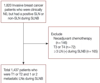
We performed a retrospective chart review of 1,820 invasive breast cancer patients who were clinically N0, but had a posi- tive SLN or non-SLNs (NSLNs) during SLNB. All patients underwent ALND between January 2005 and January 2015 at Samsung Medical Center. Patients were excluded if they had stage T3 or higher disease and had undergone neoadjuvant chemotherapy (NAC) due to high false-negative rate of SLNB after NAC [5]. Those with only 1 or 2 metastatic LNs during SLNB were included. A total of 1,437 patients were ultimately included (Figure 1). All electronic medical records and pa- thology reports were reviewed. This study adhered to the te- nets of the Declaration of Helsinki. This study was approved by the Institutional Review Board of Samsung Medical Center (IRB number: 2016-09-117).
Sentinel lymph node mapping
We conducted lymphatic mapping using technetium-99m (99mTc) sulfur colloid diluted in normal saline solution and/or vital blue dye (1% indigo carmine). The site and timing of ad- ministration of the mapping agent were at the physician’s dis- cretion. The radiolabeled colloid was injected 1 to 6 hours preoperatively, and/or 1 to 3 mL 1% indigo carmine was in- jected periareolarly and massaged into the breast tissue for 5 minutes. A handheld gamma detection probe (Neoprobe 2000; Neoprobe Corp., Dublin, USA) was used to scan the ax-
illa transcutaneously and identify the most radioactive area.
All radioactive and/or vital blue dye mapped LNs were ex- cised and submitted as SLNs while palpable LNs without up- take of radioactive and/or vital blue dye were excised and sub- mitted as NSLNs.
Histopathological evaluation
The dissected LNs were measured. Serial sections of 2-mm thickness were prepared from a portion of the frozen, dissect- ed LNs. The remaining tissue was fixed in 10% formalin, em- bedded in paraffin blocks, and stained with hematoxylin and eosin (H&E). Pathologic evaluation of the sections was per- formed. If the H&E-stained sections were negative for malig- nancy, additional sections were immunohistochemically stained for keratin using the monoclonal, anti-human cyto- keratin antibody (clone AE1/AE3; Dako, Carpinteria , USA).
Data analysis
We retrospectively evaluated the following clinicopathol- ogic factors: age, menopausal status, body mass index (BMI), family history of breast cancer, type of surgery, laterality, mul- tiplicity, histopathological type of breast cancer, estrogen re- ceptor (ER) status, progesterone receptor status, human epi- dermal growth factor receptor 2 (HER2) status, lymphovascu- lar invasion (LVI), perinodal extension (PNE), extensive in- traductal component, nuclear grade, Ki-67 status, pathological T stage according to the seventh edition of the AJCC classifi- cation, subtype; luminal A-like was group showing positive ER and negative HER2 expression. Luminal B-like was group showing not only positive for both ER and HER2 expression but also positive ER and negative HER2 expression and more than 14% ki-67. HER2 was group showing negative ER and positive HER2 expression. Triple-negative breast cancer (TNBC) was group showing negative for both ER and HER2 expression, method of SLN mapping (radiolabeled colloid and/or vital blue dye), number of removed LNs, and number of metastatic LNs observed on SLNB. Proportion analysis was conducted using the chi-square or Fisher exact tests. The as- sociation of potential risk factors with outcomes (N2 or N3) was tested using logistic regression analysis. In the case of rare events, we applied a logistic regression model using the Firth’s penalized maximum likelihood estimation method. Variables with p<0.05 in a univariate analysis were included in a multi- variate analysis. Stepwise selection methods were applied to identify covariates for the logistic regression model. Variables with p<0.05 in the multivariate analysis were included in the nomogram. The adjusted area under the receiver operating characteristic (ROC) curve (AUC) was calculated in order to quantify the ability to rank patients by risk. Internal perfor-
1,820 Invasive breast cancer patients who were clinically N0, but had a positive SLN or
non-SLN during SLNB
Total 1,437 patients who were T1 or T2 and 1 or 2 metastatic LNs during SLNB
Exclude
N eoadjuvant chemotherapy (n=146)
T3 or T4 (n=72)
≥3 LN (+) during SLNB (n=165)
Figure 1. Flow chart of the patient inclusion process.
SLN=sentinel lymph node; SLNB=sentinel lymph node biopsy; LN=
lymph node.
mance validation was performed using the 5-fold cross vali- dation method. Statistical analyses were performed using the statistical software SAS version 9.4 (SAS Institute Inc., Cary, USA). A nomogram was formulated based on the results of the stepwise multivariate analysis using the software R 3.0.3 (R foundation for Statistical Computing, Vienna, Austria; http://
www.R-project.org/).
RESULTS
Proportion of N2 or N3 stages
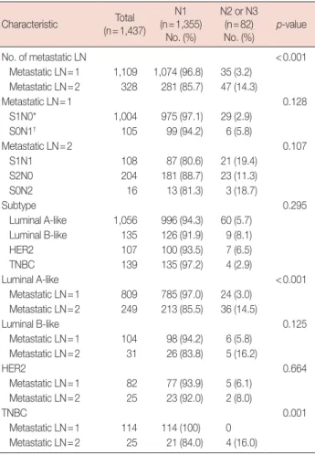
Among the 1,437 patients, 1,355 patients had stage N1 dis- ease (94.3%), while 82 had stage N2 or N3 disease (5.7%) (Ta- ble 1). The proportion of stages N2 or N3 was higher (14.3%)
when there were two metastatic LNs (3.2%), as opposed to when there was just one (p<0.001). There were no significant differences by breast cancer subtype.
The proportional differences with regard to the number of metastatic LNs are also listed in Table 1. In luminal A-like type, the proportion of stages N2 or N3 was higher when there were two metastatic LNs as opposed to when there was just one. In case of TNBC, there were no N2 or N3 cases ob- served, when there was only one metastatic LN; however, there were N2 or N3 cases when there were two metastatic LNs. For the luminal B-like subtype and HER2(+), the num- ber of metastatic LNs did not affect the proportion of N stages.
We performed a sub-classification of the N stage ratios based on all the cases of SLNs and NSLNs (Table 1). The ratio of N2 or N3 tended to be higher in the S0N1 group than in the S1N0 group among cases with 1 metastatic LN, although the difference was not significant. Among cases with two met- astatic LNs, the proportion of N2 or N3 tended to be higher in cases in which the NSLNs were metastatic than in which they were nonmetastatic. However, this difference was also not sig- nificant.
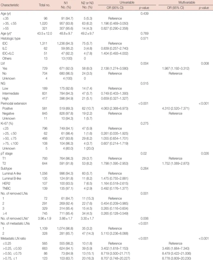
Patient characteristics and factors associated with N2 or N3 stages
Patient characteristics and N stage proportions are listed in Table 2. The following parameters had no association with N stage: age, BMI, menopause, family history, surgery type, lat- erality, multiplicity, histologic tumor type, extensive intra- ductal component, nuclear grade, Ki-67 status, subtype, and SLNs detection method.
Multivariate stepwise logistic regression showed LVI (p=
0.008), PNE (p<0.001), T2 stage (p=0.026) and metastatic LN rate (p<0.001) were independent predictors of N2 or N3.
There was an association between the number of metastatic LNs and the metastatic LN rate. Therefore, we included only the metastatic LN rate in the multivariate analysis.
Nomogram
This nomogram was developed based on the following four variables: metastatic LN ratio, PNE, LVI, and T stage (Figure 2). The total sum of each variable was located on a total point line. This line was drawn with a negative slope to calculate the probability of N2 or N3 stage. The AUC of the ROC graph was 0.8050 in the model set (Figure 3A) and 0.8246 in the test set (Figure 3B). We also developed another nomogram using size of metastatic tumor (Figure 4). The AUC of the ROC graph was 0.8129 in the model set and 0.7905 in the test set.
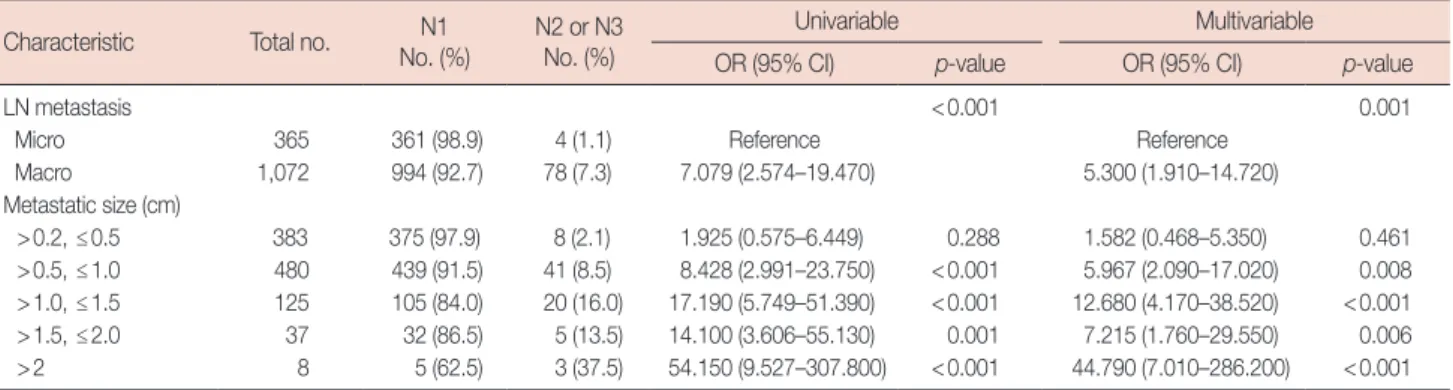
Uni- and multi-variable analyses of metastasis size related to nodal stage are shown in Table 3.
Table 1. Proportion of N stage in T1–2 invasive breast cancer patients with clinical N0 but metastatic lymph node as sentinel lymph node bi- opsy
Characteristic Total (n=1,437)
N1 (n=1,355)
No. (%)
N2 or N3 (n=82)
No. (%) p-value
No. of metastatic LN <0.001
Metastatic LN=1 1,109 1,074 (96.8) 35 (3.2) Metastatic LN=2 328 281 (85.7) 47 (14.3)
Metastatic LN=1 0.128
S1N0* 1,004 975 (97.1) 29 (2.9)
S0N1† 105 99 (94.2) 6 (5.8)
Metastatic LN=2 0.107
S1N1 108 87 (80.6) 21 (19.4)
S2N0 204 181 (88.7) 23 (11.3)
S0N2 16 13 (81.3) 3 (18.7)
Subtype 0.295
Luminal A-like 1,056 996 (94.3) 60 (5.7) Luminal B-like 135 126 (91.9) 9 (8.1)
HER2 107 100 (93.5) 7 (6.5)
TNBC 139 135 (97.2) 4 (2.9)
Luminal A-like <0.001
Metastatic LN=1 809 785 (97.0) 24 (3.0) Metastatic LN=2 249 213 (85.5) 36 (14.5)
Luminal B-like 0.125
Metastatic LN=1 104 98 (94.2) 6 (5.8) Metastatic LN=2 31 26 (83.8) 5 (16.2)
HER2 0.664
Metastatic LN=1 82 77 (93.9) 5 (6.1) Metastatic LN=2 25 23 (92.0) 2 (8.0)
TNBC 0.001
Metastatic LN=1 114 114 (100) 0 Metastatic LN=2 25 21 (84.0) 4 (16.0)
N1=nodal stage 1; N2=nodal stage 2; N3, nodal stage3; LN=lymph node;
S, sentinel node; N, non-sentinel node; HER2=human epidermal growth fac- tor receptor 2; TNBC=triple-negative breast cancer.
*S1N0, 1 metastatic sentinel lymph node and no metastatic non-sentinel lymph node during sentinel lymph node biopsy; †S0N1, no metastatic sentinel lymph node and 1 metastatic non-sentinel lymph node during sentinel lymph node biopsy.
Table 2. Univariable and multivariable analysis of related factors to nodal stage in T1–2 breast cancer patients with no lymphadenopathy
Characteristic Total no. N1
No. (%) N2 or N3 No. (%)
Univariable Multivariable
OR (95% CI) p-value OR (95% CI) p-value
Age (yr) 0.439
≤35 96 91 (94.7) 5 (5.3) Reference
>35, ≤55 1,020 957 (93.8) 63 (6.2) 1.196 (0.469–3.050)
>55 321 307 (95.6) 14 (4.3) 0.827 (0.290–2.358)
Age (yr)* 43.5±12.0 48.8±9.7 49.2±9.7 0.769
Histologic type 0.571
IDC 1,311 1,236 (94.3) 75 (5.7) Reference
ILC 62 59 (95.2) 3 (4.8) 0.839 (0.257–2.740)
IDC+ILC 51 47 (92.2) 4 (7.8) 1.404 (0.493–4.002)
Others 13 13 (100) 0
LVI 0.004 0.008
Yes 729 671 (92.0) 58 (8.0) 2.138 (1.274–3.580) 1.987 (1.192–3.312)
No 704 680 (96.5) 24 (3.5) Reference Reference
Unknown 4 4 (100) 0
NG 0.515
Low 189 175 (92.6) 14 (7.4) Reference
Intermediate 831 784 (94.3) 47 (5.7) 0.749 (0.403–1.393)
High 417 396 (94.9) 21 (5.1) 0.659 (0.327–1.327)
Perinodal extension <0.001 <0.001
Positive 581 519 (89.3) 62 (10.7) 4.063 (2.368–6.973) 4.310 (2.520–7.371)
Negative 845 826 (97.8) 19 (2.2) Reference Reference
Unknown 11 10 (94.3) 1 (5.7)
Ki-67 (%) 0.275
≤25 796 749 (94.1) 47 (5.9) Reference
>25, ≤50 62 61 (98.4) 1 (1.6) 0.261 (0.035–1.925) >50, ≤75 466 437 (93.8) 29 (6.2) 1.055 (0.654–1.701) >75, ≤100 108 104 (96.3) 4 (3.7) 0.607 (0.214–1.719)
Unknown 5 4 (80.0) 1 (20.0)
pT stage 0.02 0.026
T1 793 764 (96.3) 29 (3.7) Reference Reference
T2 644 591 (91.8) 53 (8.2) 1.798 (1.095–2.953) 1.752 (1.069–2.870)
Subtype 0.264
Luminal A-like 1,056 996 (94.3) 60 (5.7) Reference
Luminal B-like 135 124 (91.8) 11 (8.2) 1.475 (0.755–2.881)
HER2 107 100 (93.5) 7 (6.5) 1.164 (0.518–2.615)
TNBC 139 135 (97.1) 4 (2.9) 0.492 (0.176–1.377)
No. of removed LNs 0.001
1 72 61 (84.7) 11 (15.3) Reference
2 291 269 (92.4) 22 (7.6) 0.454 (0.209–0.985)
3 329 314 (95.4) 15 (4.5) 0.265 (0.116–0.604)
≥4 745 711 (95.4) 34 (4.5) 0.265 (0.128–0.549)
No. of removed LNs* 3.96±1.9 3.86±1.7 3.30±1.7 0.006
No. of metastatic LNs <0.001
1 1,109 1,074 (96.8) 35 (3.2) Reference
2 328 281 (85.7) 47 (14.3) 5.110 (3.236–8.068)
Metastatic LN ratio <0.001 <0.001
≤0.25 565 555 (98.2) 10 (1.8) Reference Reference
>0.25, ≤0.50 663 624 (94.1) 39 (5.9) 3.402 (1.618–7.153) 3.495 (1.664–7.340) >0.50, ≤0.75 86 73 (84.9) 13 (15.1) 8.719 (3.500–21.717) 8.479 (3.422–21.006)
>0.75, ≤1 123 103 (83.7) 20 (16.3) 8.707 (3.748–20.227) 8.778 (3.809–20.230)
N1=nodal stage 1; N2=nodal stage 2; N3=nodal stage3; OR=odds ratio; CI=confidence interval; IDC=invasive ductal carcinoma; ILC=invasive lobular carcinoma;
LVI=lymphovascular invasion; NG=nuclear grade; HER2=human epidermal growth factor receptor 2; TNBC=triple-negative breast cancer; LNs=lymph nodes.
*Mean±SD.
Figure 2. Nomogram to predict likelihood of N2 or N3 stage in clinical T1–2N0M0 breast cancer patients using four variables (metastasis node ratio, perinodal extension, lymphovascular invasion, and T stage).
Figure 4. Nomogram to predict likelihood of N2 or N3 stage in clinical T1–2N0M0 breast cancer patients including metastasis size data (metastasis size, metastasis node ratio, lymphovascular invasion, and T stage).
0 10 20 30 40 50 60 70 80 90 100
0 10 20 30 40 50 60 70 80 90 100 0 20 40 60 80 100 120 140 160 180 200 220 240
0 20 40 60 80 100 120 140 160 180
0.1 0.2 0.3
0.1 0.2 0.3 0.4 0.5 0.6 T2
Positive T1
Negative Negative
T1 Negative
0–0.25 0–0.25
Micrometastasis
Positive
T2
Positive
0.25–0.5 0.5–
0.25–0.5
1.5–2.0 0.2–0.5
0.75–1
2.0–
0.5–0.75
0.5–1.0 1.0–1.5 Points
Points
Metastasis node ratio
Metastasis size (cm) Perinodal extension
Metastasis node ratio Lymphovascular invasion
T stage T stage
Lymphovascular invasion Total points
Total points Predicted value
Predicted value
Figure 3. The area under the curve of the receiver operating characteristic graph. (A) In the model set (0.8050) and (B) in the test set (0.8246).
1.00
0.75
0.50
0.25
0
1.00
0.75
0.50
0.25
0
0 0.25 0.50 0.75 1.00 0 0.25 0.50 0.75 1.00
1-Specificity 1-Specificity
Sensitivity Sensitivity
A B
Machine learning analysis
We also developed a predictive model using the support vector classifier (SVC) by scikit-learn, a machine learning li- brary [6]. We analyzed the same data used in logistic regres- sion analysis. Among the 1,437 cases, 932 cases were used for modeling and 505 cases used for testing. The average accuracy of prediction was 0.840 (Table 4). The predictive model using the SVC was not superior to the nomogram evaluated using logistic regression analysis.
Prospective validation
We conducted a prospective validation with 100 patients who developed ALND after SLNB and T1–2 stage in 2016, at Samsung Medical Center. When we applied a predictive value
of 0.3 to our nomogram, the accuracy was 0.96 (Table 5).
DISCUSSION
Among the 1,437 cases, there were 82 cases (5.7%) of N2 or N3 stages, with 73 cases of N2 and nine of N3. These cases made up a low percentage of the total number of cases; none- theless, it was important to analyze them carefully because T1–2 with N2 breast cancer belongs in stage IIIA, while any T with N3 belongs in stage IIIC.
There were several studies about nomogram for prediction of axillary lymph node metastasis [7,8]. Previous studies re- viewed the records of breast cancer patients who underwent cALND and developed nomogram for prediction of four or Table 3. Univariable and multivariable analysis of metastatic size related to nodal stage in T1–2 breast cancer patients with no lymphadenopathy
Characteristic Total no. N1 No. (%)
N2 or N3 No. (%)
Univariable Multivariable
OR (95% CI) p-value OR (95% CI) p-value
LN metastasis <0.001 0.001
Micro 365 361 (98.9) 4 (1.1) Reference Reference
Macro 1,072 994 (92.7) 78 (7.3) 7.079 (2.574–19.470) 5.300 (1.910–14.720)
Metastatic size (cm)
>0.2, ≤0.5 383 375 (97.9) 8 (2.1) 1.925 (0.575–6.449) 0.288 1.582 (0.468–5.350) 0.461 >0.5, ≤1.0 480 439 (91.5) 41 (8.5) 8.428 (2.991–23.750) <0.001 5.967 (2.090–17.020) 0.008 >1.0, ≤1.5 125 105 (84.0) 20 (16.0) 17.190 (5.749–51.390) <0.001 12.680 (4.170–38.520) <0.001 >1.5, ≤2.0 37 32 (86.5) 5 (13.5) 14.100 (3.606–55.130) 0.001 7.215 (1.760–29.550) 0.006 >2 8 5 (62.5) 3 (37.5) 54.150 (9.527–307.800) <0.001 44.790 (7.010–286.200) <0.001 N1=nodal stage 1; N2=nodal stage 2; N3=nodal stage3; OR=odds ratio; CI=confidence interval; LN=lymph node; Micro=micrometastasis, meta size ≤0.2 cm; Macro=macrometastasis, meta size >0.2 cm.
Table 4. Prediction by machine learning, support vector classification (total 505 patients)*
Category True
Precision† Recall‡ F-1 score§
N1 N2 or N3
Predicted
N1 384 40 0.910 0.910 0.910
N2 or N3 39 42 0.520 0.510 0.520
Average 0.840 0.840 0.840
N1=nodal stage 1; N2=nodal stage 2; N3=nodal stage 3.
*Among 1,437 cases, 932 cases were used for modeling and 505 cases for testing; †Precision, positive predictive value=true positive/true positive+false positive;
‡Recall, sensitivity=true positive/true positive+false negative; §F-1 score, accuracy.
Table 5. Prospective validation (total 100 cases)
Predicted value True False
Specificity* Sensitivity† Precision‡ Accuracy§
Positive Negative Positive Negative
Predicted value 0.2ǁ
No. of cases 69 25 1 5 0.960 0.930 0.980 0.940
Predicted value 0.3ǁ
No. of cases 71 25 2 2 0.920 0.970 0.970 0.960
*Specificity, true negative rate=true negative/true negative+false positive; †Sensitivity, true positive rate=true positive/true positive+false negative; ‡Precision, posi- tive predictive value=true positive/true positive+false positive; §Accuracy=true positive+true negative/total; ǁEach predicted value of our nomogram was applied.
more metastatic lymph node [7,8]. Our nomogram was dif- ferent from previous nomogram in that prediction of N2 or N3 stage patients among cases fulfilling the ACOSOG Z0011 or AMAROS criteria.
In the ACOSOG Z0011 trial, cases with 1 to 2 metastatic SLNs were included. In contrast, we included cases with 1 to 2 metastatic LNs (SLNs or NSLNs) identified during SLNB, be- cause the accuracy of SLN detection is not always ideal, and NSLNs are occasionally metastatic. The false-negative rate of SLN detection is approximately 4% to 11% [9]. In the B-32 trial, which was designed to establish whether SLNs resection could achieve the same therapeutic goals as cALND, 3.9% of cases were identified by palpation alone (515 of 13,171) [10].
If we had enrolled only 1 to 2 metastatic SLNs cases in our sample pool, there would be 179 N2 or N3 cases (N2, 151 and N3, 28), i.e., 12.5% of the total 1,426 cases.
We collected data from both patients treated with breast- conserving surgery (BCS) and those treated with total mas- tectomy (TM), unlike the ACOSOG Z0011 trial, which only included patients treated with BCS. We intended to determine the actual percentage of N2 or N3 cases in breast cancer with T1–2 (and clinically N0). In this study, the T1–2 settings were the same as that in the ACOSOG Z0011 study. In addition, the type of surgery (BCS or TM) was not a significant factor influencing N2 or N3 stages.
In the ACOSOG Z0011 trial, there were no differences in survival or disease-free survival at 5 years [2]. According to new long-term results from the ACOSOG Z0011 trial (a me- dian follow-up of 9.25 years), there were also no significant differences in local recurrence-free survival [11]. Several stud- ies have appraised the results of the ACOSOG Z0011 trial [12- 14]. Of all the enrolled patients, approximately 70% had T1 disease, 83% had ER-positive, and 35% had only micro metas- tases to the SLN [12]. There also may have been bias related to patient selection. Leitch et al. [13] reported that 69% of pa- tients with node-positive disease who did not enroll in the Z0011 trial were treated with cALND. Additional axillary me- tastases were identified in 27% of patients who underwent ALND, indicating that 73% of patients who underwent ALND had only 1 to 2 metastatic LNs [14]. These delibera- tions are important for interpreting the Z0011 data and using them in clinical practice. If these trials had included more cases with a higher disease burden, survival results would have been different.
The metastatic node ratio, LVI, PNE, and metastasis size, as established by several mathematical models, are important factors for predicting additional nodal disease [15,16]. In par- ticular, the T2 stage has been shown to be a valuable predictor in previous studies. For example, Hwang et al. [17] found that
size of the primary tumor >2 cm was an independent predic- tor of positive NSLNs (odds ratio, 4.1; 95% confidence inter- val, 1.42–11.89; p=0.009).
In addition, metastasis size has been found to be an impor- tant factor that predicts additional nodal involvement in pre- vious studies [18,19]. We did analyze metastasis size with more segmented grouping than previous studies. In particu- lar, groups of 1.0 to 1.5 cm and more than 2.0 cm were closely correlated with N2 or N3 stage. We assumed that the relatively lower odds ratio of the 1.5 to 2.0 cm group than that of the 1.0 to 1.5 cm group was caused by the small number of 1.5 to 2.0 cm cases. If there were more number of cases, the prediction accuracy of our nomogram would have improved.
The SVC has shown comparable performance in the few studies on medical data sets [20,21]. In our results, the predic- tive model using the SVC was not superior to those obtained with the nomogram, using logistic regression analysis. Logis- tic regression models are useful to test the statistical signifi- cance of the coefficients in the model and it has a linear nature [22]. Currently, the SVC builds optimal separating boundaries between data sets and produce dichotomous results [22]. Lo- gistic regression is more suitable for the prediction of N stage.
However, the SVC also showed reasonable predictive results (the average accuracy was 0.840).
Our study has several limitations. First, we used results from permanent tissue for analysis. In previous studies, there is no mention of clear references regarding the results of LVI from preoperative biopsies or permanent tissues [15,23]. In order to apply our nomogram clinically, we would need data from pre- or intra-operative biopsies. Some authors have found that core needle biopsy (CNB) is unable to accurately detect LVI [24,25]. In contrast, in a study of 500 cases of inva- sive carcinoma diagnosed by CNB, Harris et al. [25] found a 69% concordance rate between CNB and surgical specimens, in LVI detection. PNE on frozen section is not only used in breast cancer [26], but also in prostate cancer research [27].
Therefore, it is possible that the PNE results from frozen sec- tions during SLNB apply to our nomogram too. In the future, we plan to develop a new nomogram that includes only pre- or intraoperative biopsy results. Second, we did not collect complication data including wound infections, axillary sero- mas, lymphedema, and paresthesias that could have been in- creased by ALND than by SLNB [28]. Third, we did not con- duct external validation. We tried to obtain external data but we could not find detailed data that included metastatic results of SLN or NSLN. Hence, further external validation is needed for improving the value of the nomogram in clinical use.
There is a growing tendency to avoid ALND in patients with clinical T1–2 N0M0 stage breast cancer. In addition, SLNB is
not acceptable in stage IIIA or IIIC patients as much as it is in early-stage breast cancer patients. Our nomogram could be used for the selection of N2 or N3 stage patients, among cases fulfilling the ACOSOG Z0011 or AMAROS criteria.
CONFLICT OF INTEREST
The authors declare that they have no competing interests.
REFERENCES
1. Bevilacqua JL, Kattan MW, Fey JV, Cody HS 3rd, Borgen PI, Van Zee KJ. Doctor, what are my chances of having a positive sentinel node? A validated nomogram for risk estimation. J Clin Oncol 2007;25:3670-9.
2. Giuliano AE, McCall L, Beitsch P, Whitworth PW, Blumencranz P, Leitch AM, et al. Locoregional recurrence after sentinel lymph node dissection with or without axillary dissection in patients with sentinel lymph node metastases: the American College of Surgeons Oncology Group Z0011 randomized trial. Ann Surg 2010;252:426-32.
3. Donker M, van Tienhoven G, Straver ME, Meijnen P, van de Velde CJ, Mansel RE, et al. Radiotherapy or surgery of the axilla after a positive sentinel node in breast cancer (EORTC 10981-22023 AMAROS): a randomised, multicentre, open-label, phase 3 non-inferiority trial. Lancet Oncol 2014;15:1303-10.
4. Esteva FJ, Hortobagyi GN. Locally advanced breast cancer. Hematol Oncol Clin North Am 1999;13:457-72.
5. Han A, Moon HG, Kim J, Ahn SK, Park IA, Han W, et al. Reliability of sentinel lymph node biopsy after neoadjuvant chemotherapy in breast cancer patients. J Breast Cancer 2013;16:378-85.
6. Lee Y, Lee CK. Classification of multiple cancer types by multicategory support vector machines using gene expression data. Bioinformatics 2003;19:1132-9.
7. Katz A, Smith BL, Golshan M, Niemierko A, Kobayashi W, Raad RA, et al. Nomogram for the prediction of having four or more involved nodes for sentinel lymph node-positive breast cancer. J Clin Oncol 2008;26:
2093-8.
8. Unal B, Gur AS, Beriwal S, Tang G, Johnson R, Ahrendt G, et al. Pre- dicting likelihood of having four or more positive nodes in patient with sentinel lymph node-positive breast cancer: a nomogram validation study. Int J Radiat Oncol Biol Phys 2009;75:1035-40.
9. Harlow SP, Krag DN, Julian TB, Ashikaga T, Weaver DL, Feldman SA, et al. Prerandomization Surgical Training for the National Surgical Ad- juvant Breast and Bowel Project (NSABP) B-32 trial: a randomized phase III clinical trial to compare sentinel node resection to conven- tional axillary dissection in clinically node-negative breast cancer. Ann Surg 2005;241:48-54.
10. Krag DN, Anderson SJ, Julian TB, Brown AM, Harlow SP, Ashikaga T, et al. Technical outcomes of sentinel-lymph-node resection and con- ventional axillary-lymph-node dissection in patients with clinically node-negative breast cancer: results from the NSABP B-32 randomised phase III trial. Lancet Oncol 2007;8:881-8.
11. Giuliano AE, Ballman K, McCall L, Beitsch P, Whitworth PW, Blumencranz P, et al. Locoregional recurrence after sentinel lymph node dissection with or without axillary dissection in patients with sentinel lymph node
metastases: long-term follow-up from the American College of Sur- geons Oncology Group (Alliance) ACOSOG Z0011 randomized trial.
Ann Surg 2016;264:413-20.
12. Caudle AS, Hunt KK, Kuerer HM, Meric-Bernstam F, Lucci A, Bedrosian I, et al. Multidisciplinary considerations in the implementation of the findings from the American College of Surgeons Oncology Group (ACOSOG) Z0011 study: a practice-changing trial. Ann Surg Oncol 2011;18:2407-12.
13. Leitch AM, McCall L, Beitsch P, Whitworth P, Reintgen D, Blumencranz P, et al. Factors influencing accrual to ACOSOG Z0011, a randomized phase III trial of axillary dissection vs. observation for sentinel node positive breast cancer. J Clin Oncol 2006;24:601.
14. Gatzemeier W, Mann GB. Which sentinel lymph-node (SLN) positive breast cancer patient needs an axillary lymph-node dissection (ALND):
ACOSOG Z0011 results and beyond. Breast 2013;22:211-6.
15. Van Zee KJ, Manasseh DM, Bevilacqua JL, Boolbol SK, Fey JV, Tan LK, et al. A nomogram for predicting the likelihood of additional nodal metastases in breast cancer patients with a positive sentinel node biopsy.
Ann Surg Oncol 2003;10:1140-51.
16. Scomersi S, Da Pozzo F, Torelli L, Zanconati F, Tonutti M, Dore F, et al.
Clinicopathologic factors predicting involvement of nonsentinel axil- lary lymphnodes in breast cancer patients: is axillary dissection always indicated? Ann Ital Chir 2010;81:335-41.
17. Hwang RF, Krishnamurthy S, Hunt KK, Mirza N, Ames FC, Feig B, et al. Clinicopathologic factors predicting involvement of nonsentinel ax- illary nodes in women with breast cancer. Ann Surg Oncol 2003;10:
248-54.
18. Schrenk P. Predicting the risk for additional axillary metastases in pa- tients with breast carcinoma and positive sentinel lymph node biopsy.
Eur Surg 2005;37:175-7.
19. Straver ME, Meijnen P, van Tienhoven G, van de Velde CJ, Mansel RE, Bogaerts J, et al. Sentinel node identification rate and nodal involve- ment in the EORTC 10981-22023 AMAROS trial. Ann Surg Oncol 2010;17:1854-61.
20. Chang RF, Wu WJ, Moon WK, Chou YH, Chen DR. Support vector machines for diagnosis of breast tumors on US images. Acad Radiol 2003;10:189-97.
21. Dreiseitl S, Ohno-Machado L, Kittler H, Vinterbo S, Billhardt H, Binder M. A comparison of machine learning methods for the diagnosis of pigmented skin lesions. J Biomed Inform 2001;34:28-36.
22. Dreiseitl S, Ohno-Machado L. Logistic regression and artificial neural network classification models: a methodology review. J Biomed Inform 2002;35:352-9.
23. Viale G, Maiorano E, Pruneri G, Mastropasqua MG, Valentini S, Galimberti V, et al. Predicting the risk for additional axillary metastases in patients with breast carcinoma and positive sentinel lymph node bi- opsy. Ann Surg 2005;241:319-25.
24. Usami S, Moriya T, Kasajima A, Suzuki A, Ishida T, Sasano H, et al.
Pathological aspects of core needle biopsy for non-palpable breast le- sions. Breast Cancer 2005;12:272-8.
25. Harris GC, Denley HE, Pinder SE, Lee AH, Ellis IO, Elston CW, et al.
Correlation of histologic prognostic factors in core biopsies and thera- peutic excisions of invasive breast carcinoma. Am J Surg Pathol 2003;
27:11-5.
26. van der Loo EM, Sastrowijoto SH, Bril H, van Krimpen C, de Graaf
PW, Eulderink F. Less operations required due to perioperative frozen section examination of sentinel nodes in 275 breast cancer patients.
Ned Tijdschr Geneeskd 2001;145:1986-91.
27. Cheng L, Pisansky TM, Ramnani DM, Leibovich BC, Cheville JC, Slezak J, et al. Extranodal extension in lymph node-positive prostate cancer. Mod Pathol 2000;13:113-8.
28. Lucci A, McCall LM, Beitsch PD, Whitworth PW, Reintgen DS, Blumencranz PW, et al. Surgical complications associated with sentinel lymph node dissection (SLND) plus axillary lymph node dissection compared with SLND alone in the American College of Surgeons Oncology Group Trial Z0011. J Clin Oncol 2007;25:3657-63.