Correlation Between p53 and p21 Proteins Expression and Prognostic Factors Related with Colon Cancer
Tai-Jeon Kim1 and Tae-Geun Kim2
Department of Biomedical Laboratory Sciences, College of Health Sciences, Eulji University, Sungnam, Gyeonggi-do 461-713, Korea1
Department of Pathology, Hanyang University Guri Hospital, Guri, Gyeonggi-do 471-701, Korea2
This study was designed to investigate the correlation between the expression rate of p53 and p21 proteins by immunohistochemical staining and tumor prognostic factors including the tumor size, histological differentiation and Dukes' stage of tumor prognostic factors in colon cancer, and to acquire necessary data for the presumption of diagnosis, treatment and prognosis of colon cancer patients. From January 2000 to January 2003 at Hanyang University Guri Hospital, the paraffin blocks of 35 patients diagnosed with colon cancer whose pathologic reports were possible to review were selected. Harris hematoxylin & eosin (H&E) staining and immunohistochemical staining by ABC (Avidin Biotin Conjugate) method were performed. The histological differentiation grade and stage were classified according to the classification of the World Health Organization (WHO) and modified Dukes's stage from H&E staining. The expression rate of p53 and p21 proteins were analyzed by immunohistochemical staining. The results was analyzed statistically by SPSS (Windows version 8.0). As a result, the expression rate of p53 protein was 11.4% (4 cases) in clear differentiation, 48.6% (17 cases) in moderate differentiation, and 17.1% (6 cases) in poor differentiation. In other words, the poorer the differentiation, the higher the expression rate of p53 protein (p<0.05). The expression rate of p21 was 17.1% (6 cases) in clear differentiation, 40.0%(14 cases) in moderate differentiation, and 8.6% (3 cases) in poor differentiation, According to the progression of histological malignant degeneration, the expression rate of p21 protein decreased distinctively (p<0.05). However, the correlation between the two above mentioned proteins and the tumor-size and Dukes' stage was not of statistical significance. In the comparison of the expression rate of p53 protein with that of p21 protein, in 10 cases, p53 protein expression was positive while p21 protein expression was negative, and in 6 cases, p53 protein expression was negative whereas p21 protein expression was positive. Consequently a statistically significant inverse correlation between the expression rate of p53 protein and that of p21 protein was observed (p<0.05). In conclusion, we found a significant correlation between histological differentiation and the expression rate of p53 and p21 proteins (p<0.05), and a significant inverse correlation between the expression rate of p53 protein and that of p21 protein (p<0.05). Also, it could be confirmed that the over expression of p53 and p21 proteins is closely associated with the occurrence of colon cancer and its progress. Therefore, it is thought that this study may be greatly beneficial to the presumption of diagnosis, treatment and prognosis of colon cancer patients.
Key Words : Polymorphism chromosome, Multiple congenital anomalies
Corresponding author : Tae-Geun Kim, Department of Pathology, Hanyang University Guri Hospital, 249-1
Gyomoon-dong, Guri 471-701, Korea.
Tel : 031) 560-2648
E-mail : blueyaya@hanmail. net
I. INTRODUCTION
Researchers have studied actively pathological pro- gnostic factors related with treatment or causes of colon cancer and genes as influential on prognostic factors (Elkablawy et al, 2000). Especially, they were concerned about p53 and p21 oncogenes associated with cell division.
P53 gene can inhibit cell proliferation and induce the recovery of damaged DNA. However, it induces apoptosis if the DNA could not be recovered (Kerr et al, 1994).
When some mutations occur in normal p53 gene, it may cause problems such as inhibition of cell proliferation, induction of apoptosis in cellular function. So the damage to the DNA may not be repaired. Subsequently, the cell with damaged DNA would be transformed to carcinogenesis.
The p21 protein known as universal inhibitor of CDK (Cyclin Depedent Kinase) is induced by normal p53 protein. The inhibition of the activation of cyclin/CDK complex by this p21 protein inhibits the phosphorylation of Rb(Retinoblast), and so cell is blocked to proceed into the S phase of the cell cycle, and apoptosis is induced.
This process undergone by p21 protein may reflect indirectly the function of normal p53 protein. But, in the study using cell line of colon cancer, it was found that TGF-β1, independently from p53, could activate p21 protein and induce apoptosis (Yamamoto et al, 1996;
Akagi et al, 1996). It appears that p21 protein can be induced by another p53-independent pathway, which is different from the p53-dependent pathway. Also, it was proven that there was correlation between p21 protein and the prognostic factor in various carcinoma (Yasui et al, 1997; Ferrandina et al, 2000). The loss of p21 protein may be related with malignant progression and p21 protein became known as a prognostic factor (Zirbes et al, 2000).
In this study, immunohistochemical staining for p53 and p21 proteins in colon cancer was performed. We made efforts to acquire necessary data for the presumption of treatment and prognosis of colon cancer by this study
on the relationships between these proteins and the tumor size, histological differentiation, and Dukes' stage of tumor prognostic factors.
II. MATERIALS AND METHODS,
1. Materials
After researching histology samples of patients diagnosed with colon cancer, from Jan. 2000 to Jan. 2003 at Hanyang University Guri Hospital, whose pathologic reports were possible to review, the paraffin blocks of 35 cases that were well fixated and preserved were selected.
The patients didn't undergo chemotherapy or any conservative therapies before surgical colectomy. The 35 cases, for which the pathologic reports were possible to review, were enrolled for this study. 5 cases, which consisted of normal tissue located over 5 cm from the adenocarcinoma, were used as a control group.
2. Methods
1) Pathological classification
Patients' age and tumor size of the samples were confirmed by the review of the pathologic reports. H&E staining was performed and the histological differentiation grade and Dukes' stage of the samples were classified by WHO (World Health Organization) classification and modified Dukes' staging, respectively.
2) Immunohistochemical staining
After 4 μm tissue ribbon cut by microtome was adhered to slide coated with 3-Aminopropyl triethoxy- silane (Sigma Chemical, Germany), the expression of p53 protein, p21 protein was assessed by ABC (Avidin Biotin Conjugate) staining method as a kind of immuno- histochemical staining. The staining method is as follows.
Each slide ribbon, deparaffined and hydrated for antigen
retrieval of p53 protein, and p21 protein, was soaked into a citric acid buffer solution (pH 6.0, 10 mM). Then the slides were worked with heat treatment for 10 minutes:
p53 and p21 proteins in autoclave (121℃). The activation of endogenous factors in the slide was inhibited by 10%
H2O2-methanol and these slides were washed by Tris buffer solution for 10 minutes. After the preparation of primary antibodies with p53 protein (DAKO, USA, clone DO-7 mouse monoclonal antibody, 1 : 100), p21 protein (DAKO, USA, clone S×53 G 8 mouse monoclonal antibody, 1 : 100), they were dropped on each slide and reacted for 30 minutes at room temperature. Subsequently, they were reacted with secondary antibody (Anti-Mouse and Anti-Rabbit Ig; LSAB 2kit, DAKO, USA) bound with biotin for 10 minutes at room temperature. These were washed by Tris buffer solution for 10 minutes and reacted with ABC for 10 minutes at room temperature. They were washed by Tris buffer solution for 10 minutes again.
Next, the slides went through color reaction by using 3,3’-diaminobenzidine tetrachloride (DAB) and counter staining by Mayer‘s hematoxylin solution. Lastly, the slides were mounted and observed by light microscope.
3) Evaluation of immunohistochemical staining and statistical analysis
Criteria for the positive in immunohistochemical staining are as follows: granules stained brown in the nucleus for p53 protein and p21 protein. Results were divided into 5 groups for statistical analysis by using semi-quantitative method: Group I consists of result data with 0-10% positive cell of tumor cells; group II consists of result data with 11~30% positive cell; group III consists of result data with 31~50% positive cell; group IV consists of result data with 51~75% positive cell; group V consists of result data with 76% or above positive cell.
The results were analyzed statistically by SPSS(Windows version 8.0) and were regarded as statistically significant data if the p value was less than 0.05.
III. RESULTS
1. Pathological classification
The age of the 35 patients diagnosed with colon cancer ranged from 36 to 83 years. 19 cases were less than 60 years and 16 cases were more than 60 years. The mean age was 57.7 years. The tumor size ranged from 1.5 to 13 cm and the mean size was 6.5 cm. 20 cases were less than 7.3 cm and 15 cases were more than 7.3 cm (Table 1).
Parameters Classification No. of
patient(%) Mean
Age(year) < 60 19 (54.3) 57.7
≥ 60 16 (45.7)
Total 35 (100)
Tumor size < 7.3 20 (57.1) 06.5
≥ 7.3 15 (42.9)
Total 35 (100)
Table 1. Distribution of age and tumor size in 35 cases of colon cancer
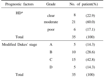
Prognostic factors Grade No. of patient(%)
HD* clear 8 (22.9)
moderate 21 (60.0)
poor 6 (17.1)
Total 35 (100)
Modified Dukes' stage A 5 (14.3)
B 10 (28.6)
C 15 (42.8)
D 5 (14.3)
Total 35 (100)
* Histological differentiation
Table 2. Distribution of histological differentiation and Dukes' stage in 35 cases of colon cancer
As results of the histological differentiation, there were 8 cases (22.9%) in clear differentiation, 21 cases (60.0%) in moderate differentiation, 6 cases (17.1%) in poor differ- entiation (Table 2). Less nuclear pleomorphism and more clearly formed tubules were observed in the clear differ- entiation. More nuclear pleomorphism and less clearly formed tubules were observed in poor differentiation.
According to modified Dukes' stage, it was found that there were 5 cases (14.3%) in stage A, 10 cases (28.6%) in stage B, 15 cases (42.8%) in stage C, and 5 cases (14.3%) in stage D. Stage B and C got the majority of cases. Therefore, it was shown that the detection rate of colon cancer in advanced stage was higher than that in early stage (Table 2).
2. Assessment of immunohistochemistry
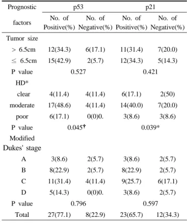
1) Correlation between the expression rate of p53 protein and pathologic prognostic factors
The p53 protein expression was positive in 27 cases (77.1%: group II, 11 cases; group III, 11 cases; group IV, 3 cases; group V, 2 cases) of all 35 cases and negative in 8 cases (22.9%: group I). p53 protein was strongly stained brown at the nuclei in 22 cases of all the 27 positive cases, but some over stains in the cytoplasm were observed in the other 5 cases (Fig. 1. A, B).
In the p53 protein expression, it was found that there were 4 positive cases (11.4%) in clear differentiation, 17 positive cases (48.6%) in moderate differentiation and 6 positive cases (17.1%) in poor differentiation. Therefore, the poorer differentiation showed the higher expression rate of p53 protein (p<0.05, Table 3). We did not find a statistically significant correlation between the expression rate of p53 protein and the tumor size, and the positive rate among tumor stages by Dukes' staging (Table 3).
2) Correlation between the expression rate of p21 protein and pathologic prognostic factors
The p21 protein expression was positive in 23 cases
(65.7%: group II, 9 cases; group III, 6 cases; group IV, 7 cases; group V, 1 case) of all 35 cases and negative in 12 cases (34.3%: group I) (Table 3). p21 protein was clearly observed as being brown mostly at the nuclei (Fig. 1. C, D).
In the examination of histological differentiation, p21 protein expression was observed from 6 cases (17.1%) in clear differentiation, from 14 cases (40.0%) in moderate differentiation and from 3 cases (8.6%) in poor differentiation.
According to the progression of histological malignant degeneration, the expression rate of p21 protein decreased distinctively (p<0.05, Table 3). However, we did not find a statistically significant correlation between the expression rate of p21 protein and the tumor size, and the expression rate among tumor stages by Dukes' staging (Table 3).
Prognostic p53 p21
factors No. of Positive(%)
No. of Negative(%)
No. of Positive(%)
No. of Negative(%) Tumor size
> 6.5cm 12(34.3) 6(17.1) 11(31.4) 7(20.0)
≤ 6.5cm 15(42.9) 2(5.7) 12(34.3) 5(14.3)
P value 0.527 0.421
HD*
clear 4(11.4) 4(11.4) 6(17.1) 2(50) moderate 17(48.6) 4(11.4) 14(40.0) 7(20.0)
poor 6(17.1) 0(0)0. 3(8.6) 3(8.6)
P value 0.045† 0.039*
Modified Dukes' stage
A 3(8.6) 2(5.7) 3(8.6) 2(5.7)
B 8(22.9) 2(5.7) 8(22.9) 2(5.7)
C 11(31.4) 4(11.4) 9(25.7) 6(17.1)
D 5(14.3) 0(0)0. 3(8.6) 2(5.7)
P value 0.796 0.597
Total 27(77.1) 8(22.9) 23(65.7) 12(34.3)
* Histological differentiation
†Correlation is significant at the 0.05 level(p<0.05)
Table 3. Correlation between the expression rate of p53 and p21 proteins and prognostic factors in 35 colon cancer cases
A B
C D
E F
Fig. 1. Immunohistochemical staining for p53 and p21 proteins in colon cancer. p53 protein was stained brown in cancer cell nuclei (A, ✕100; B,✕ 400). p21 protein was stained brown on the nuclei surface of the cancer cells (C, ✕100; D, ✕400). Several cancer cell nuclei were stained brown by p53 protein staining (E, 400×) while portions of the nuclei negatively expressed in p53 protein staining were stained brown by p21 protein staining (F, 400×).
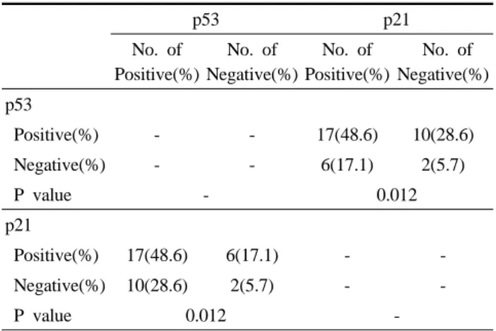
3) Correlation between the expression rate of p53 and p21 proteins
In the result that compared the expression rate of p53 protein with p21 protein, it was found that in 10 cases(28.6%), the expression of p53 protein was positive, compared to a negative expression of p21 protein and in
6 cases (17.1%), the expression of p21 protein was positive, compared to a negative expression of p53 protein. Consequently, inverse correlation between the expression rate of p53 protein and that of p21 protein was observed (p<0.05, Table 4, Fig. 1. E, F).
p53 p21 No. of
Positive(%)
No. of Negative(%)
No. of Positive(%)
No. of Negative(%) p53
Positive(%) - - 17(48.6) 10(28.6)
Negative(%) - - 6(17.1) 2(5.7)
P value - 0.012
p21
Positive(%) 17(48.6) 6(17.1) - -
Negative(%) 10(28.6) 2(5.7) - -
P value 0.012 -
Table 4. Correlation between the expression rate of p53 and p21 protein in 35 colon cancer cases
IV. DISCUSSION
Recurrence remarkably affecting the prognostic presu- mption of patients with colon cancer is very closely related with prognostic factors such as Dukes' stage and lymphnode metastasis. In addition, other prognostic factors such as patients' age, tumor size, histological differ- entiation, expression rate of oncogene, and invasion into blood vessel and neural tissue have been shown to influence on the prognosis of patients with colon cancers (Kawasaki et al, 1992).
This study examined the correlation between the expressions of p53 and p21 proteins of colon cancer- related proteins and the tumor size, the histological differentiation, and Dukes' stage, which are considered as key prognostic factors with close relation to recurrence of colon cancer.
As shown in the results, statistical significance was not proven between the expression of p53 protein and the tumor size, and Dukes' stage as pathologic prognostic factors. However, as found with histological differentiation, the more histological malignant degeneration progressed, the more the positive expression rate increased. This correlation has statistical significance (p<0.05, Table 3), and it indirectly suggested that the expression of p53
protein was related with the histological differentiation of colon cancer. This result corresponded to other reports on relationship between p53 and cancer: Greenblatt et al (1994) reported that normal p53 protein was not observed by immunohistochemical staining due to short half-life and small quantities, while the mutation of p53 gene let p53 protein be stained immunohistochemically by its increased half-life and intracelluar accumulation;
Mehregan et al (1996) reported that the mutated p53 gene might be related with prognosis and malignancy in cancers of various organs; Yamaguchi et al (1993) and Pricolo et al (1997) reported that the mutation of p53 gene in colon cancer exacerbated the malignancy of the tumor and made the prognosis poorer. Thus, normal p53 gene, as tumor suppressor gene, controls cell cycle and apoptosis, therefore keeping normal cellular functions. But the mutated p53 gene lost its functions as administrator and causes the proliferation of tumor cells, so it was considered that p53 mutation was responsible for the occurrence and the malignant degeneration of colon cancer.
p21 protein was known as a reverse regulator of cell cycle and seems to be related with apoptosis. This study could not confirm significant correlation between the expression of p21 protein and the tumor size, and Dukes' stage, which are known as pathologic prognostic factors.
However, correlation between p21 and histological differentiation was significantly confirmed because the poorer differentiation got the lower expression rate of p21 protein (p<0.05, Table 3). This corresponds to the report that p21 protein was valuable as a prognostic factor because p21 protein was lost in the progress of carcinoma (Yasui et al, 1997). Thus, it is thought that the over expression of p21 protein can inhibit the progression of colon cancer and appears to be related with the prognosis of patients. In this study, it was found that in 10 cases (28.6%), the expression of p53 protein was positive, but the expression of p21 protein was negative, and in 6 cases (17.1%), the expression of p21 protein was positive, but
the expression of p53 protein was negative. Consequently, inverse correlation between p53 and p21 proteins was observed (p<0.05, Table 4). Digiuseppe et al (1995) reported that the activation of p21 protein might be independently induced by each of two pathways:
activation of p53 gene for damaged DNA and activation of other various growth factors. Pasz-walczak et al (2000) suggested that the expression of p21 protein was related with the pathway dependent on p53 gene due to the inverse correlation between the expression of p53 protein and p21 protein. Yamamoto et al (1996) reported that TGF-β1 could activate p21 protein and induce apoptosis that was not related with the pathway dependent on p53 gene. They have not agreed about this among themselves.
It may be said that inverse correlation between the expression of p53 protein and the expression of p21 protein would be proven if the activation of p21 protein by pathway dependent on p53 gene could be confirmed.
But if the activation of p21 protein follows a pathway independent of p53 gene, the correlation between p53 and p21 proteins can't be regarded as significant. This study seems to support that the activation of p21 protein depends on the p53 gene-related pathway, and it corresponds to the studies of Pasz-walczak et al (2000) and Girlando et al (1999). However, close relationship without statistical significance was shown in 19 cases including 17 cases with positive expression of both p53 and p21 proteins, 2 cases with negative expression of both p53 and p21 proteins. These results did not coincide with the reports of Yamamoto et al (1996) and Akagi et al (1996) that the expression of p21 protein was related with a pathway independent of p53 gene. It suggests that many factors including p53 gene and TGF-β1 may be involved in the expression of p21 protein in colon cancer.
This study found that there were significant correlations between the histological differentiation of colon cancer and the expression rate of p53 and p21 proteins (p<0.05).
And a significant inverse correlation between the expression rate of p53 protein and p21 protein (p<0.05).
This could confirm that the over expression of p53 and p21 proteins is closely associated with the occurrence of colon cancer and its progress.
Therefore, it is thought that this study will be helpful for the presumption of diagnosis, treatment and prognosis of colon cancer patients, but further studies on a large number of cases are needed to clarify the valuable relationship prognostic factors with the expression of proteins related with colon cancer.
REFERENCES
1. Akagi M, Yasui W, Akama Y, Yokizaki H, Tahara H, Haruma K, Kajiyama G, Tahara E. Inhibition of cell growth by transforming growth facctor 1 in associated with p53-independent induction of p21 in gastric carcinoma cells. J Cancer Res 87:377-384, 1996.
2. Digiuseppe JA, Redston NS, Yeo CJ, Kern SE, Hruban RH. p53-independent expression of the cyclin dependent kinase in pancreatic carcinoma. Am J Pathol 147:884-888, 1995.
3. Elkablawy MA, Maxwell P, Williamson K, Anderson N, Hamilton PW. Apoptosis and cell cycle regulatory proteins in colorectal carcinoma: relationship to tumor stage and patient survival. J Pathol 194:436-443, 2000.
4. Ferrandina G, Stoler A, Fagotti A, Fanfani F, Sacco R, De Pasqua A, Mancuso S, Scambia G. p21WAF/
CIP1 protein expression in primary ovarian cancer. Int Oncol 17:1231-1235, 2000.
5. Girlando S, Slomp P, Caffo O, Amichetti M, Togni R, Dvornik G, Tomio L, Galligioni E, Dalla Palma P, Barbarreschi Ml. p21 expression in colorectal carcino- mas: a study on 103 cases with analysis of p53 gene mutation/expression and clinicopathological correla- tions. Virchows Arch 435:559-565, 1999.
6. Greenblatt MS, Bennett WP, Hollstein M, Harris CC.
Mutations in the p53 tumor suppressor gene: clues to cancer etiology and molecular pathogenesis. Cancer
Res 54:4855-4878, 1994.
7. Kawasaki Y, Monden T, Morimoto H, Murotani M, Miyoshi Y, Kobayashi T, Shimano T, Mori T.
Immunohistochemical study of p53 expression in Microwave-fixed, paraffin embedded sections of colo- rectal carcinoma and adenoma. Am J Clin Pathol 97:244-249, 1992.
8. Kerr JF, Winterfford CM, Harmon BV. Apoptosis. Its significance in cancer and cancer therapy. Cancer 83:2013-2026, 1994.
9. Mehregan D, Mehregan D. Immunohistochemistry: a prognostic as well as diagnostic tool. Semin Cutan Med Surg 15:317-325, 1996.
10. Pasz-walczak G, Kordek R, Faflik M. p21 (WIF1) expression in colorectal cancer: correlation with p53 and cyclin D1 expression, clinicopathological parameters and prognosis. Pathol Res Pract 197:683-689, 2000.
11. Pricolo VE, Finkelstein SD, Hansen K, Cole BF, Bland KI. Mutated p53 gene is an independent adverse predictor of survival in colon carcinoma. Arch
Surg 132:371-374, 1997.
12. Yamaguchi A, Nakagawara G, Kurosaka Y, Nishimura G, Yonemura Y, Miyazaki I. p53 immunoreaction in endoscopic biopsy specimens of colorectal cancer and its prognostic significance. Br J Cancer 68:399-402, 1993.
13. Yamamoto M, Maehara Y, Sakaguchi Y, Jusumoto T, Ichiyoshi Y, Sugimachi K. Transforming growth factor β1 induces apoptosis in gastric cancer cells through a p53-independent pathway. Cancer 77:1628-1633, 1996.
14. Yasui W, Akama Y, Yokozaki H, Semba S, Kudo Y, Shimamoto F, Tahara E. Expression of p21WAF/CIP1 in coloretal adenomas and adenocarcinomas and its correlation with p53 protein expression. Pathol Int 47:470-477, 1997.
15. Zirbes TK, Baldus SE, Moenig SP, Nolden S, Kunze D, Shafizaden ST, Schneider PM, Thiele J, Hoelscher AH, Dienes HP. Prognostic impact of p21WAF/CIP1 in colorectal cancer. Int J Cancer 89:14-18, 2000.