455
and Safety
Available online at http://www.foodhygiene.or.kr
https://doi.org/10.13103/JFHS.2017.32.6.455
Molecular Epidemiology of Norovirus in Asymptomatic Food Handlers in South Korea
Jeong Su Lee, Min Hee Jeong, Si Yeon Ju, Kyung Ah Kang, and In Sun Joo* Food Microbiology Division, Food Safety Evaluation Department, National Institute of Food and
Drug Safety Evaluation, Osong, Korea
(Received April 28, 2017/Revised May 20, 2017/Accepted October 11, 2017)
ABSTRACT - Norovirus (NoV) is the most common cause of acute gastroenteritis in all age groups worldwide.
In this study, prevalence of asymptomatic norovirus infection was investigated in food handler being employed at food catering facilities in South Korea. A total of 2,729 fecal specimens from asymptomatic food handlers were analyzed, and 1.06% of food handlers (29/2,729) had asymptomatic NoV infection. Of these, 17.2% (5/29) were positive for NoV GI and 82.7% (24/29) were positive for NoV GII. Especially, sequencing and phylogenetic analysis showed that GII-4 was the most prevalent genotype and a large number of asymptomatic food handlers were infested with norovi- rus GII-4 strains. The results of this study show that asymptomatic food handlers may be potential transmission sources for NoV infection. These results emphasize the need for training of food catering employees about norovirus prevention. Asymptomatic norovirus infection should receive more attention.
Key words : Asymptomatic Food Handler, Genotype, Norovirus
The positive-sense polyadenylated single-stranded RNA virus family Caliciviridae contains four genera: Vesivirus, Sapovirus, Lagovirus, and Norovirus (NoV)1). The NoV is currently classified into 6 genogroups (GI to GVI)2), and only NoV GI, GII, and GIV have been associated with human gastroenteritis3). The GI and GII genogroups of human NoV are further classified into 9 and 22 genotypes, respectively4). The virus is transmitted predominantly through ingestion of contaminated food as well as person-to-person by the fecal-oral route, airborne transmission and contact with contaminated surfaces5). In previous reports, infected food handlers have been implicated repeatedly as the source of infection in several outbreaks6,7). Asymptomatic NoV infections of food handlers may play a role in transmission8). NoV disease outbreaks are reported year-round in Korea.
Furthermore, there have been several large outbreaks of NoV since 20039,10). The NoV outbreak has exhibited a high prevalence during winter with cold temperate climates10). In other words, the environment with low temperature (average 2.1oC) and humidity (average 59.6%) may be a possible contributors to the transmission of enteric infections12). Many studies have reported the monitoring of NoV in facilities
with outbreak11,13,14). However, little research is available about circulating viral strains in asymptomatic individuals in facilities without NoV outbreaks. Recently, another epi- demiologic study of NoV outbreak in South Korea has reported that the excretion of NoV from asymptomatic food handlers may be an important portion of NoV out- break9,12,14,17). The aims of this study were to investigate the molecular epidemiological characteristics of NoV detection from asymptomatic food handlers working at food catering facilities in South Korea from October 2012 to April 2013.
Materials and Methods
Clinical samples
Rectal swab samples were collected from asymptomatic food handlers during regular physical examinations at five health centers (Tongyeong, Gyeonggi, Yeosu, Taean and Chenogju) in South Korea. Among 2,729 food handlers, the gender ratio of this study was 72% (1,965) female and 28%
(764) male. Swab samples collected from randomly selected food handlers distributed in South Korea were suspended in 2 mL of phosphate buffered saline (pH 7.2), vortexed slightly, and then centrifugated at 3,000 rpm for 10 min to separate the supernatants. Supernatants were stored at −80oC until use.
Viral RNA extraction
The QIAamp Viral RNA Mini kit (Qiagen, Hilden, Germany) was used to perform the viral RNA extraction in
*Correspondence to: In Sun Joo, Food Microbiology Division, Food Safety Evaluation Department, National Institute of Food and Drug Safety Evaluation, Osong 28159, Korea
Tel: 82-43-719-4302, Fax: 82-43-719-4300 E-mail: [email protected]
accordance with manufacturer's instructions. Viral RNA was eluted in 60µL of AVE buffer and stored at −80oC until use.
Conventional nested RT-PCR
NoV was identified using conventional nested RT-PCR as previously described17). PCR amplification was performed to detect the ORF 1-2 junction region of the NoV. 5µL of extracted RNA was used in the RT-PCR mixture with a total volume of 25µL comprised of 10 µL one-step RT-PCR premix (with AMV reverse transcriptase), 6µL of distilled water and 2µL of each NoV GI and GII primer (20 pmol) (Table 1). The cycling conditions were: 30 min at 45oC for cDNA synthesis, 5 min at 94oC for predenaturation, then 35 cycles consisting of denaturation at 94oC for 30 sec, annealing at 55oC for 30 sec, and extension at 72oC for 1 min 30 sec.
The semi-nested PCR amplification procedure was followed using the first-round amplicon. 2µL of amplicon was added to 48µL of the PCR mixture containing 5 µL of 10× reaction buffer (Bioneer, Daejeon, Korea), 4µL 10 mM dNTPs (Bio- neer, Daejeon, Korea), 2.5µL of each NoV GI and GII primer (20 pM), 1µL of Top DNA polymerase (Bioneer, Daejeon, Korea) and 33µL distilled water. The cycling conditions were: 5 min at 94oC for predenaturation, then 25 cycles consisting of denaturation at 94oC for 30 sec, annealing at 55oC for 30 sec, and extension at 72oC for 1 min 30 sec.
The amplification products were analyzed by 2% agarose gel electrophoresis and visualized with ultraviolet (UV) light after ethidium bromide staining. Samples that were NoV positive by conventional RT-PCR were further characterized (genotyped) by DNA sequencing. All PCR products were
sequenced using an ABI Prism 3500×L genetic analyzer and BigDye Terminator cycle sequencing mix (Applied Biosys- tems, Foster City, CA, USA). For genotyping of sequenced products, the sequences were compared to those in the GenBank database using the NCBI BLAST search program.
To confirm the genotype of NoV, phylogenetic analysis was performed and the phylogenetic trees were obtained using the CLUSTAL W method and MegAlign (Lasegene, DNA- star, Inc. Madison, WI, USA) software.
Real time RT-PCR
To analyze viral copy number within each sample, NoVs were amplified with a one-step real time RT-PCR kit (Ambion, Austin, TX, USA) as previously described17). The real time RT-PCR reaction mixture contained 5µL of ex- tracted RNA, 12µL of 2× RT-PCR buffer (Ambion, Austin, TX, USA), 1µL of each NoV GI and GII primer (10 pM), 0.5µL of each fluorescent probe (10 pM), 0.5 µL of 25×
enzyme mix, 1.5µL of enhancer, and 3 µL of distilled water.
5µL of extracted NoV sample was added to each well, and the final total volume was 25µL (Table 2). The cycling conditions were: reverse transcription at 45oC for 30 min, predenaturation at 95oC for 10 min, and 45 cycles of dena- turation at 95oC for 15 sec and annealing, and extension at 56oC for 1 min.
Results and Discussion
Among 2,729 food handlers comprised of 764 male and 1,965 female, asymptomatic infection was detected in 29
Table 1. Primers used for NoV detection by conventional PCR
Genogroup Primer Sequence (5’→ 3’) Size (bp) Application
I
GI-FIM CTG CCC GAA TTY GTA AAT GAT GAT
313
One-step RT PCR
GI-RIM CCA ACC CAR CCA TTR TAC ATY TG One-step RT PCR/Semi-nested PCR/
Sequencing
GI-F2 ATG ATG ATG GCG TCT AAG GAC GC Semi-nested PCR/Sequencing
II
GII-FIM GGG AGG GCG ATC GCA ATC T
310
One-step RT PCR
GII-RIM CCR CCI GCA TRI CCR TTR TAC AT One-step RT PCR/Semi-nested PCR/
Sequencing
GII-F3M TTG TGA ATG AAG ATG GCG TCG ART Semi-nested PCR/Sequencing
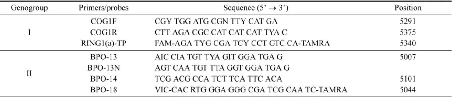
Table 2. Primers and probes used for NoV detection by real-time RT-PCR
Genogroup Primers/probes Sequence (5’→ 3’) Position
I
COG1F CGY TGG ATG CGN TTY CAT GA 5291
COG1R CTT AGA CGC CAT CAT CAT TYA C 5375
RING1(a)-TP FAM-AGA TYG CGA TCY CCT GTC CA-TAMRA 5340
II
BPO-13 AIC CIA TGT TYA GIT GGA TGA G 5007
BPO-13N AGT CAA TGT TTA GGT GGA TGA G
BPO-14 TCG ACG CCA TCT TCA TTC ACA 5101
BPO-18 VIC-CAC RTG GGA GGG CGA TCG CAA TC-TAMRA 5044
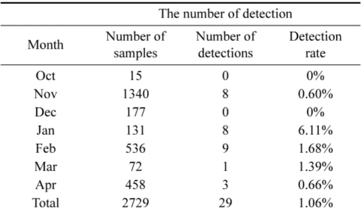
(1.06%) by conventional nested RT-PCR. The prevalence of NoV in asymptomatic food handlers was 58.62% during winter season (from December to February). Especially, there was a tendency that the prevalence of asymptomatic NoV infection in January has higher detection than other months (Table 3). Among 29 samples, 5 GI-positive samples and 24 GII-positive samples were identified by real time RT- PCR. Mean viral load in stool specimens for GI NoV was
6.1× 10⁷ viruses/g (range, 2.1× 10² to 3.0× 10⁸ viruses/g) and was 6.1× 10⁴ viruses/g (range, 1.5× 101 to 9.7× 10⁵ viruses/g) for GII (data not shown). Sequencing and phy- logenetic analysis showed that the 5 GI-positive samples were genotyped as GI-4, GI-6, GI-7, GI-9 and 24 GII- positive samples were GII-2, GII-3, GII-4, GII-6, GII-16, GII-17. Our data demonstrated that mean viral load in stool specimens of GI NoV was higher than that of GII NoV. The genotypic distribution of the 29 NoV strains was as follows:
GI-4, 6.89% (n=2); GI-6, 3.44% (n=1); GI-7, 3.44% (n=1);
GI-9, 3.44% (n=1); GII-4, 41.37% (n=12); GII-17, 20.69%
(n=6); GII-2, 10.34% (n=3); GII-3, 3.44% (n=1); GII-6, 3.44% (n=1); GII-16, 3.44% (n=1) (Table 4).
Molecular epidemiological studies of NoV strains in asymptomatic food handlers have reported that GII-4 was dominant in transmissibility1,16,18). Likewise, in our study, the NoV GII-4 strain was more prevalent than others. This is similar to the previous Japanese study investigating NoV outbreaks, in which the NoV samples were collected from asymptomatic food handlers in a hotel and confirmed belonging to NoV GII2,4). Accordingly, it is possible to assume Table 3. The number of detection (per months)
The number of detection Month Number of
samples
Number of detections
Detection rate
Oct 15 0 0%
Nov 1340 8 0.60%
Dec 177 0 0%
Jan 131 8 6.11%
Feb 536 9 1.68%
Mar 72 1 1.39%
Apr 458 3 0.66%
Total 2729 29 1.06%
Table 4. The genotypic distribution of detection strains
Prevalence of genotypes
Total GI-4 GI-6 GI-7 GI-9 GII-2 GII-3 GII-4 GII-6 GII-16 GII-17
Total 29 2 1 1 1 3 1 12 1 1 6
Fig. 1. Phylogenetic tree of NoV detected in asymptomatic food handler. Neighbor-joining phylogenetic tree based on nucleotide sequences of the capsid region of the NoV genome (A, norovirus GI; B, norovirus GII). The numbers in the branches indicate the bootstrap values. Refer- ence strains of NoV selected from Genbank are indicated by accession numbers. The scaled indicates nucleotide substitutions per position.
that GII is the dominant genogroup in an asymptomatic food handler population. This might be related to the fact that genogroup II strains (especially GII-4) are more transmissible than the others4,9). The clinical manifestations (e.g., increased vomiting) of GII-4 strain infections or physical charac- teristics (e.g., environmental persistence) of this strain might facilitate spreading. In South Korea, previous study that the vehicle of transmission in this outbreak was dried radish salad prepared by the food handler and served to students in the elementary school22). Furthermore, approximately 50%
of all NoV outbreaks in the United States are linked to ill food-handlers20). In this regard, asymptomatic carriers of Nov are main cause related to the occurrence of NoV out- breaks9,15,21).
In this study, phylogenetic analysis was used to evaluate the relatedness of NoV strains detected and to compare their partial capsid sequence with those of GI and GII genogroup reference strains (Fig. 1). In phylogenetic analysis with GI genogroup, FH-R4-KR8 (KF773988) and FH-R5-KR1 (KF- 773980) strains were identified as genotype GI-4. 3 strains, FH-R1-KR5 (KF773995), FH-R1-KR10 (KF774000) and FH-R4-KR12 (KF774002) were analyzed as GI-6, GI-7 and GI-9, respectively. The FH-R4-KR8 (KF773988) and FH- R5-KR1 (KF773980) strains were clustered into the GI-4 NV34 (KF049146) with 97.0% and 89.9% identity, re- spectively. Sequence analysis revealed that strain FH-R1- KR5 (KF773995) shared the greatest identity with strain GI- 6 BS-5 (AF093797) (92.5%). Strains FH-R1-KR10 (KF- 774000) and FH-R4-KR12 (KF774002) were found to be related most closely to GI-7 Miyagi-JP (AB758449) (90.3%) and GI-9 Vancouver730 (HQ637267) (94.5%), respectively.
The NoV GII strains were 87.3 to 97.6% homologous with the reference GII-4, 89.9 to 92.1% with the reference GII- 17, 90.0 to 96.7% with the reference GII-2, 91.6% with the reference GII-3, 93.5% with the reference GII-6 and 95.3%
with the reference GII-16. FH-R4-KR3 (KF773983), FH- R4-KR6 (KF773986), FH-R4-KR5 (KF773985), FH-R3- KR4 (KF773978), FH-R4-KR4 (KF773984), FH-R1-KR7 (KF773997), FH-R1-KR8 (KF773998), FH-R1-KR3 (KF7- 73993), FH-R1-KR4 (KF773994), FH-R3-KR5 (KF773979), FH-R3-KR1 (KF773975), and FH-R4-KR2 (KF773982) stains showed 87.3% to 97.6% sequence identity to the GII- 4 genotype, suggesting that GII-4 is the most prevalent genotype in an asymptomatic Nov infection.
The results of this study showed that asymptomatic employees as well as symptomatic food handlers may con- tribute infection as a potential transmission source in NoV outbreaks. To reduce food contamination, strict general hygiene practices should be implemented. More attention should be paid to facilities for food workers to reinforce hand hygiene practices and prevent foodborne outbreak.
Nucleotide sequence accession numbers
The nucleotide sequence data have been submitted to GenBank and assigned accession numbers KF773975 to KF774002.
Acknowledgements
We thank the staffs at 5 centers for collecting stool spe- cimens of food handler. This study was supported by a grant (121612KFDA033) from Ministry of Food and Drug Safety in 2013.
국문요약
노로바이러스는 전세계적으로 모든 연령에게 급성 장염 을 일으키는 주요 원인체이다. 본 연구는 국내 식품업계 의 식품종사자를 대상으로 무증상 노로바이러스 감염의 유행성을 조사하였다. 2,729명의 식품종사자에서 29명 (1.06%)이 무증상 노로바이러스로 확인되었고 이 중 5명 (17.24%)은 노로바이러스 GI 양성이었고 24명(82.76%)은 노로바이러스 GII 양성이었다. 특히 유전자 염기서열 분 석과 계통 분석에서 GII-4 유전자형이 가장 유행성이 높 은 것으로 나타났으며 많은 수의 무증상 식품종사자는 노 로바이러스 GII-4 에 감염되어있었다. 따라서 본 연구의 결과는 무증상 식품종사자가 노로바이러스 감염에 잠재적 인 전파 원인일 가능성이 있음을 보여준다. 또한 이러한 결과는 노로바이러스 예방에 대한 식품업체의 교육의 필 요성을 강조한다. 무증상 노로바이러스 감염에 더 많은 주 의를 기울여야 할 것이다.
References
1. Ozawa, K., Oka, T., Takeda, N., Hansman, G.S.: Norovirus infections in symptomatic and asymptomatic food handlers in Japan. J. Clin. Microbiol, 45, 3996-4005 (2007).
2. White, P.A.: Evolution of norovirus. Clin. Microbiol. Infect, 20, 741-745 (2014).
3. Trujillo, A.A., McCaustland, K.A., Zheng, D.P., Hadle, L.A., Vaughn, G., Adams, S.M., Ando, T., Glass, R.I., Monroe, S.S.: Use of TaqMan real-time reverse transcription-PCR for rapid detection, quantification, and typing of norovirus. J.
Clin. Microbiol, 44, 1405-1412 (2006).
4. Lu, J., Sun, L., Fang, L., Yang, F., Mo, Y., Lao, J., Zheng, H., Tan, X., Lin, H., Rutherford, S., Guo, L., Ke, C., Hui, L.:
Gastroenteritis Outbreaks Caused by Norovirus GII.17, Guang- dong Province, China, 2014-2015. Emerg. Infect. Dis, 21, 1240-1242 (2015).
5. Cho M.G., Jeong H.M., Ahn J.B.: Detection of feline calicivi- rus as norovirus surrogate in food and water sources using filtration and real-time RT-PCR. Food Sci. Biotechnol, 20,
1475 (2011).
6. García, C., DuPont, H.L., Long, K.Z., Santos, J.I., Ko, G.:
Asymptomatic norovirus infection in Mexican children. J.
Clin. Microbiol, 44, 2997-3000 (2006).
7. Qi, R., Ye, C., Chen, C., Yao, P., Hu, F., Lin, Q.: Norovirus prevention and the prevalence of asymptomatic norovirus infection in kindergartens and primary schools in Changzhou, China: Status of the knowledge, attitudes, behaviors, and re- quirements. Am. J. Infect. Control, 43, 833-838 (2015).
8. Sukhrie, F.H., Teunis, P., Vennema, H., Copra, C., Thijs, M.F., Bogerman, J., Koopmans, M.: Nosocomial transmission of norovirus is mainly caused by symptomatic cases. Clin. Infect.
Dis, 54, 931-937 (2012).
9. Gong Y.W., Oh B.Y., Kim H.Y., Lee M.Y., Kim Y.H., Go J.M., Lee J.M., Jeong H.S., Cheon D.S.: Molecular epidemi- ologic investigation of Norovirus infections in Incheon City, Korea, from 2005 to 2007. J. Bacteriol. Virol, 38, 249-257 (2008).
10. Koh S.J., Cho H.G., Kim B.H., Choi B.Y.: An outbreak of gastroenteritis caused by norovirus-contaminated groundwater at a waterpark in Korea. J. Korean Med. Sci, 26, 28-32 (2011).
11. Lowther, J.A., Gustar, N.E., Powell, A.L., Hartnell, R.E., Lees, D.N.: Two-year systematic study to assess norovirus contam- ination in oysters from commercial harvesting areas in the United Kingdom. Appl. Environ. Microb, 78, 5812-5817 (2012).
12. Yu J.H., Kim N.Y., Lee E.J., Jeon I.S.: Norovirus infections in asymptomatic food handlers in elementary schools without norovirus outbreaks in some regions of Incheon, Korea. J.
Korean Med. Sci, 26, 734-739 (2011).
13. Ayukekbong, J.A., Andersson, M.E., Vansarla, G., Tah, F., Nkuo-Akenji, T., Lindh, M., Bergström, T.: Monitoring of seasonality of norovirus and other enteric viruses in Cameroon by real-time PCR: an exploratory study. Epidemiol. Infect, 142, 1393-402 (2014).
14. Park D.J., Kim J.S., Park J.Y., Kim H.S., Song W., Hur M., Lee K.M.: Epidemiological analysis of norovirus infection between March 2007 and February 2010. Korean J. Lab.
Med, 30, 647-653 (2010).
15. Barrabeig, I., Rovira, A., Buesa, J., Bartolomé, R., Pintó, R., Prellezo, H., Domínguez, A.: Foodborne norovirus outbreak:
the role of an asymptomatic food handler. BMC Infect. Dis, 10, 269 (2010).
16. Gallimore, C.I., Cubitt, D., du Plessis, N., Gray, J.J.: Asymp- tomatic and symptomatic excretion of noroviruses during a hospital outbreak of gastroenteritis. J. Clin. Microbiol, 42, 2271-2274 (2004).
17. Cheon D.S., Jeong H.S., Jeong A., Lee K.B., Lee M.H., Tahk H., Choi C.: Seasonal prevalence of asymptomatic norovirus infection in Korean children. Foodborne Pathog. Dis, 7, 1427-1430 (2010).
18. Shinkawa, N., Noda, M., Yoshizumi, S., Tokutake, Y., Shiraishi, T., Arita-Nishida, T., Nishio, O.: Molecular epidemiology of noroviruses detected in food handler-associated outbreaks of gastroenteritis in Japan. Intervirology, 51, 422-426 (2008).
19. Barreira, D.M., Ferreira, M.S., Fumia, T.M., Checon, R., de Sadovsky, A.D.: Viral load and genotypes of noroviruses in symptomatic and asymptomatic children in Southeastern Bra- zil. J. Clin. Virol, 47, 60-64 (2010).
20. Widdowson, M.A., Sulka, A., Bulens, S.N., Beard, R.S., Chaves, S.S., Hammond, R., Salehi, E.D.: Norovirus and foodborne disease, United States, 1991-2000. Emerg. Infect.
Dis, 11, 95-102 (2005).
21. Chen, M.Y., Chen, W.C., Chen, P.C., Hsu, S.W., Lo, Y.C.: An outbreak of norovirus gastroenteritis associated with asymp- tomatic food handlers in Kinmen, Taiwan. BMC Public Health, 16, 372 (2016).
22. Yu J.H., Kim N.Y., Koh Y.J., Lee H.J.: Epidemiology of food- borne Norovirus outbreak in Incheon, Korea. J. Korean Med.
Sci, 25, 1128-1133 (2010).