Phosphorylation Properties of Recombinant OsCPK11, a Calcium-dependent Protein Kinase from Rice
Il-Sang Cho
1, Su-Hee Lee
2, Chung-Mo Park
3and Sung-Ha Kim
4*
1Somyong Girls' High School, 571bun-gil, Booil-ro, Bucheon-si, Gyunggi-do 14647, Korea
2Iwol Middle School, 753 Jingwang-ro, Iwol-myeon, Jincheon-gun, Chungbuk 27813, Korea
3Department of Chemistry, Seoul National Univirsity, 1 Gwanak-ro, Gwanak-gu, Seoul 08826, Korea
4Department of Biology Education, Korea National Univirsity of Education, 250 TaesungTabyeon-ro, Gangnae-myeon, Heungdeok-gu, Cheongju-si, Chungbuk 28173, Korea
Received September 22, 2016 /Revised November 1, 2017 /Accepted November 13, 2017
In plants, calcium (Ca
2+)-dependent protein kinases (CDPKs) are important sensors of Ca
2+signals.
Previous research demonstrated the expression of the OsCPK11 gene in various tissues at the tran- scription level, but its developmental and biochemical functions at the protein level were not determined. This study was aimed to identify biochemical characteristics of OsCPK11. GST- OsCPK11 was expressed in E. coli and used for an in vitro kinase assay. Biochemical analyses identified OsCPK11 as a CDPK. OsCPK11 autophosphorylated itself and transphosphorylated histone III-s and MBP as substrates in the presence of Ca
2+. The activity of the recombinant OsCPK11 was influenced by Mg
2+, with optimum activity detected at pH 7.0-7.5. OsCPK11 activity was not affected by Mg
2+, Mn
2+, or Na
+in the presence of a high level of Ca
2+. Autophosphorylation of OsCPK11 decreased Ca
2+sensitivity of OsCPK11. An anti-OsCPK11 rabbit antibody recognized 95.5 kD of GST-OsCPK11, as shown by an immunoblot analysis. These results shed light on the function of OsCPK11 in Ca
2+-medi- ated signaling in rice.
Key words : Autophosphorylation, Ca2+
-mediated signaling, CDPKs, GST-OsCPK11, transphosphorylation
*Corresponding author
*Tel : +82-43-230-3738, Fax : +82-43-232-7176
*E-mail : [email protected]
This is an Open-Access article distributed under the terms of the Creative Commons Attribution Non-Commercial License (http://creativecommons.org/licenses/by-nc/3.0) which permits unrestricted non-commercial use, distribution, and reproduction in any medium, provided the original work is properly cited.
Journal of Life Science 2017 Vol. 27. No. 12. 1393~1402 DOI : https://doi.org/10.5352/JLS.2017.27.12.1393
Introduction
Calcium is a ubiquitous second messenger during signal transduction in eukaryotes. In plants, calcium is regulated by many exogenous signals such as hormone, light, physical stimuli, and pathogens [11, 36, 39, 40]. The molecular de- coders of calcium signals are the calcium-binding proteins, which include protein kinases regulated by calcium [42].
Calcium sensors that recognize specific calcium signals are activated and transfer the signal to downstream substrate [36, 39]. In animals, protein kinase C (PKC) and calm- odulin-dependent kinases(CaMKs) are main response or to calcium signals [20]. On the other hand, calcium-dependent protein kinases (CDPKs or sometimes called as CPKs) are interspersed in plants, but they are not observed in animals
except for several protozoan [5, 16]. CDPKs widely exist in different plant species and are encoded by a multi-gene family. 34 CDPK genes were identified in Arabidopsis thaliana [3]. And 31 CDPK genes were confirmed recently in rice [46].
The CDPK gene family comprises Ser/Thr protein kinases organized in four subgroups [7, 16]; families of varying size have been characterized in Arabidopsis, rice, wheat, maize, and poplar [5, 16]. Transcripts of CDPKs have been found in every studied plant organ. In many cases, CDPKs are strongly expressed in proliferating tissues. For example, to- bacco CDPKs are expressed in rapidly growing tissues such as root tip, lateral root primordia, and vascular tissue in leaf and anther, suggesting that they might be related to cell dif- ferentiation and particular metabolic function [23, 49].
CDPKs exhibit at multiple locations including the cytosol, nucleus, plasma membrane, endoplasmic reticulum, perox- isomes, mitochondrial outer membrane and oil bodies [18].
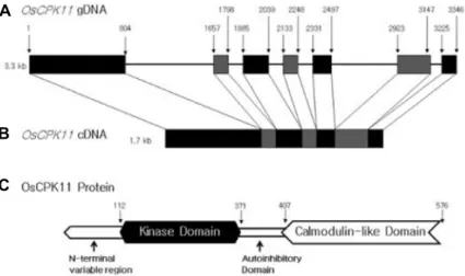
Calcium-dependent protein kinase (CDPK) is a kind of calcium sensor which binds to calcium and phosphorylases substrate peptide shaving Ser or Thr residues [39]. CDPK has N-terminal variable domain, kinase domain, auto in- hibitory domain, and calmodulin-like domain [7, 17, 20].
Calmodulin-like domain is organized in N-terminal and
A
B C
Fig. 1. Structure of OsCPK11 gene and protein.
A genomic DNA (A) of OsCPK11 and its cDNA (B) were presented as well as func- tional domains (C) of OsCPK11 protein.
Numbers in (C) indicated the sequence number of amino acids.
C-terminal EF lobes and may display different Ca
2+-binding affinities [27]. CDPKs bind to calcium directly for the kinase activity, while calmodulin does not affect their kinase activity. In other words, its different from calcium/calm- odulin-dependent protein kinase (CCaMK) binding to a cal- cium-calmodulin complex [33]. Amino acid alignment of 34
Arabidopsis thaliana CDPKs showed that their kinase, autoinhibitory and calmodulin-like domains have high con- sensus sequences except for N-terminal variable domain [14]. OsCPKs are composed of the four domains (Fig. 1) and Fig. 1C shows the structure of OsCPK11 protein. The specific function of N-terminal variable domain was not known well, but this domain is supposed to contain information of sub- cellular targeting [15]. Under some conditions, glycine resi- due of N-terminal variable region binds covalently to myr- istic acid and increase the interaction between protein and protein or between protein and membrane [22]. The N-termi- nal variable domain differs in length and amino acid se- quence even within the same species. Palmitoylation also seems to be required for the stability of binding membrane and 24 AtCPKs have been shown to have potential palmitoy- lation sites including Cys residue [29]. The autoinhibitory domain contains a pseudo-substrate site that binds to the catalytic center in the absence of Ca
2+, resulting in an inactive state of the kinase. However, the binding of Ca
2+to the calm- odulin-like domain can induce a conformational change for the release the pseudo-substrate domain from the active site and kinase activation [15, 17, 49]. Calmodulin-like domain has EF-hand sites binding calcium. EF-hand composed of a loop of 13 amino acid residues adjacent to two α-helix and each Ca
2+binds to each EF-hand one by one [48]. Most CDPKs in Arabidopsis thaliana have 4 EF-hands, but some have fewer sites. In the experiment with a deleted EF-hand
sequentially, the number of EF-hand found to be important for regulation of CDPK activity by calcium [19]. In addition, study using site-directed mutagens is showed that EF-hand strongly regulated CDPK activity as it is located nearby au- toinhibitory domain [50]. Therefore, it is proposed that the number and location of EF-hands determine allosteric prop- erty and active form of this enzyme. With the recent avail- ability of crystal structures from apicomplexan CDPKs, en- compassing the kinase and autoinhibitory domain, this mod- el has been refined [44, 45]. It has been proposed that both the apo- and calcium-bound forms of the enzyme are stabi- lized by distinct contact sites between the kinase and regu- latory domains [45]. It is tempting to speculate that, in partic- ular, some of the amino acids involved in forming these con- tact sites may be subjected to post-translational mod- ifications during the activation process [28], modulating the stability of the active or inactive form.
CDPKs phosphorylate proteins that are involved in nitro-
gen and carbon metabolism, defense-related processes, pro-
tein degradation, cytoskeletal organization and ABA signal-
ing processes [31]. CDPKs in rice have an effect on its toler-
ance to the cold, salt, and drought stress. [38]. Transgenic
rice constitutively expressing OsCPK7 and OsCPK13
showed enhanced tolerance to cold, salt and drought stress
[24, 38]. And the tissue-specific expression of CDPKs which
is developmentally regulated, suggests their involvement in
early developmental processes such as embryogenesis, seed
development and germination [1, 2]. In addition, some
CDPKs mediate the accumulation of storage starch and pro-
tein in maturing seeds [4]. Activity of CDPKs is regulated
not only by calcium, but also by phospholipids, 14-3-3 pro-
tein, various cations and hormones [7]. Regulation by hor-
mone is important for growth and development and it is
closely related with changing Ca
2+concentration. Kinase ac- tivity has been determined in the absence or presence of dif- ferent Ca
2+concentrations, and the effect of further regu- latory components such as lipids [10] or 14-3-3 proteins [25]
was investigated. CDPKs are known to modulate cal- cium-dependent plant responses caused by phytohormones, mostly gibberellins (GA), cytokinins and auxins. For exam- ple, in order to compete with the natural substrates for the protein kinases, syntide-2 was microinjected into the barley aleurone protoplasts and subsequently GA-induced amylase expression, protoplast vacuolation, and amylase secretion are selectively inhibited [32].
Both native and recombinant CDPKs found to autophos- phorylate themselves [6, 16, 37]. However, it is not clear whether autophosphorylation is essential for the activities of CDPKs. For example, in vitro autophosphorylation acti- vates a groundnut (Arachis hypogea) CDPK but inhibits one in winged bean (Psophocarpus tetragonolobus) [6, 37]. In other case, autophosphorylation of CDPK has no effect on the cal- cium-dependent activities of ground nut and soybean [6, 16].
Furthermore, activation of CDPK may be modulated by oth- er protein kinases. For instance, tobacco (Nicotiana tabacum) CDPK (NtCDPK2) needs both calcium and direct phosphor- ylation by upstream protein kinase for its full activation [34, 35]. But it is still unknown how they work in vivo. With the identification of stress- or pathway-specific CDPK iso- forms, the biochemical characterization is now extended to in vivo kinase activation, mediated by stimulus-induced, post-translational modification/phosphorylation of the CDPK, which may occur at all enzyme domains [28].
Dephosphorylation process is as important as phosphor- ylation in controlling signal pathways. A soluble phospho- serine phosphatase from winged bean shoots dephosphor- ylates an inactivated, autophosphorylated winged bean CDPK1 (Wb CDPK1) in vitro [12]. It is thought that this ac- tion releases an inhibitory effect of autophosphorylation and suggests existence of a regulatory region. Therefore, some CDPK activities are involved in regulating processes be- tween protein kinases and phosphatases [7]. Earlier reports showed that recombinant CDPKs exhibit calcium -stimulated protein kinase activity for casein, MBP, histone III-s and syntide as substrates [21, 26, 43, 47]. But there is little information concerning the regulation of OsCPKs in response to various cations, pH, substrates and autophosphorylation. In this study, recombinant GST-linked OsCPK11 was purified from E. coli and its biochemical prop-
erties as kinase was determined.
Materials and Methods
Expression and purification of recombinant OsCPK11 Cloned pET41a(+)-OsCPK11 in E.coli strain BL21 was ex- pressed and purified by GST-affinity column as describe in [8]. Transformed colony was inoculated in 5 ml of LB me- dium containing 50 ug/ml kanamycin and cultured over- night at 37℃ to obtain a saturated culture. 1 ml of a cultured solution was reinoculated with 1 l of LB medium containing 100 ug/ml kanamycin in a 2 l flask and incubated with shak- ing at 37℃ until the culture has reached the mid-log phase of growth (A
600=0.6~0.8). Isopropyl-β-D-thiogalacto-pyrano- side(IPTG) was added to a final concentration of 0.2 mM to induce the expression of GST-OsCPK11. The cells were cultured for 5 hrs at 30℃ or overnight at 24℃ and they were harvested by centrifugation at 7,000× g for 5 min at 4℃.
Pellet was resuspended in 10 ml of TN buffer (20 mM Tris pH 7.4, 150 mM NaCl, 1 mM EDTA, 10 uM β-mercaptoetha- nol, 1 mM PMSF) per 100 ml of cell culture and the suspen- sion was frozen with liquid nitrogen and thawed in 25℃
several times to break open the cells. Lysozyme was added to the cell suspension to a final concentration of 1 mg/ml and this mixture was incubated on ice for 1 hr. The insoluble debris was removed by centrifugation at 13,000× g for 1 hr at 4℃. Supernatant was collected in a fresh tube and cell lysate was mixed with an appropriate amount of 50% slurry of glutathione-agarose resin in TN buffer. In order to bind GST-OsCPK11 protein with the resin, the mixture including both the supernatant and the resin was shaken gently over- night at 4℃. It was loaded onto the poly-prep chromatog- raphy column (Bio-Rad, USA) under gravity flow. Unbound proteins were washed away from the resin by adding 10 bed volumes of TN buffer except for PMSF to the pellet.
Bound GST fusion protein was eluted from the resin using 1 bed volume of Glutathione Elution Buffer containing 10 mM reduced glutathione and 50 mM Tris-HCl, pH 8.0. All steps for the expression and purification of the OsCPK11 were followed as procedure modified from [39].
SDS-Polyacrylamide Gel Electrophoresis (SDS- PAGE)
Discontinuous SDS-PAGE was performed as described in [27] using minislab gels containing 10% or 12% acrylamide.
Equal amount of total proteins from each collected samples
Fig. 2. Ca2+ dependency of OsCPK11 for autophophor- ylation. In vitro kinase assay was carried out using 95.5 kD of GST-OsCPK11 for autophosphorylation with different Ca2+ concentration. 1 μg of GST- OsCPK11 was used on each lane. It showed that OsCPK11 is calcium-dependent and the highest activity was shown in the presence of 100 uM Ca2+.
were loaded on each lane. After electrophoresis, gel was
stained with 0.1% Coomassie brilliant blue R-250 in 40%
methanol and 10% acetic acid for several hours. Then, the gel was destained with 40% methanol and 10% acetic acid for 1 hr and repeated same destaining procedure twice.
Molecular weight was identified by the method of [41].
Kinase assay
Autophosphorylation assay
In vitro kinase assay was performed as [9] with some
modification. For the autophosphorylation reaction, 1 ug of GST–OsCPK11 protein was mixed with reaction buffer con- taining 20 mM HEPES, pH 7.4, 200 nM ATP, 1 mM Na
3VO
4, 0.5 mM PMSF, 2 mM EDTA, 2 mM DTT and 0.5 UCi [γ-
32P]
ATP (New England Nuclear) in the absence or presence of 100 uM MgCl
2. 0 to 1 mM CaCl
2or 1 mM EGTA was added into the reaction mixture and total volume was adjusted to 10 ul. After incubation for 30 min at 30℃, reaction was ter- minated by adding 2.5 ul of SDS-PAGE sample buffer. All samples are boiled for 5 min at 100℃. 10 ul of each sample was loaded and separated on 10% or 12% SDS-PAGE gels.
The gel was stained and destained according to previous description. It was dried with Bio-Rad Model 583 Gel Dryer (BioRad) for 30 min at 80℃ and subjected to auto- radiography by exposing the gel to BioMax film (Kodak) overnight at -80℃ or for 1-5 days at room temperature. The intensity of radioactivity was calculated by Image J software (National Institutes of Health, USA).
Transphosphorylation assay
1 ug of substrate (histone III-s, myelin basic protein, or casein) was added to the reaction mixture prior to assay.
For determining optimal pH, the buffers contained 20 mM HEPES with pH ranging from 5.0 to 10.0. To identify effect of several cations, reaction buffers were adjusted with 100 uM of NaCl, MgCl
2, or MnCl
2. And all other procedure were performed same as autophosphorylation assay. To identify effect of pre-autophosphorylation on transphosphorylation
reaction, pre-autophosphorylation samples were prepared by incubation of 1 ug of GST-OsCPK11 containing 1-100 uM CaCl
2in the presence/absence of 100 uM MgCl
2for 10min at 30℃. And samples were incubated for 30 min at 30℃
after 1 ug of MBP was added into the mixture. Control sam- ples were prepared by incubation of buffer only containing 1-100 uM CaCl
2in the presence/absence of 100 uM MgCl
2for 10 min at 30℃. And samples were incubated for 30 min at 30℃ after both 1 ug of MBP and 1 ug of GST-OsCPK11 were added into the mixture. All the reactions were termi- nated by adding 2.5 ul of SDS-PAGE sample buffer and fol- lowing procedures were the same as described previously.
Immunoblotting with polyclonal anti-OsCPK11 antibody
19 amino acid sequence (54-72) of PSEHSSHHSSRSTDPS TPT from OsCPK11 protein was determined. C-terminal of the peptide was added with Cys and its N-terminal was acetylated. Peptide was synthesized by the Fmoc solid-phase method by using an automated peptide synthesizer (Peptron III-R24, Peptron, Daejeon, Korea) and antibody was raised and immunoblotting was performed as described in [8].
Results
Autophosphorylation of recombinant OsCPK11 Calcium dependence of recombinant OsCPK11 protein was investigated using in vitro kinase assay. Autophosphor- ylation activity of GST-OsCPK11 was measured in the pres- ence/absence of calcium. As a result, it was confirmed that GST-OsCPK11 presented calcium dependence, a major fea- ture of CDPK (Fig. 2). These followed the prediction that EF-hands of calmodulin-like domains of CDPKs bind to cal- cium and that conformational change causes to release active sites. But it was not detected in the absence of calcium.
Autophosphorylation activity of GST-OsCPK11 in the pres-
ence of 1 nM calcium was observed weakly and activity was
Fig. 3. Effect of Mg2+ on Ca2+ dependence of auto- phosphorylation. It was determined that GST- OsCPK11 does not show any autophosphor- ylation in the absence of Mg2+ by in vitro kin- ase assay. Strong signals were shown in the presence of both Ca2+ and Mg2+ together.
A
B
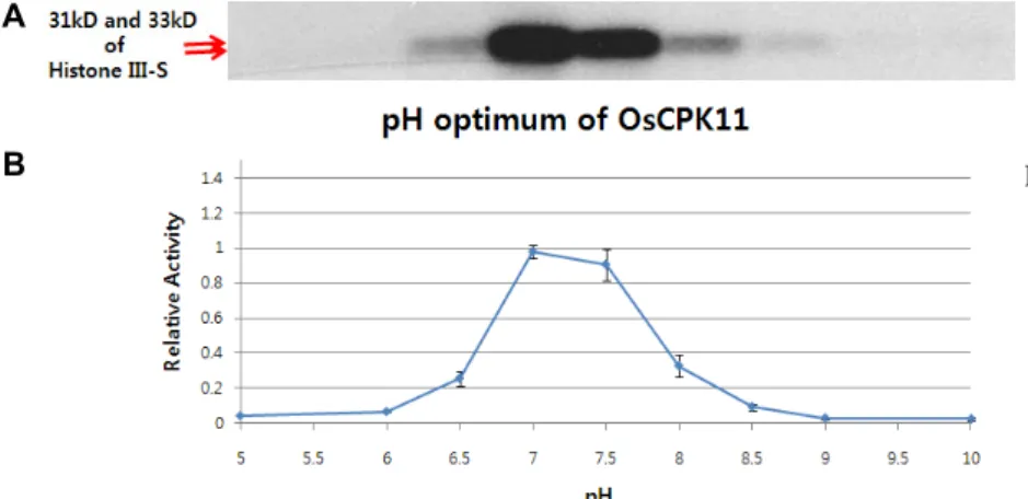
Fig. 5. Optimal pH for the transphosphor-ylation of OsCPK11. 1 ug of GST- OsCPK11 and 1 μg of Histone III-s were included in the buffer with dif- ferent pH ranging from 5.0 to 10.0. All buffers contained 1 uM Ca2+ and 100 uM Mg2+. Optimum pH for the trans- phosphorylation of OsCPK11 seemed to be pH 7.0 to 7.5.
Fig. 4. Searching for the substrates of OsCPK11. 1 ug of GST- OsCPK11 and each 1 ug of Histone III-s, MBP, and ca- sein as a substrate were used for the assay and all buffers contained 100 uM Mg2+. Histone III-s and MBP found to be good substrates for the phosphorylation reaction in the Ca2+ dependent manner.
strongest with 100 uM calcium. Though calcium-dependency results were shown in the presence of Mg
2+, it was not de- tected with 100 uM calcium in the absence of Mg
2+(Fig. 3).
GST-OsCPK11 did not show a kinase activity for itself with Mg
2+only, but its activity was shown when 100 nM Ca
2+is present with Mg
2+.
Transphosphorylational property of recombinant OsCPK11
In order to determine the transphosphorylational prop- erty of GST-OsCPK11, 1 μg of histone III-S, MBP, and casein as a substrate. Histone III-S and MBP were clearly phos- phorylated with Ca
2+dependent manner. But casein showed
no signal with or without Ca
2+(Fig. 4). So histone III-S and MBP could be used for later assays. In vitro kinase assay was performed with varying pH from 5 to 10 using GST- OsCPK11 (Fig. 5). Activity of GST-OsCPK11 was optimal at pH 7.0-7.5. Calcium-dependency for each substrate was identified by the same procedure as autophosphorylation method with or without Mg
2+(Fig. 6 and Fig. 7). GST- OsCPK11 in the absence of Mg
2+was activated only in the presence of 100 uM Ca
2+, but GST-OsCPK11 with Mg
2+also showed an strong signal in the presence of 1 uM Ca
2+. In the presence of 100 uM Ca
2+, activity of GST-OsCPK11 was confirmed regardless of any cation present (Fig. 8). Activity of GST-OsCPK11 in the absence of Ca
2+seemed to be weaker compared with one in the presence of 100 uM Ca
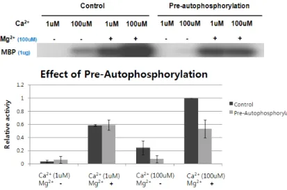
2+. It was determined whether autophosphorylation of OsCPK11 af- fected its transphosphorylation (Fig. 9).
Pre-autophosphorylation was carried out by incubating with MBP for 30 min after incubation for 10 min with GST-OsCPK11. Control samples were prepared by incubat- ing with MBP and GST-OsCPK11 for 30 min after incubation for 10 min with buffer only. Mg
2+enhanced the phosphor- ylation activity in the control sample and it was Ca
2+- dependent. Mg
2+also enhanced the phosphorylation activity in the pre-autophosphorylation sample but it did not seem to be Ca
2+-dependent. As a result, pre-autophosphorylation showed that signal intensity was not increased when Ca
2+concentration increased from 1 uM to 100 uM.
Fig. 6. Ca2+ dependence of transphosphorylation in the absence of Mg2+. 1 ug of Histone III-s was used as a transphosphorylation substrate for the 1 ug of GST-OsCPK11. In the absence of Mg2+, Histone III-s was barely phosphorylated up to 10 uM Ca2+, but was strongly phosphorylated in the presence of 100 uM Ca2+.
Fig. 7. Effect of Mg2+ on Ca2+ dependence of transphosphorylation. 1 ug of GST-OsCPK11 and 1 ug of Histone III-s as substrate were used for the assay and buffers contained either no Mg2+ (left panel) or with 100 uM Mg2+ (right panel). In the absence of Mg2+, results were the same as Fig. 6. In the presence of Mg2+, GST-OsCPK11 phosphorylated Histone III-s in the presence of Ca2+ as low as 1 uM.
Fig. 8. Effect of several cations on Ca2+ dependence of transphosphorylation. GST-OsCPK11 was incubated with Histone III-s as a substrate. In the presence of 100 uM Ca2+, strong phosphorylation activity of OsCPK11 was shown regardless of any cation present. In the absence of Ca2+, Histone III-s was a little bit phosphorylated with 100 uM Mg2+.
Immunoblotting analysis
Serum obtained from a immunized rabbit with OVA-con- jugated 19 amino acids (PSEHSSHHSSRSTDPSTPT) deduced from OsCPK11 gene was assayed to determine an antibody activity. Immune serum was specific to the peptide deduced from OsCPK11 gene. Immunoblotting results showed that GST-OsCPK11 was detected by immune serum while pre- immune serum did not show any clear band (Fig. 10).
Discussion
Calcium is an important factor in preventing physio-
logical disorders in plant tissues during growth and devel- opment [30]. Calcium ions are the most important regulator of CDPKs activity and CDPKs are different in their affinity for Ca
2+. For example, soy bean CDPK isoforms α and γ dis- play Ca
2+activation thresholds that differ by more than ten-fold (isoform α shows a very low threshold around 60 nM calcium). Thus, low level Ca
2+may selectively activate CDPK α and much higher level of Ca
2+would activate both isoforms [26]. This study showed that both Ca
2+and Mg
2+are important for the autophosphorylation of OsCPK11. In
almost all studied cases, autophosphorylation was observed
in CDPKs and it was Ca
2+-dependent except for WbCDPK
Fig. 9. Effect of pre-autophosphorylation of OsCPK11 on its transphosphorylation. Pre-autophos- phorylation samples were prepared by in- cubating with MBP for 30 min after in- cubation with GST-OsCPK11 for 10 min. Con- trol samples were prepared by incubating with MBP and GST-OsCPK11 for 30 min after incubation for 10 min with buffer only. Mg2+
enhanced the phosphorylation activity in the control sample and could be calcium-depend- ent. Mg2+ also enhanced the phosphorylation activity in the pre-autophosphorylation sam- ple, but it did not seem to be calcium-depend- ent.
Fig. 10. Immunoblotting analysis of GST-OsCPK11 using im- mune serum. GST-OsCPK11 samples were subjected to SDS-PAGE on 10% polyacrylamide gel, transferred on- to nitrocellulose membrane and processed to immuno- blotting. Lane 1, stained with Coomassie Brilliant blue (numbers indicated the molecular weights of the pro- tein markers); lane 2, treated with pre-immune serum;
lane 3, treated with immune serum.
[23]. Unlike the substrate phosphorylation reaction, auto- phosphorylation of WbCDPK is Ca
2+-independent and is not inhibited by the calmodulin antagonist [37]. It was identified that autophosphorylation of OsCPK11 showed Ca
2+-depend- ent and that Mg
2+was essential for the reaction. Recombi- nant OsCPK11 exhibited Ca
2+requirement for the substrate phosphorylation activity. In the presence of Ca
2+OsCPK11 phosphorylated Histone III-s and MBP but not casein. In the presence of 100 uM Ca
2+, Mg
2+was required for autophos- phorylation, but Mg
2+was not always essential for the transphosphorylation. It was expected that activity condition
of autophosphorylation is different from one of trans- phosphorylation in vivo. Soybean CDPKs showed broad pH optimum in a range of pH 6.0-10.0 [26]. Unlikely, the pH optimum of OsCPK11 were found to be in a range between pH 7.0 and pH 7.5 and it is similar to the green alga,
Dunaliella salina CDPK that showed a peak activity at pH7.5 [13]. Cations such as Mg
2+, Mn
2+, or Na
+did not seem to affect the activity of OsCPK11 in the presence of 100 uM Ca
2+. However, in vitro kinase assay also showed that pre-au- tophosphorylation of GST-OsCPK11 arrested the sensitivity to Ca
2+. This result was similar to one of [12] that autophos- phorylation inhibits kinase activity of WbCDPK1 in winged bean. Various stimuli could change intracellular ionic con- dition including increase of Ca
2+. OsCPK11 might be auto- inhibited with a low Ca
2+concentration and activated by high Ca
2+concentration. However, if substrates were not suf- ficient with high Ca
2+concentration, autophosphorylation could occur more frequently than transphosphorylation.
And OsCPK11 might become insensitive to increasing amount of substrates. Anti-OsCPK11 rabbit antibody was produced against a specific N-terminal variable region of OsCPK11. GST-OsCPK11 was detected with immune serum.
It can be used as a tool to determine expression and cellular localization of OsCPK11 at the protein level. These results in conjunction with one described in [27] should help better understanding and navigating the function of OsCPK11 in Ca
2+-mediated signaling in rice. It will be necessary to de- termine the proteomic data of various OsCPKs for further study.
Acknowledgement
This research was supported by Basic Science Research
Program through the National Research Foundation of Korea (NRF) funded by the Ministry of Education [NRF- 2011-0025592] to S.-H. Kim.
References
1. Anil, V. S. and Rao, K. S. 2000. Calcium-mediated signaling during sandalwood somatic embryogenesis. Role for exoge- nous calcium as second messenger. Plant Physiol. 123, 1301- 1311.
2. Anil, V. S. and Rao, K. S. 2001. Purification and character- ization of a Ca2+-dependent protein kinase from sandalwood (Santalum album L.): evidence for Ca2+-induced conforma- tional changes. Phytochemistry 58, 203-212.
3. Arabidopsis Genome Initiative 2000. Analysis of the genome sequence of the flowering plant Arabidopsis thaliana. Nature 408, 796-815.
4. Asano, T., Kunieda, N., Omura, Y., Ibe, H., Kawasaki, T., Takano, M., Sato, M., Furuhashi, H., Mujin, T., Takaiwa, F., Wu, C., Tada, Y., Satozawa, T., Sakamoto, M. and Shimada, H. 2002. Rice SPK, a calmodulin-like domain protein kinase, is required for storage product accumulation during seed development: phosphorylation of sucrose synthase is a pos- sible factor. Plant Cell 14, 619-628.
5. Asano, T., Tanaka, N., Yang, G., Hayashi, N. and Komatsu, S. 2005. Genome-wide identification of the rice calcium- dependent protein kinase and its closely related kinase gene families: Comprehensive analysis of the CDPKs gene family in rice. Plant Cell Physiol. 46, 356-366.
6. Chaudhuri, S., Seal, A. and Dasgupta, M. 1999. Autophos- phorylation -dependent activation of a calcium-dependent protein kinase from ground nut. Plant Physiol. 120, 859-866.
7. Cheng, S. H., Willmann, M. R., Chen, H. C. and Sheen, J.
2002. Calcium signaling through protein kinases. The Arabidopsis calcium dependent protein kinase gene family.
Plant Physiol. 129, 469-485.
8. Cho, I. S. 2010 Biochemical characterization of the recombi- nant OsCPK11, a calcium-dependent protein kinase from rice. Thesis for master's degree. Korea National University of Education, Chungbuk, Korea.
9. Dasgupta, M. 1994. Characterization of a calcium-dependent protein kinase from Arachis hypogea (groundnut) seeds. Plant Physiol. 104, 961-969.
10. Dixit, A. K. and Jayabaskaran, C. 2012. Molecular cloning, soluble expression and characterization of autophosphor- ylation in recombinant calcium dependent protein kinase 1(CaCDPK1) from Cicer arietinum. Appl. Microbiol. Biotechnol.
97, 3429-3439.
11. Evans, N H., McAinsh, M. R. and Hetherington, A. M. 2001.
Calcium oscillations in higher plants. Curr. Opin. Plant Biol.
4, 415-420.
12. Ganguly, S. and Singh, M. 1999. Purification and character- ization of a protein phosphatase from winged bean. Phyto- chemistry 52, 239-246.
13. Guo, Y. L. and Roux, S. J. 1990. Partial purification and char-
acterization of a Ca2+-dependent protein kinase from the green alga, Dunaliella salina. Plant Physiol. 94, 143-150.
14. Hanks, S. K. and Hunter, T. 1995. The eukaryotic protein kinase super family: kinase (catalytic) domain structure and classification. FASEB J. 9, 546-596.
15. Harmon, A. C., Yoo, B. C. and McCaffery, C. 1994. Pseudo- substrate inhibition of CDPK, a protein kinase with a calm- odulin-like domain. Biochemistry 33, 7278-7287.
16. Harmon, A. C., Gribskov, M. and Harper, J. F. 2000. CDPKs:
a kinase for every Ca2+ signal? Trends Plant Sci. 5, 154-159.
17. Harper, J. F., Sussman, M. R., Schaller, G. E., Putnam-Evans, C., Charbonneau, H. and Harmon, A. C. 1991. A calcium- dependent protein kinase with a regulatory domain similar to calmodulin. Science 252, 951-954.
18. Harper, J. F., Breton, G. and Harmon, A. 2004. Decoding Ca2+ signals through plant protein kinases. Annu. Rev. Plant Physiol. Plant Mol. Biol. 55, 263-288.
19. Hong, Y., Takano, M., Liu, C. M., Gasch, A., Chye, M. L.
and Chua, N. H. 1996. Expression of three members of the calcium-dependent protein kinase gene family in Arabidopsis thaliana. Plant Mol. Biol. 30, 1259-1275.
20. Hrabak, E. M., Chan, C. W., Gribskov, M., Harper, J. F., Choi, J. H., Halford, N., Kudla, J., Luan, S., Nimmo, H. G., Sussman, M. R., Thomas, M., Walker-Simmons, K., Zhu, J.
K. and Harmon, A. C. 2003. The Arabidopsis CDPK-SnRK superfamily of protein kinases. Plant Physiol. 132, 666-680.
21. Huang, Q. S., Wang, H. Y., Gao, P., Wang, G. Y. and Xia, G. X. 2008. Cloning and characterization of a calcium de- pendent protein kinase gene associated with cotton fiber development. Plant Cell Rep. 27, 1869-875.
22. Johnson, D. R., Bhatnagar, R. S., Knoll, L. J. and Gordon, J. I. 1994. Genetic and biochemical studies of protein N- myristoylation. Annu. Rev. Biochem. 63, 869-914.
23. Klimecka, M. and Muszynska, G. 2007. Structure and func- tions of plant calcium-dependent protein kinases. Acta Biochim. Pol. 54, 219-233.
24. Komatsu, S., Yang, G., Khan, M., Onodera, H., Toki, S. and Yamaguchi, M. 2007. Over-expression of calcium-dependent protein kinase 13 and calreticulin interacting protein 1 con- fers cold tolerance on rice plants. Mol. Genet. Genomics 277, 713-723.
25. Lachaud, C., Prigent, E., Thuleau, P., Grat, S., Da Silva, D., Brière, C., Mazars, C. and Cotelle, V. 2013. 14-3-3-regulated Ca2+-dependent protein kinase CPK3 is required for sphin- golipid-induced cell death in Arabidopsis. Cell Death Differ.
20, 209-217.
26. Lee, J. Y., Yoo, B. C. and Harmon, A. C. 1998. Kinetic and calcium-binding properties of three calcium-dependent pro- tein kinase isoenzymes from soybean. Biochemistry 37, 6801- 6809.
27. Lee, S. H. 2009. Functional characterization of OsCPK11, a calcium-dependent protein kinase gene from rice and its cDNA cloning. Thesis for master's degree. Korea National University of Education, Chungbuk, Korea.
28. Liese, A and Romeis, T. 2013. Biochemical regulation of in vivo function of plant calcium-dependent protein kinases
(CDPK). Biochim. Biophys. Acta. 1833, 1582-1589.
29. Martin, M. L. and Busconi, L. 2000. Membrane localization of a rice calcium-dependent protein kinase (CDPK) is medi- ated by myristoylation and palmitoylation. Plant J. 24, 429- 435.
30. Millaway, R. and Wiersholm, L. 1979. Calcium and metabol- ic disorders. Commun. Soil Sci. Plant Anal. 10, 1-28.
31. Reddy, V. S. and Reddy, A. S. 2004. Proteomics of cal- cium-signaling components in plants. Phytochemistry 65, 1745-1776.
32. Ritchie, S. and Gilroy, S. 1998. Calcium-dependent protein phosphorylation may mediate the gibberellic acid response in barley aleurone. Plant Physiol. 116, 765-776.
33. Roberts, D. M. and Harmon, A. C. 1992. Calcium-modulated proteins: targets of intracellular calcium signals in higher plants. Annu. Rev. Plant Physiol. Plant Mol. Biol. 43, 375-414.
34. Romeis, T., Piedras, P. and Jones, J. D. 2000. Resistance gene-dependent activation of a calcium-dependent protein kinase in the plant defense response. Plant Cell 12, 803-815.
35. Romeis, T., Ludwig, A. A., Martin, R. and Jones, J. D. 2001.
Calcium-dependent protein kinases play an essential role in a plant defense response. EMBO J. 20, 5556-5567.
36. Rudd, J. J. and Franklin-Tong, V. E. 2001. Unravelling re- sponse-specificity in Ca2+ signalling pathways in plant cells.
New Phytol. 151,7-33.
37. Saha, P. and Singh, M. 1995. Characterization of a winged bean (Psophocarpus tetragonolobus) protein-kinase with calm- odulin-like domain regulation by autophosphorylation. Bio- chem. J. 305, 205-210.
38. Saijo, Y., Hata, S., Kyozuka, J., Shimamoto, K. and Izui, K.
2000. Over-expression of a single Ca2+-dependent protein kinase confers both cold and salt/drought tolerance on rice plants. Plant J. 23, 319-327.
39. Sambrook, J. and Russell, D. W. 2001. Molecular cloning:
a laboratory manual (3rd ed.). NY: CSHL press. Sanders, D., Brownlee, C., and Harper, J. F. 1999. Communicating with calcium. Plant Cell 11, 691-706.
40. Sanders, D., Pelloux, J., Brownlee, C. and Harper, J. F. 2002.
Calcium at the cross roads of signaling. Plant Cell 14, S401- S417.
41. See, Y. and Jackowski, G. 1989. Protein structure: A practical approach (Creighton, T. E. ed.). Oxford: IRL Press.
42. Szczegielniak, J., Klimecka, M., Liwosz, A., Ciesielski, A., Kaczanowski, S., Dobrowolska, G., Hormon, A. C. and Muszyńka, G. 2005. A wound-responsive and phospholi- pid-regulated maize calcium-dependent protein kinase.
Plant Physiol. 139, 970-983.
43. Urao, T., Katagiri, T., Mizoguchi, T., Yamaguchi-Shinozaki, K., Hayashida, N. and Shinozaki, K. 1994. Two genes that encode Ca2+-dependent protein kinases are induced by drought and high-salt stresses in Arabidopsis thaliana. Mol.
Gen. Genet. 244, 331-340.
44. Wernimont, A. K., Amani, M., Qiu, W., Pizarro, J. C., Artz, J. D., Lin, Y. H., Lew, J., Hutchinson, A. and Hui, R. 2011.
Structures of parasitic CDPK domains point to a common mechanism of activation. Proteins 79, 803-820.
45. Wernimont, A. K., Artz, J. D., Finerty, P. Jr., Lin, Y. H., Amani, M., Allali-Hassani, A., Senisterra, G., Vedadi, M., Tempel, W., Mackenzie, F., Chau, I., Lourido, S., Sibley, L.
D. and Hui, R. 2010. Structures of apicomplexan calcium- dependent protein kinases reveal mechanism of activation by calcium. Nat. Struct. Mol. Biol. 17, 596-601.
46. Ye, S., Wang, L., Xie, W., Wan, B., Li, X. and Lin, Y. 2009.
Expression profile of calcium-dependent protein kinase (CDPKs) genes during the whole life span and under phyto hormone treatment conditions in rice (Oryza sativa L. ssp.
indica). Plant Mol. Biol. 70, 311-325.
47. Yoon, G. M., Cho, H. S., Ha, H. J., Liu, J. R. and Lee, H.
S. 1999. Characterization of NtCDPK1, a calcium-dependent protein kinase gene in Nicotiana tabacum, and the activity of its encoded protein. Plant Mol. Biol. 39, 991-1001.
48. Zhang, M. and Yuan, T. 1998. Molecular mechanisms of calmodulin’s functional versatility. Biochem. Cell Biol. 76, 313-323.
49. Zhang, M., Liang, S. and Lu, Y. T. 2005. Cloning and func- tional characterization of NtCPK4, a new tobacco calcium- dependent protein kinase. Biochim. Biophys. Acta 1729, 174- 185.
50. Zhao, Y., Pokutta, S., Maurer, P., Lindt, M., Franklin, R. M.
and Kappes, B. 1994. Calcium-binding properties of a cal- cium-dependent protein kinases from Plasmodium falciparum and the significance of individual calcium-binding sites for kinase activation. Biochemistry 33, 3714-3721.
초록:벼의 칼슘-의존적 단백질 카이네즈인 재조합 OsCPK11의 인산화 특성 조일상
1․이수희
2․박충모
3․김성하
4*
(1소명여자고등학교, 2이월중학교, 3서울대학교 화학과, 4한국교원대 생물교육과)