www.jpis.org
pISSN 2093-2278 eISSN 2093-2286 Copyright © 2012 Korean Academy of PeriodontologyThis is an Open Access article distributed under the terms of the Creative Commons Attribution Non-Commercial License (http://creativecommons.org/licenses/by-nc/3.0/).
Novel analysis model for implant
osseointegration using ectopic bone formation
via the recombinant human bone morphogenetic
protein-2/macroporous biphasic calcium
phosphate block system in rats: a
proof-of-concept study
Jung-Chul Park1, Jong-Bin Lee1, Guy Daculsi2, Sang-Yeop Oh1, Kyoo-Sung Cho1, Gun-Il Im3, Byung-Soo Kim4,
Chang-Sung Kim1,* 1Department of Periodontology, Research Institute for Periodontal Regeneration, Yonsei University College of Dentistry, Seoul, Korea 2Research Center on Materials of Biological Interest, Dental faculty, Nantes University, Nantes, France 3Department of Orthopedics, Dongguk University Ilsan Hospital, Goyang, Korea 4School of Chemical and Biological Engineering, Seoul National University, Seoul, Korea
Purpose: The osseointegration around titanium mini-implants installed in macroporous biphasic calcium phosphate (MBCP)
blocks was evaluated after incubation with recombinant human bone morphogenetic protein-2 (rhBMP-2) in an ectopic sub-cutaneous rat model.
Methods: Mini-implants (ϕ1.8×12 mm) were installed in MBCP blocks (bMBCPs, 4×5×15 mm) loaded with rhBMP-2 at 0.1
mg/mL, and then implanted for 8 weeks into subcutaneous pockets of male Sprague-Dawley rats (n=10). A histomorphomet-ric analysis was performed, and the bone-to-implant contact (BIC) and bone density were evaluated.
Results: Significant osteoinductive activity was induced in the rhBMP-2/bMBCP group. The percentage of BIC was 41.23±
4.13% (mean±standard deviation), while bone density was 33.47±5.73%. In contrast, no bone formation was observed in the bMBCP only group.
Conclusions: This model represents a more standardized tool for analyzing osseointegration and bone healing along the
implant surface and in bMBCPs that excludes various healing factors derived from selected animals and defect models.
Keywords: Animals, Dental implants, Osseointegration.
INTRODUCTION
Recombinant human bone morphogenetic protein-2 (rh-BMP-2) is a member of the bone morphogenetic protein
(BMP) family that is involved in de novo bone induction. In both preclinical and clinical settings, rhBMP-2 has been dem-onstrated to induce bone formation in a variety of conditions including supra-alveolar periodontal defects [1], three-wall Received: Apr. 21, 2012; Accepted: Jun. 12, 2012
*Correspondence: Chang-Sung Kim
Department of Periodontology, Research Institute for Periodontal Regeneration, Yonsei University College of Dentistry, 50 Yonsei-ro, Seodaemun-gu, Seoul 120-752, Korea
III alveolar defects [4], calvarial defects [5,6], and ectopic sub-cutaneous pockets [7]. The advent of dental implants, which are gradually replacing conventional prosthetic treatments, has recently expanded the application of rhBMP-2 to alveolar ridge augmentation [8-10] and sinus floor elevation [11], for which inadequate bone volume might adversely affect the long-term prognosis of dental implants [12].
Currently, various animal models exist for both implant os-seointegration and rhBMP-2-enhanced bone regeneration including the ectopic rat model [13,14], supra-alveolar peri-im-plant model [15-20], and mandible/tibia model [21,22]. Among these, the ectopic rat model is one of the most extreme in vivo osteoinduction models that completely exclude the osteo-conductive factors from the surrounding native bone, there-by enabling the evaluation of the effect of rhBMP-2 alone. This model is also considered to be one of the best models for evaluating the bone-forming potential of rhBMP-2 in combination with various implant surface treatments. Previ-ous studies have already demonstrated that rhBMP-2 can in-duce significant new bone regeneration around titanium implants transplanted into the subcutaneous pouches of im-munocompromised mice [13,14]. However, such studies have ignored the crucial role of the scaffold, which allows cell in-vasion for osteoinduction and retains rhBMP-2 at the im-plantation site; tissue regeneration should always be consid-ered in terms of basic factors such as cells, signals, and scaf-folds [23]. Furthermore, the biomaterials used as a scaffold could duplicate the clinical characteristics of implants installed in alveolar bony ridges as well as in microenvironments in-volving osteoconduction or osteoinduction.
Various materials have been utilized in combination as car-rier materials for rhBMP-2 including hydroxyapatite, absorb-able collagen sponge, β-tricalcium phosphate, and macropo-rous biphasic calcium phosphate (MBCP). We previously evaluated the potential of MBCP block (bMBCP) for the ap-plication of rhBMP-2 [24] and demonstrated significant new bone formation. In the present study, we evaluated bone healing and osseointegration around titanium mini-implants installed in bMBCPs treated with Escherichia coli-expressed rhBMP-2 to induce osseointegration in an ectopic subcuta-neous rat model from a genetically homogesubcuta-neous strain. This approach constituted a proof-of-concept study of osseo-integration and ectopic bone formation in the rhBMP-2/bM-BCP system that used a standardized experimental model to evaluate the sole effect of rhBMP-2 on both osseointegration and bone formation. To our knowledge, this is the first study to demonstrate ectopic osseointegration and bone formation using the rhBMP-2/bMBCP system.
Expression of rhBMP-2 in E. coli
As previously described, rhBMP-2 was expressed in E. coli at the Research Institute of Cowellmedi (Busan, Korea) [25]. Brief-ly, total RNA from human osteosarcoma cells was reverse-transcribed with reverse transcriptase (Gibco BRL, Grand Is-land, NY, USA). The cDNA encoding of the mature form of the BMP-2 protein was amplified via polymerase chain reac-tion. The cDNA of human BMP-2 (hBMP-2) was then sub-cloned into a pRSET(A) vector (Invitrogen, Paisley, UK) to produce the pRSET(A)/hBMP-2 expression vector, which was used to transform the E. coli BL21(DE3) strain. A high-cell density cultivation of E. coli was crushed twice in a French press and then centrifuged. The pellet was then resuspended at 25 mg wet weight/mL in a suspension buffer (20 mM Tris-HCl [pH 8.5], 0.5 mM ethylenediaminetetraacetic acid [EDTA], and 2% v/v Triton X-100), and then centrifuged again. The inclusion bodies (pellets) were resuspended and incubated overnight.
For the in vitro dimerization, the solubilized rhBMP-2 was incubated in a renaturation buffer (0.5 M guanidine-HCl, 50 mM Tris-HCl [pH 8.5], 0.75 M N-cyclohexyl-2-aminoethane-sulfonic acid, 1 M NaCl, 5 mM EDTA, and 3 mM total gluta-thione). Purification of the active rhBMP-2 (dimer) was per-formed with a heparin column (Heparin Sepharose 6 Fast Flow, GE Healthcare, Milwaukee, WI, USA). The active rh-BMP-2 protein was eluted and separated using a stepped NaCl gradient (0.15, 0.3, and 0.5 M). Finally, this rhBMP-2 was reconstituted and diluted in a buffer to a concentration of 0.1 mg/mL.
Kinetics of rhBMP-2 release from the bMBCP carrier An experiment to determine the release kinetics of rhBMP-2
in vitro was conducted using a slight modification of a
previ-ously described method [26]. Briefly, bMBCPs (n=5) loaded with 0.1 mL of rhBMP-2 solution were placed into 2 mL mi-crocentrifuge tubes containing 1.0 mL of phosphate-buffered saline solution (pH 7.4) and 0.02% (w/v) sodium azide. The tubes were incubated at 37°C with continuous agitation. The supernatant medium was collected and completely replaced with a fresh buffer solution at the scheduled time points. The amount of rhBMP-2 in the supernatant was determined by a competitive indirect enzyme-linked immunosorbent assay (ELISA) for rhBMP-2 (Human BMP-2 R&D Systems, Minne-apolis, MN, USA) according to the manufacturer’s protocol. The absorbance of the samples was read at 450 nm using an ELISA plate reader (Model 680, Bio-Rad Laboratories Inc., Hercules, CA, USA). The amount of rhBMP-2 was calculated with the aid of a calibration curve (Fig. 1).
bMBCP construct with a mini-implant
Titanium-surfaced mini-implants (1.8 mm in diameter and 12 mm long; Cheil Pharma and Instrument, Seoul, Korea) were placed into the bMBCPs (4×5×15 mm; Biomatlante, Vi-gneux de Bretagne, France). The blocks had one longitudinal hole to allow for implant placement and three horizontal holes to facilitate bone healing (Fig. 2). The diameter of each horizontal hole was 2 mm, while that of the vertical hole was 1.5 mm. Implants were installed manually along the vertical hole, with subsequent confirmation that they did not rotate after fixation. The bMBCP was treated overnight with either a phosphate buffer solution (control group) or rhBMP-2 solu-tion at 0.1 mg/mL (test group) before transplantasolu-tion. Animals
Ten male Sprague-Dawley rats (body weight 250 to 300 g) were assigned to either the test or control group (n=5 animals per group). The animals were maintained in plastic cages in a room with a 12-hour day/night cycle, an ambient tempera-ture of 21°C, and ad libitum access to water as well as a
stan-dard laboratory pellet diet. Animal selection, management, surgical protocol, and preparation followed routines approved by the Institutional Animal Care and Use Committee of Yon-sei Medical Center in Seoul, Korea.
Surgical protocol
The animals were placed under general anesthesia using an intramuscular injection (5 mg/kg body weight) of ketamine hydrochloride (Ketalar, Yuhan, Seoul, Korea). The surgical site was shaved and scrubbed with iodine, and a vertical incision was then made in the skin of the back. After flap reflection, a subcutaneous pocket was prepared by blunt dissection, and a bMBCP with a titanium implant, treated or untreated with rhBMP-2, was implanted into the pocket. The skin was su-tured for primary closure with 4-0 Vicryl sutures (Polyglactin 910 braided absorbable suture, Ethicon, Johnson & Johnson, Edinburgh, UK). The animals were sacrificed after a healing period of 8 weeks, and samples were later obtained for histo-logical analysis.
Histological analysis
After the bMBCP transplants were retrieved from the ani-mals after necropsy, clinical photographs were taken (Fig. 3). Block samples were fixed in a 10% formalin solution, dehy-drated in an ascending graded ethanol series (70%, 80%, 90%, 95%, and 100%) and pure acetone, and then impreg-nated and embedded in glycol methylmethacrylate. Polym-erization was achieved by increasing the temperature from 20°C to 80°C. Each resin block was cut into two sections (15 µm thickness) along the longitudinal axis of the biopsy sample using an internal circular diamond saw (Leitz, Wetzlar, Ger-many) [25]. After staining with hematoxylin and eosin (H&E), all of the samples were examined with the aid of light and polarized-light microscopy (BX50, Olympus Co., Tokyo, Japan), as well as scanning electron microscopy (SEM; S-4300, Hita-chi, Tokyo, Japan). The SEM observations were performed af-ter the sections were polished and sputaf-tered with gold
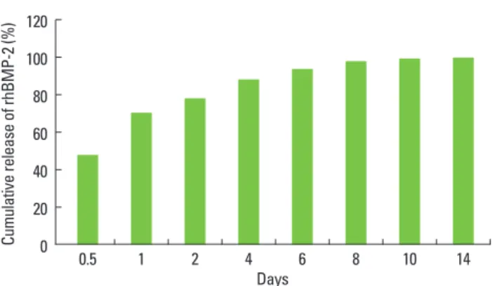
palla-Figure 1. Kinetics of recombinant human bone morphogenetic
pro-tein-2 (rhBMP-2) release from macroporous biphasic calcium phos-phate block observed in vitro. Sustained release of rhBMP-2 was ob-served for up to 2 weeks.
100 80 60 40 20 0 0.5 1 2 4 6 8 10 14 Cu m ul at ive re le as e of rh BM P-2 ( Days
Figure 2. Clinical photograph of a macroporous biphasic calcium
phosphate block (bMBCP) and titanium mini-implant installation. One vertical hole and three horizontal holes are present in the bM-BCP. The titanium mini-implant was installed manually.
A B C
Figure 3. Clinical photograph of a macroporous biphasic calcium
phosphate block (bMBCP) retrieved after necropsy. (A) High-density connective tissue formation was observed with infiltration of mi-crovessels in the test group (recombinant human bone morphoge-netic protein-2/bMBCP). (B) Loose connective tissue, which was easily wiped off, dominantly formed over the bMBCP in the control group (phosphate buffer solution/bMBCP).
(Leo 1450VP, Zeiss, Germany). The most central section from each block was selected to compare the findings between the groups. New bone formation and osseointegration in H&E-stained histological slides were analyzed quantitatively using an image analysis system (Image-Pro Plus; Media Cybernet-ics, Silver Spring, MD, USA). The following parameters were measured: 1) Bone-to-implant contact (BIC): The length of osseointegration that was along the thread profile and in di-rect contact with the newly formed bone was calculated for both sides of the implant on each of the two central sections obtained from each implant; 2) Bone density within the im-plant threads: The area of the newly formed bone within the implant threads was measured. The value of each was ex-pressed as a percentage.
RESULTS
The rhBMP-2 release kinetics of bMBCP were evaluated in
vitro (Fig. 1). Approximately 80% of the loaded rhBMP-2 was
released during the first 4 days. However, rhBMP-2 was con-tinuously released for up to 2 weeks. Clinical healing was un-eventful. In the test group, the horizontal holes became com-pletely filled with hard collagenous tissues that were firmly attached to the block (Fig. 3A). Small diameter blood vessels were integrated within the dense fibrous tissue. The collage-nous tissue could not be easily rubbed off from the bMBCP surface, suggesting highly increased tissue integration. Con-versely, loosely formed connective tissue was formed along the bMBCP block of the control group, which was easily washed off (Fig. 3B).
Histomorphometric analysis using H&E staining
The general pattern of bone formation was favorable in the test group, which was in line with the results from previous studies [24,27,28]. Newly formed bone stained with H&E ex-hibited a high-density, predominantly lamellated bone pat-tern along the implant surface and within the micropores of the bMBCP. This pattern was different from the control group, in which no mineralized tissue had formed. The distribution of the newly formed bone was equally observed along the implant and bMBCP surfaces, while the bridging pattern be-tween the implant and bMBCP surfaces was clearly evident. The newly formed bone did not appear to be homogeneous and exhibited cement lines separating areas of bone that had been deposited at different times. The new bone appeared to be well mineralized and had numerous lacunae. Numerous osteocytes were observed, as well as a few osteoblasts that were aligned along the surface of the newly formed bone.
The results of the histomorphometric analysis are shown
in Table 1. The percentage of direct BIC was 41.23±4.13% (mean±standard deviation) in the test group (n=5), while the control group exhibited no bone formation along the titani-um mini-implants (n=5). The bone density in the test group was 33.47±5.73%.
Light microscopy, polarized-light microscopy, and SEM analysis
Polarized-light microscopy was used to evaluate the pat-terns of lamellation in the newly formed bone, which is con-sidered to be an important hallmark of bone maturation. In the treated group, groups of mineralized collagen fibers with numerous osteocytes were clearly observed under polarized light (Fig. 4C), while there appeared to be new bone forma-tion in direct contact with the bMBCP and implant surfaces. Conversely, no mineralized tissue was found in the control group (Fig. 4F).
The back-scattered SEM images confirmed that the area of newly formed bone was significantly larger in the rhBMP-2-treated group than in the unrhBMP-2-treated control group 8 weeks after transplantation (Fig. 5). Newly formed bone was gener-ally observed within the bMBCP pores and along the implant surface, appearing as light-gray areas. However, no mineral-ized tissue was observed in the sections from the control group. Collectively, mineralized bony tissue on the implant and bMBCP surfaces (including osteocytes, a Haversian sys-tem, and cement lines) was observed exclusively in the test group both during polarized light microscopy and in back-scattered SEM images.
DISCUSSION
To the best of our knowledge, this is the first report of an ectopic osseointegration analysis model based on titanium mini-implants and the rhBMP-2/bMBCP system, which closely simulated clinical parameters. We also evaluated the true osseointegration potential of rhBMP-2 by excluding other contributing factors. This study confirmed the feasibil-ity of this evaluation model and the new bone formation po-tential of rhBMP-2. The results suggest that candidates for
of the newly formed bone.
BIC (%) Bone density (%) Test group (rhBMP-2/bMBCP) 41.23±4.13 33.47±5.73 Control group (phosphate buffer solution/ bMBCP) 0 0 Values are presented as mean±standard deviation. rhBM-2: recombinant human bone morphogenetic protein-2, bMBCP: macroporous biphasic calcium phosphate block.
bone induction and techniques for new bone formation and osseointegration can be analyzed without experimental er-rors using this model.
The present study achieved clinically relevant amounts of BIC and an adequate density of newly formed bone within a relatively short period of time. This finding is in agreement with previous results, suggesting that the mechanism of new bone formation involves initiation of the osteoinduction cas-cade by MBCP at the local site and support for osteoinduc-tive capacity by rhBMP-2 [24,27,29]. The potential of BMPs to induce new bone formation is crucially dependent on the characteristics of the carrier [30,31]. Moreover, it has been re-ported that the surface microstructure plays an important
role in the osteoinductive effect of biomaterials [32]; the mi-crostructure may allow biological apatite to precipitate and induce cells to attach to the surfaces of biomaterials. In par-ticular, micropores with diameters smaller than 5 µm might serve as nucleation sites for biological apatite precipitation because they can facilitate ionic exchange with body fluids [33]. Additionally, micropores at the MBCP surface may favor the adsorption and entrapment of rhBMP-2, which has a high affinity for calcium phosphates [34]. As such, this micro-environment can enhance the adhesion and differentiation of progenitor cells into osteoblasts. Our results demonstrate that bMBCP can potentially mimic the clinical healing envi-ronment of an implant installed in the alveolar ridge.
In this study, newly formed bone was generally observed on implant surfaces and within the vertical and horizontal holes. A possible mechanism underlying this phenomenon appears to be related to the specific microenvironment of bMBCP. It has been reported that microparticles or ionic molecules are released from MBCP during the initial healing period [35] and that the released microparticles may induce the local release of inflammatory cytokines. This release of cytokines stimulates circulating stem cells to repopulate on the MBCP surface and differentiate into osteoblasts, thereby producing bone tissue. In our study, the sustained release of rhBMP-2 in the bMBCP for a relevantly long period may have induced these osteoblastic differentiation cascades. Even though bone formation and osseointegration were evenly distributed, more bone formation was observed in the vertical hole of the implant apex. We postulated that the
os-Figure 4. Histological overview of the retrieved titanium mini-im-plants and recombinant human bone morphogenetic protein-2/ macroporous biphasic calcium phosphate blocks (rhBMP-2/bMB-CPs) or phosphate buffer solution/bMBCPs (H&E staining). (A, B) The rhBMP-2-treated test group presenting significant new bone formation along the implant surface and within the bMBCP (A, ×40; B, ×200). (C) The newly formed bone exhibited a lamellated pattern under polarized-light microscopy, suggesting the occurrence of waves of differentiated bone formation over time (×200). (D, E) The untreated control group demonstrated no mineralized tissue for-mation along the implant surface or within the bMBCP (D, ×40; E,
×200). (F) No mineralized tissue was observed under polarized-light microscopy in the control tissue (×200). Ti: titanium mini-implant, M: bMBCP, NB: new bone.
Figure 5. (A) Scanning electron microscopy (SEM) images of the test group (recombinant human bone morphogenetic protein-2/macro-porous biphasic calcium phosphate blocks [bMBCPs]) 8 weeks after subcutaneous implantation in rats (×50). A higher magnification of the boxed area is shown in B. New bone formation is evident along the implant surface. The bMBCP is revealed as dark-gray areas, while MBCP and the titanium mini-implant appear as light-gray and white, respectively. (B) A higher magnification view of the boxed area clearly shows newly formed bone tissue (×100). The yellow ar-rowheads indicate the obvious formation of NB. (C) The SEM imag-es of the control group (phosphate buffer solution/bMBCP) (×50). (D) Polarized-light microscopy showing the absence of mineralized tissue (×100). Ti: titanium mini-implant, M: bMBCP, NB: new bone.
Te st g ro up Co nt ro l g ro up A D C F B E M M Ti Ti NB NB M M Ti Ti Te st g ro up Co nt ro l g ro up A B C D M M Ti Ti NB
cific microenvironment in this area, although additional studies are required to fully address this phenomenon.
Previous studies have demonstrated that carrier type can influence the effectiveness of rhBMP-2-induced bone for-mation, with several types of carrier systems having been suggested for the clinical use of rhBMPs [7,27,32,33,36,37]. MBCP is reportedly one of the most effective carrier systems for rhBMPs [29] and is composed of hydroxyapatite and tri-calcium phosphate at a ratio of 60:40. MBCP also exhibits an excellent capacity for bone conduction and induction [34,35], which is accelerated when incorporated with rhBMP-2 [38,39]. Although we did not observe any ectopic bone formation in the control group in this study, other reports have described MBCP-induced ectopic new bone formation [34,35]. Particu-larly, excellent bone induction was observed when a pow-dered type of MBCP was used as an rhBMP-2 carrier, which shows its potential for future clinical application.
Research into the effects of rhBMPs on implant osseointe-gration has focused mainly on the alveolar bone defect mod-el in canines. However, an orthotopic modmod-el in canines is difficult to standardize because of the individuality of the animals as well as the different healing patterns and varia-tions in both bone quantity and quality, which may be associ-ated with different healing vectors. Furthermore, most defect morphologies cannot exclude the contribution of each pa-tient’s innate healing potential. Therefore, it has been difficult to analyze the isolated induction capacity of rhBMP-2 on os-seointegration and bone regeneration. The results of the present study, which involved a genetically identical rat strain, revealed that favorable ectopic new bone formation and os-seointegration were possible using an rhBMP-2/bMBCP sys-tem. In addition, minimal bone formation was observed in the control group, indicating that this evaluation model was very standardized and reliable as a result of the exclusion of innate bone forming potential; this suggests that our stan-dardized model was a good candidate for the analysis of or-thotopic implant osseointegration.
Within the limitations of this study, the histology results, the polarized-light microscopy, and SEM analyses all dem-onstrated that impregnation of rhBMP-2 into a bMBCP sig-nificantly induced osteoinductive activity and enhanced os-seointegration in subcutaneous rat tissues at 8 weeks. The ti-tanium mini-implant and rhBMP-2/bMBCP system can be used to evaluate isolated healing characteristics of bone re-generation and osseointegration resulting from rhBMP-2, while excluding the innate healing factors from the host. We postulate that this specific model could be widely used in the process of developing new implant surfaces.
In conclusion, this model represents a more standardized
the implant surface and bMBCP that excludes various heal-ing factors derived from selected animals and defect models.
CONFLICT OF INTEREST
No potential conflict of interest relevant to this article was reported.
ACKNOWLEDGEMENTS
This study was supported by a grant of the Korea Health technology R&D Project, Ministry of Health & Welfare, Re-public of Korea (A100443). We thank Ms. J. M. Lee for provid-ing us with excellent graphics.
REFERENCES
1. Wikesjo UM, Guglielmoni P, Promsudthi A, Cho KS, Trom-belli L, Selvig KA, et al. Periodontal repair in dogs: effect of rhBMP-2 concentration on regeneration of alveolar bone and periodontal attachment. J Clin Periodontol 1999;26: 392-400.
2. Blumenthal NM, Koh-Kunst G, Alves ME, Miranda D, So-rensen RG, Wozney JM, et al. Effect of surgical implanta-tion of recombinant human bone morphogenetic pro-tein-2 in a bioabsorbable collagen sponge or calcium phos-phate putty carrier in intrabony periodontal defects in the baboon. J Periodontol 2002;73:1494-506.
3. Boyne PJ, Nath R, Nakamura A. Human recombinant BMP-2 in osseous reconstruction of simulated cleft palate defects. Br J Oral Maxillofac Surg 1998;36:84-90.
4. Barboza EP, Duarte ME, Geolas L, Sorensen RG, Riedel GE, Wikesjo UM. Ridge augmentation following implanta-tion of recombinant human bone morphogenetic pro-tein-2 in the dog. J Periodontol 2000;71:488-96.
5. Jung JH, Yun JH, Um YJ, Jung UW, Kim CS, Choi SH, et al. Bone formation of Escherichia coli expressed rhBMP-2 on absorbable collagen block in rat calvarial defects. Oral Surg Oral Med Oral Pathol Oral Radiol Endod 2011;111: 298-305.
6. Jang JW, Yun JH, Lee KI, Jang JW, Jung UW, Kim CS, et al. Osteoinductive activity of biphasic calcium phosphate with different rhBMP-2 doses in rats. Oral Surg Oral Med Oral Pathol Oral Radiol 2012;113:480-7.
7. Kim CS, Kim JI, Kim J, Choi SH, Chai JK, Kim CK, et al. Ec-topic bone formation associated with recombinant human bone morphogenetic proteins-2 using absorbable colla-gen sponge and beta tricalcium phosphate as carriers. Biomaterials 2005;26:2501-7.
Weber FE. Effect of rhBMP-2 on guided bone regenera-tion in humans. Clin Oral Implants Res 2003;14:556-68. 9. Smeets R, Maciejewski O, Gerressen M, Spiekermann H,
Hanisch O, Riediger D, et al. Impact of rhBMP-2 on re-generation of buccal alveolar defects during the osseoin-tegration of transgingival inserted implants. Oral Surg Oral Med Oral Pathol Oral Radiol Endod 2009;108:e3-12. 10. Wikesjo UM, Sorensen RG, Wozney JM. Augmentation of
alveolar bone and dental implant osseointegration: clini-cal implications of studies with rhBMP-2. J Bone Joint Surg Am 2001;83 Suppl 1(Pt 2):S136-45.
11. Nevins M, Kirker-Head C, Nevins M, Wozney JA, Palmer R, Graham D. Bone formation in the goat maxillary sinus induced by absorbable collagen sponge implants impreg-nated with recombinant human bone morphogenetic protein-2. Int J Periodontics Restorative Dent 1996;16:8-19. 12. Lekholm U, Adell R, Lindhe J, Branemark PI, Eriksson B,
Rockler B, et al. Marginal tissue reactions at osseointegrat-ed titanium fixtures. (II) A cross-sectional retrospective study. Int J Oral Maxillofac Surg 1986;15:53-61.
13. Hall J, Sorensen RG, Wozney JM, Wikesjo UM. Bone for-mation at rhBMP-2-coated titanium implants in the rat ectopic model. J Clin Periodontol 2007;34:444-51.
14. Herr G, Hartwig CH, Boll C, Kusswetter W. Ectopic bone formation by composites of BMP and metal implants in rats. Acta Orthop Scand 1996;67:606-10.
15. Wikesjo UM, Qahash M, Polimeni G, Susin C, Shanaman RH, Rohrer MD, et al. Alveolar ridge augmentation using implants coated with recombinant human bone morpho-genetic protein-2: histologic observations. J Clin Peri-odontol 2008;35:1001-10.
16. Lee J, Decker JF, Polimeni G, Cortella CA, Rohrer MD, Wozney JM, et al. Evaluation of implants coated with rh-BMP-2 using two different coating strategies: a critical-size supraalveolar peri-implant defect study in dogs. J Clin Periodontol 2010;37:582-90.
17. Susin C, Qahash M, Polimeni G, Lu PH, Prasad HS, Rohrer MD, et al. Alveolar ridge augmentation using implants coated with recombinant human bone morphogenetic protein-7 (rhBMP-7/rhOP-1): histological observations. J Clin Periodontol 2010;37:574-81.
18. Wikesjo UM, Susin C, Qahash M, Polimeni G, Leknes KN, Shanaman RH, et al. The critical-size supraalveolar peri-implant defect model: characteristics and use. J Clin Peri-odontol 2006;33:846-54.
19. Tatakis DN, Koh A, Jin L, Wozney JM, Rohrer MD, Wikesjo UM. Peri-implant bone regeneration using recombinant human bone morphogenetic protein-2 in a canine model: a dose-response study. J Periodontal Res 2002;37:93-100.
Wozney JM, et al. Evaluation of implants coated with re-combinant human bone morphogenetic protein-2 and vacuum-dried using the critical-size supraalveolar peri-implant defect model in dogs. J Periodontol 2010;81:1839-49.
21. Becker J, Kirsch A, Schwarz F, Chatzinikolaidou M, Ro-thamel D, Lekovic V, et al. Bone apposition to titanium implants biocoated with recombinant human bone mor-phogenetic protein-2 (rhBMP-2). A pilot study in dogs. Clin Oral Investig 2006;10:217-24.
22. Jones AA, Buser D, Schenk R, Wozney J, Cochran DL. The effect of rhBMP-2 around endosseous implants with and without membranes in the canine model. J Periodontol 2006;77:1184-93.
23. Langer R, Vacanti JP. Tissue engineering. Science 1993; 260:920-6.
24. Park JC, So SS, Jung IH, Yun JH, Choi SH, Cho KS, et al. In-duction of bone formation by Escherichia coli-expressed recombinant human bone morphogenetic protein-2 us-ing block-type macroporous biphasic calcium phosphate in orthotopic and ectopic rat models. J Periodontal Res 2011;46:682-90.
25. Rohrer MD, Schubert CC. The cutting-grinding technique for histologic preparation of undecalcified bone and bone-anchored implants. Improvements in instrumentation and procedures. Oral Surg Oral Med Oral Pathol 1992;74: 73-8.
26. Jeon O, Rhie JW, Kwon IK, Kim JH, Kim BS, Lee SH. In vivo bone formation following transplantation of human adipose-derived stromal cells that are not differentiated osteogenically. Tissue Eng Part A 2008;14:1285-94.
27. Lee YJ, Jung SW, Chae GJ, Cho KS, Kim CS. The effect of recombinant human bone morphogenetic protein-2/ macroporous biphasic calcium phosphate block system on bone formation in rat calvarial defects. J Korean Acad Periodontol 2007;37(Suppl):397-407.
28. Lee JH, Kim CS, Choi KH, Jung UW, Yun JH, Choi SH, et al. The induction of bone formation in rat calvarial defects and subcutaneous tissues by recombinant human BMP-2, produced in Escherichia coli. Biomaterials 2010;31:3512-9. 29. Bessho K, Konishi Y, Kaihara S, Fujimura K, Okubo Y,
Iizu-ka T. Bone induction by Escherichia coli -derived recom-binant human bone morphogenetic protein-2 compared with Chinese hamster ovary cell-derived recombinant human bone morphogenetic protein-2. Br J Oral Maxillo-fac Surg 2000;38:645-9.
30. Haidar ZS, Hamdy RC, Tabrizian M. Delivery of recombi-nant bone morphogenetic proteins for bone regeneration and repair. Part A: current challenges in BMP delivery.
31. Seeherman H, Wozney JM. Delivery of bone morphoge-netic proteins for orthopedic tissue regeneration. Cyto-kine Growth Factor Rev 2005;16:329-45.
32. Kawakatsu N, Oda S, Kinoshita A, Kikuchi S, Tsuchioka H, Akizuki T, et al. Effect of rhBMP-2 with PLGA/gelatin sponge type (PGS) carrier on alveolar ridge augmentation in dogs. J Oral Rehabil 2008;35:647-55.
33. Schliephake H, Weich HA, Dullin C, Gruber R, Frahse S. Mandibular bone repair by implantation of rhBMP-2 in a slow release carrier of polylactic acid: an experimental study in rats. Biomaterials 2008;29:103-10.
34. Le Nihouannen D, Saffarzadeh A, Gauthier O, Moreau F, Pilet P, Spaethe R, et al. Bone tissue formation in sheep muscles induced by a biphasic calcium phosphate ceram-ic and fibrin glue composite. J Mater Sci Mater Med 2008; 19:667-75.
35. Le Nihouannen D, Guehennec LL, Rouillon T, Pilet P, Bil-ban M, Layrolle P, et al. Micro-architecture of calcium
tissue engineering. Biomaterials 2006;27:2716-22.
36. Hong SJ, Kim CS, Han DK, Cho IH, Jung UW, Choi SH, et al. The effect of a fibrin-fibronectin/beta-tricalcium phos-phate/recombinant human bone morphogenetic pro-tein-2 system on bone formation in rat calvarial defects. Biomaterials 2006;27:3810-6.
37. Abarrategi A, Moreno-Vicente C, Ramos V, Aranaz I, Sanz Casado JV, Lopez-Lacomba JL. Improvement of porous beta-TCP scaffolds with rhBMP-2 chitosan carrier film for bone tissue application. Tissue Eng Part A 2008;14:1305-19. 38. Alam I, Asahina I, Ohmamiuda K, Enomoto S. Compara-tive study of biphasic calcium phosphate ceramics im-pregnated with rhBMP-2 as bone substitutes. J Biomed Mater Res 2001;54:129-38.
39. Oda S, Kinoshita A, Higuchi T, Shizuya T, Ishikawa I. Ecto-pic bone formation by biphasic calcium phosphate (BCP) combined with recombinant human bone morphogenet-ic protein-2 (rhBMP-2). J Med Dent Sci 1997;44:53-62.
![Figure 5. (A) Scanning electron microscopy (SEM) images of the test group (recombinant human bone morphogenetic protein-2/macro-porous biphasic calcium phosphate blocks [bMBCPs]) 8 weeks after subcutaneous implantation in rats (×50)](https://thumb-ap.123doks.com/thumbv2/123dokinfo/5096734.78118/5.892.74.434.153.622/scanning-electron-microscopy-recombinant-morphogenetic-phosphate-subcutaneous-implantation.webp)
