42(8), 1197~1203(2013) http://dx.doi.org/10.3746/jkfn.2013.42.8.1197
지구성 운동과 Ginsenoside Rb
1가 쥐 골격근의 AMP-Activated Protein Kinase(APMK), Phosphatidylinositol 3-Kinase(PI3K) 발현 및 Glucose Uptake에 미치는 영향
정현령․신영호․강호율†
경북대학교 운동대사 실험실
Effects of Endurance Exercise and Ginsenoside Rb
1on AMP-Activated Protein Kinase, Phosphatidylinositol 3-Kinase Expression and
Glucose Uptake in the Skeletal Muscle of Rats
Hyun-Lyung Jung, Young Ho Shin, and Ho-Youl Kang†
Exercise Metabolism Laboratory, Kyungpook National University, Daegu 702-701, Korea
ABSTRACT This study investigated the effects of endurance exercise and ginsenoside Rb1 on AMP-activated protein kinase (AMPK), phosphatidylinositol 3-kinase (PI3K) protein expression and glucose uptake in the skeletal muscle of rats. A total of 32 rats were randomly divided into four groups: CON (Control group, n=8), Ex (Exercise group;
25 m/min for 1 h, 6 days/week, 2 weeks, n=8), Rb1 (Ginsenoside Rb1 group; n=8), and Rb1/Ex (Rb1+Exercise group, n=8). The Rb1 and Rb1/Ex groups were incubated in ginsenoside Rb1 (KRBP buffer, 100 μg/mL) for 60 min after a 2-week experimental treatment. After 2 weeks, the expression of phosphorylated AMPKα Thr172, total AMPKα, the p85 subunit of PI3K, pIRS-1 Tyr612, and pAkt Ser473 were determined in the soleus muscle. Muscle glucose uptake was measured using 2-deoxy-D-[3H] glucose in epitroclearis muscle. Muscle glucose uptake was significantly higher in the three experimental groups (Ex, Rb1, Rb1/Ex) compared to the CON group (P<0.05). The expression of tAMPKα and pAMPKα Thr172 was significantly higher in the Ex, Rb1, and Rb1/Ex groups compared to the CON group (P<0.05).
The expression of pAkt Ser473 was significantly higher in the Rb1 group compared to the CON and EX groups. However, the expression of pIRS-1 Tyr612 and the p85 subunit of PI3K were not significantly different between the four groups.
Overall, these results suggest that ginsenoside Rb1 significantly stimulates glucose uptake in the skeletal muscle of rats through increasing phosphorylation in the AMPK pathway, similar to the effects of exercise.
Key words: exercise, ginsenoside Rb1, muscle glucose uptake, AMPK
Received 8 July 2013; Accepted 22 July 2013
†Corresponding author.
E-mail: [email protected], Phone: 82-53-950-5945
서 론
현대인의 신체활동 부족과 고지방 섭취에 의한 에너지 불 균형은 비만(obesity), 인슐린 저항(insulin resistance), 고 인슐린증(hyperinsulinemia)을 야기하며, 제2형 당뇨병 (type 2 diabetes mellitus)의 발병률을 증가시키고 있다 (1,2). 제2형 당뇨병을 유발하는 근육 세포 내 인슐린 신호전 달과정의 이상에 대한 연구들은 많이 보고되고 있으며(3,4), 비만과 제2형 당뇨병의 예방과 치료를 위한 약물적 접근 및 운동을 통한 근육 내 인슐린 저항 완화에 대한 연구들은 지 속적으로 연구되고 있다(5-7). 특히 제2형 당뇨병은 인슐린 저항의 증가로 인해 식이로 섭취된 탄수화물이 근육 세포내 로 이동/저장되는 과정에서 문제가 발생되어 유발하는 질병 으로서(4), 인슐린 저항을 완화시켜 섭취한 탄수화물을 근육
내로 혈당 이동을 용이하게 하는 것이 중요하다. 근육 내 혈당 이동은 일반적으로 크게 두 경로(signal pathway)로 나눌 수 있는데 첫째, 인슐린 비의존성 신호경로(non-insulin dependent signal pathway)로서 운동에 의한 근육수축을 통해 AMP-activated protein kinase(AMPK)의 활성화시켜 세포 내 GLUT-4 소포체(vesicle)를 세포막과 T세관(T- tubule)으로 이동시켜 혈당을 근육 내로 이동(GLUT-4 translocation)시키는 것이고(8-10), 둘째는 인슐린 의존성 신호경로(insulin dependent signal pathway)로서 인슐린 이 세포막 수용체에 결합하면 세포막에 있는 phosphatidy- linositol 3-kinase(PI3K)의 활성도가 증가되어 세포 내 GLUT-4 소포체를 세포막으로 이동(GLUT-4 transloca- tion)시켜 혈당을 조절하는 것이다(4,11). AMPK는 세포 및 조직의 에너지 항상성에 중요한 역할을 하는 효소로 영양상 태와 외부환경의 변화(exercise, stress)에 대한 세포의 적 응을 중재하는 에너지 감지기 역할을 하는 것으로 알려져 있다(12-14). 특히 골격근에서는 지방산의 산화와 glucose uptake를 촉진하며, 인슐린 분비를 증가시킨다고 보고하고
있다(3,15). 다른 많은 선행연구에서는 지구성 운동, 전기적 자극에 의한 근육 수축 등을 통해 AMPK 활성화를 증가되면 GLUT-4 translocation이 증가되어 glucose uptake가 증 가되는 것으로 보고되고 있다(16-21). 또한 제약 회사에서 는 AMPK signal pathway가 제2형 당뇨병과 비만의 원인 에 관여하는 것으로 알려지면서 그에 관련된 치료제를 개발 을 위한 많은 연구가 이루어지고 있다(1,22,23).
최근에는 인삼 추출물 중 ginsenoside Rb1은 동물의 체중 감소, 혈당내성 개선, 인슐린 활동을 증가시키며(24), Shang 등(25)의 연구에서는 3T3-L1 adipocytes에서 glucose uptake를 증가시킨다고 보고하고 있다. 또한 in vitro(Min6 cell) 연구에서는 ginsenoside Rb1이 췌장의 인슐린 분비를 증가시킨다고 보고하고 있다(26). 이처럼 최근 선행 연구에 서 ginsenoside Rb1과 항비만 그리고 항당뇨 효과에 대한 연구가 보고되고 있으나, 제2형 당뇨병 유발에 크게 기여하 는 골격근 인슐린 저항성에 대한 연구는 보고되지 않고 있 다. 따라서 본 연구의 목적은 ginsenoside Rb1과 운동처치 가 근육내 insulin signal pathway 및 glucose uptake에 미치는 영향을 조사하는데 있다.
재료 및 방법
실험대상
본 연구는 4주령의 SD 수컷 쥐 32마리로 하였다. 쥐는 한 케이지(20.7×35×17 cm)에 1마리씩 넣어 사육하였으 며, 사육실의 온도는 21°C로 유지하였다. 명기와 암기를 각 각 12시간으로 조절하였으며 암기는 7시~19시로 하였다.
1주간의 환경 적응 기간을 거친 후 2주간 유산소 운동을 실시하였고, 무작위로 네 집단으로 구분하였다. 집단 분류는 다음과 같다.
• 비교집단(Control group, CON; n=8)
• 운동비교집단(Exercise group, Ex; n=8)
• Ginsenoside Rb1집단(Rb1 group, Rb1; n=8)
• Rb1+운동집단(Rb1+Exercise group, Rb1/Ex; n=8)
실험설계
식이는 일반 사료(Harlan Teklad, Indianapolis, IN, USA) 를 제공하고 사료와 물의 양은 자유롭게 섭취하도록 하였다.
비교집단과 ginsenoside Rb1 집단을 제외한 집단은 2주간 운동을 실시하였다. 운동은 전동식 실험동물용 트레드밀 (Quinton Instrument, Seattle, WA, USA)을 이용하여 8%
경사도에서 주 6회, 2주 동안 지구성 유산소 운동을 실시하 였다. 트레드밀 속도와 지속시간은 25 m/min, 30분에서 운 동적응 후 2일 간격으로 점증적으로 증가하여, 운동 실시 2주에는 25 m/min, 60분 동안 달리도록 하였다. 모든 집단 은 체중 100 g당 6.5 mg의 sodium pentobarbital(Hanlim Pharm. Co., Ltd., Yongin, Korea)을 복강내 주사로 마취한 다음 근조직 적출하였다. 적출한 soleus muscle은 ginse-
noside Rb1에 incubation한 후에 phosphorylated AMPKα Thr172, total AMPKα, PI3K p85, pIRS-1 Tyr612, pAkt Ser473 분석 전까지 액체질소에 냉각시켜 -70°C(DF-9007, IlShin Lab. Co., Busan, Korea)에 보관하였다. Epitro- chlearis muscle은 glucose transport rate를 분석하기 위 해 incubation 후 즉시 분석하였다.
Muscle incubation(ginsenoside Rb1)
Epitroclearis와 soleus muscle은 운동과 ginsenoside Rb1의 효과를 알아보기 위해 2주간의 운동 후 근육배양 (muscle incubation)을 실시하였다. 적출한 근육은 Krebs Ringer Bicarbonate Buffer(KRBP, 117 mM NaCl, 4.7 mM KCl, 2.5 mM CaCl2, 1.2 mM KH2SO4, 1.2 mM MgSO4, 24.6 mM LaHCO3, 2 mM pyruvate)에 배양하였고, KRBP 는 37°C를 유지하며 95% O2와 5% CO2의 가스를 계속적으 로 주입하였다. 근육은 KRBP에 40분간 pre incubation한 후 ginsenoside Rb1(93537, Sigma-Aldrich Co., St.
Louis, MO, USA)의 효과를 알아보기 위해 근육을 나누어 신선한 KRBP에 ginsenoside Rb1(absence or presence, 100 μg/mL)과 함께 60분간 배양하여 즉시 액체질소에 얼려 분석 전까지 -70°C에서 보관하였다.
Muscle homogenization
근육은 차가운 완충액(20 mM tris-HCl pH 7.4, 1%
Triton X-100, 50 mM NaCl, 250 mM sucrose, 5 mM Na4P2O7, 50 mM NaF, 0.5 mM PMSF, 2 mM DTT, 0.1 mM benzamidine, 4 mg/L leupeptin, 50 mg/L trypsin in- hibitor)과 함께 1:20 비율로 혼합한 다음 polytron homog- enizer(Kinematica, Littau, Switzerland)로 균질화 하였 다. 균질화한 근조직과 기준액은 16,000×g에서 4°C, 40분 간 원심분리 하여(VS-550, Vision Scientific Co., LTD, Bucheon, Korea) 상층액을 분리하였다.
Western blotting
균질화된 근육의 분리된 상층액을 이용하여 DC protein assay(Bio Rad Laboratores, Hercules, CA, USA)를 이용 하여 단백질 정량을 하였다. Laemmli sample buffer(125 mM tris, 20% glycerol, 2% SDS, 0.008% bromophenol blue, pH 6.8)로 1:2 희석하여 95°C에서 5분간 중탕한 후 22,000×g에서 1분간 원심분리 하여 상층액을 분리하였다.
단백질이 함유한 상층액를 10% SDS-polyacrylamide gel electrophoresis(PAGE)에 주입한 다음 전기영동(Mini Trans-Blot Electrophoretic Transfer Cell, Bio Rad Laboratores)한 후 transfer buffer를 이용하여 PVDF membrane으로 이동시켰다. Membrane은 T-TBS(3번×
10분)로 세척하고 10% skin milk를 이용하여 60분간 blocking한 후 1차 항체(#2531, #2532, #4257, #4060, Cell Signaling Technology, Beverly, MA, USA; ab66153,
Table 1. Changes of body weight (g)
Pre Post
CON Ex Rb1
Rb1/Ex
98.6±1.2 95.8±1.0 97.9±1.2 98.6±1.7
210.9±2.3 192.3±2.7*# 207.7±1.4 191.1±4.2*# Values are means±SE. CON, Control group; EX, Exercise group;
Rb1, Rb1 group; Rb1/Ex, Rb1+Exercise group. *P<0.05 vs CON;
#P<.005 vs Rb1.
0.0 1.0 2.0 3.0 4.0 5.0
CON EX Rb1 Rb1/EX CON EX Rb1 Rb1/EX
submaximal insulin concentration non insulin condition
μmol/ml/h .
*
*
*
Rb1 Rb1/EX Rb1 Rb1/EX
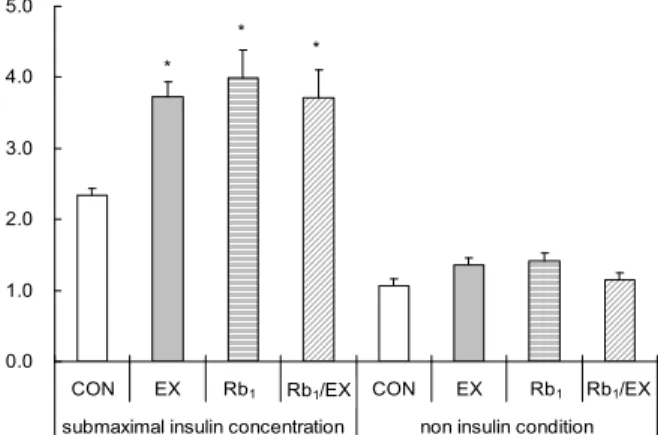
Fig. 1. Glucose transport rate under submaximal insulin concen- trations and non-insulin concentrations in epitrochlearis muscle.
CON, Control group; EX, Exercise group; Rb1, Rb1 group; Rb1/Ex, Rb1+Exercise group. Values are means±SE, n=8. *P<0.05 vs CON.
Abcam, Cambridge, UK; 1:1,000 dilution)에 넣어 in- cubation(4°C with gentle shaking, overnight)하였다. T- TBS를 이용하여 세척한 후, membrane은 2차 항체(#7074, Cell Signaling Technology, 1:2,000 dilution)와 함께 60 분 동안 incubation하고 다시 세척하였다. Protein은 X-ray film에 ECL Western blot detection kit(RPN2106, Am- ersham, Arlington Heights, IL, USA)를 사용하여 시각화 하였다. X-ray film 시각화된 밴드는 스캔 후 Image J (http://wsrnih.gov, Image Processing and Analysis in Java) 프로그램을 이용하여 densitometry analysis(gel internal density and gel peak analysis) 방법으로 분석하 였다.
Muscle glucose transport rate
포도당 운반률은 비대사물질인 glucose analog 2-de- oxy-D-glucose(American Radiolabeled Chemicals Inc., St. Louis, MO, USA)를 이용하여 측정하였다. 측정을 위해 좌우측 epitrochlearis muscle을 적출하였다. 근육내 glu- cose를 제거하기 위해 KHB(32 mM mannitol, 8 mM glu- cose, 0.1% BSA)에 30~60분간 세척하였다. 세척 후 32 mM mannitol, 8 mM glucose, 0.1% BSA(1,000 μIU/mL insulin, absence or presence)가 함유된 KHB에 20분간 배양하였다. 다시 10분간 36 mM mannitol, 2 mM pyr- uvate, 0.1% BSA에 세척 후 deoxy-D-glucose, 2-[1,2-
3H](2.5 μCi/mL)와 16 mM[14C] mannitol(0.3 μCi/mL)이 함유된 1.5 mL KHB(36 mM mannitol, 4 mM deoxy glu- cose, 0.1% BSA)(1,000 μIU/mL insulin, absence or presence)에 30°C, 20분간 하였다. 세포 내외의 2-DG의 활성도는 scintillation counter(Wallac 1220 QUANTULUS, PerkinElmer Inc., Waltham, MA, USA)를 이용하여 10분 간 측정하였다.
통계처리
각 측정항목에 대한 결과는 평균과 표준오차(mean±SE) 를 산출하였으며, 통계적 분석은 SPSS 18.0 통계프로그램 (SPSS Inc., Chicago, IL, USA)을 이용하였다. 통계처리는 집단 간 처치 후 변화를 알아보기 위해 일원분산분석법 (one-way ANOVA)을 이용하였고 사후검정은 Tukey 방법 을 이용하였다. 통계적 유의수준은 α=0.05로 하였다.
결과 및 고찰
체중의 변화
체중의 변화는 1일 1회 운동 2시간 전 저울(METTER PJ6, Metter Toledo International Inc., Seoul, Korea)을 이용하여 쥐의 체중을 측정하였으며 2주간의 실험처치 후 네 집단의 체중은 Table 1과 같다. 체중은 운동집단(Ex, Rb1/Ex)이 비운동집단(CON, Rb1)에 비해 유의한 차이가 있
는 것으로 나타났다(F=13.083, p=0.000).
Muscle glucose uptake
2주간의 실험 처치 후 non-insulin condition과 sub- maximal 인슐린 농도(1,000 μIU/mL)에서 muscle glucose uptake를 측정하였다(27). Submaximal condition에서 muscle glucose uptake는 Ex(3.73±0.36 μmol/mL/h), Rb1(3.99±0.21 μmol/mL/h), Rb1/Ex(3.71±0.39 μmol/
mL/h) 집단이 CON(2.34±0.10 μmol/mL/h) 집단에 비해 유의하게 높게 나타났다(F=6.611, P=0.002, Fig. 1). 그러 나 non-insulin condition에서는 네 집단간 muscle glu- cose uptake는 유의한 차이가 없는 것으로 나타났다(F=
1.681, P=0.194, Fig. 1).
선행연구에 의하면 인삼 섭취는 혈당 조절에 효과적인 것 으로 보고되고 있으며(24) 제2형 당뇨병, 정상인, 당뇨 쥐의 혈당을 감소시킨다고 보고하고 있다(28). 특히 최근에는 ginsenoside를 이용한 혈당 조절에 대한 연구가 진행되고 있다(24-26). Xiong 등(24)은 고지방식이 비만 쥐를 대상 으로 in vivo 조건에서 4주간 복강 내 Rb1을 투여한 결과 공복 시 혈당 감소와 혈당 내성이 개선되었다고 보고하였다.
또한 본 실험처럼 in vitro 연구에서 ginsenoside Rb1은 3T3-L1 adipocytes와 C2C12 myotubes cell에서 glu- cose uptake를 증가시킨다고 보고하고 있다. 이는 인슐린
CON EX Rb1 Rb1/Ex t-AMPKα
p-AMPKα Thr172 GAPDH
0 20 40 60 80 100
% of heart standard .
CON Ex
Rb1 Rb1/EX
* * *
Rb1 Rb1/EX
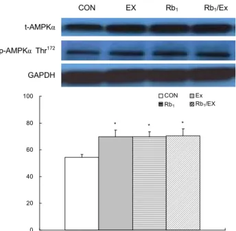
Fig. 2. Expression of total AMPKα in soleus muscle. CON, Control group; EX, Exercise group; Rb1, Rb1 group; Rb1/Ex, Rb1+Exercise group. Values are means±SE, n=8. *P<0.05 vs CON.
0 20 40 60 80
% of heart standard .
CON EX
Rb1 Rb1/EX
* *
*
Rb1 Rb1/EX
Fig. 3. Expression of pAMPKα Thr172 in soleus muscle. CON, Control group; EX, Exercise group; Rb1, Rb1 group; Rb1/Ex, Rb1+Exercise group. Values are means±SE, n=8. *P<0.05 vs CON.
신호체계 활성화에 의한 GLUT-1과 GLUT-4 protein ex- pression과 세포내 소포체 형태의 GLUT-4가 세포막으로 translocation 증가가 insulin-sensitive cell의 혈당 운반을 자극한 것이라고 보고하였다(25). 또한 Kong 등(29)의 연 구에서도 저산소증(hypoxic)의 cardiomyocyte cell에 다 양한 농도의 ginsenoside Rb1을 처치(incubation)한 결과 glucose uptake와 GLUT-4 expression이 증가한 것으로 보고하였다. 본 연구의 결과 2주간의 운동 후 epitroclearis 근육에 ginsenoside Rb1(100 μg/mL)을 배양(incubation) 한 결과 비교집단에 비해 근육 내 glucose uptake가 유의하 게 증가한 것을 알 수 있었다(Fig. 1). 이는 선행연구의 결과 처럼 운동과 ginsenoside Rb1이 근육 내 GLUT-4 trans- location을 증가시켜 glucose uptake를 증가시킨 것으로 사 료된다. 하지만 본 연구에서는 골격근 배양 후 골격근의 세 포막(sarcolema)를 분리하여 GLUT-4를 직접 측정하는 것 이 타당하다고 생각하지만, incubation한 근육의 무게가 약 20~40 mg으로서 다른 분석과 동시에 membrane purifi- cation을 하기에는 어려움이 있었다. 따라서 추후 연구에서 는 sarcolema 분리에 의한 GLUT-4 대한 연구가 이루어져 야 할 것으로 판단된다. 또한 본 연구에서 복합처치를 받은 Rb1/Ex 집단의 glucose uptake가 단독처치 집단인 Ex와 Rb1 집단과 비교하여 유의한 차이가 없는 것으로 나타났다.
이것은 첫째, 선행연구 Rb1 실험에서는 본 실험에 비해 상대 적 incubation을 장시간 실시했으나(24,25), 근육세포에서 는 cell culture처럼 장시간 incubation을 하기에는 실질적 으로 실험적 한계가 있으므로 이것이 본 실험에서 복합처치 효과가 나타나지 않은 것에 대한 설명이 될 수 있을 것으로 사료된다. 그러므로 추후 이러한 실험의 제한점을 보완한 실험이 실시되어 incubation 시간에 대한 가설을 검정할 필 요가 있을 것으로 사료된다. 둘째, 실험처치는 다르지만 본 연구와 실험설계가 비슷한 Borst와 Snellen(30)의 연구에 서 exercise와 metformin 단독처치는 glucose uptake를 유의하게 증가하였으나 exercise와 metformin 복합처치에 서 glucose uptake에서 additive effect는 나타나지 않았 다. 이것은 세포 내 여러 단계의 signal pathway에서 차이가 있더라도 최종 단계 biological effect인 glucose uptake 에는 유의한 차이가 없게 나타난 것처럼 본 연구도 이와 유 사한 이유에 의해 복합처치 효과가 나타나지 않은 것으로 여겨진다.
Total AMPKα, phosphorylated AMPKα Thr172의 발현량 2주간의 실험 처치 후 total AMPKα는 CON(54.3±
0.2.2%)집단에 비해 Ex(69.8±4.9%), Rb1(69.8±3.9%), Rb1/Ex(70.5±5.2%) 집단이 유의하게 높게 나타났다(F=
3.516, P=0.028, Fig. 2). pAMPKα Thr172에서는 CON (39.2±2.0%) 집단보다 세 집단(Ex: 52.9±4.2%, Rb1: 53.1
±3.4%, Rb1/Ex: 51.3±3.2%)에서 유의하게 높게 나타났다 (F=4.154, P=0.015, Fig. 3).
Glucose uptake에 관여하는 AMPK는 골격근 내 glu- cose uptake를 촉진하며, 췌장의 β-cell에서는 인슐린 분 비를 증가시킨다고 보고하고 있다(3,15). 선행연구에 의하면 AMPK의 활성화는 GLUT-4 발현을 직접 조절하고, 근육 세포 내 glucose uptake를 증가시켜 근육 내 혈당조절에 효과가 있다고 보고하고 있으며(22,29,31,32). 트레이닝에 의한 골격근의 GLUT-4 발현은 근육 내 AMPK의 활성을 증가시킨다고 보고하고 있다(16-21). 최근 ginsenoside Rb1과 AMPK에 관한 선행연구를 살펴보면, hepatic cell을 이용한 농도별(5~20 μM) ginsenoside Rb1을 처치한 결과 10과 20 μM 농도에서 pAMPK가 증가하였다고 보고하였 고, 10 μM Rb1 처치 4시간 후 pAMPK가 증가하였다고 보고 하였다(33). 또한 고지방 비만 쥐를 대상으로 ginsenoside
CON EX Rb1 Rb1/Ex pIRS-1 Tyr612
PI3K p85
pAkt Ser473 GAPDH
0 20 40 60 80 100
% of heart standard .
CON EX
Rb1 Rb1/EX Rb1 Rb1/EX
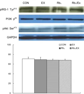
Fig. 4. Expression of pIRS-1 Tyr612 in soleus muscle. CON, Control group; EX, Exercise group; Rb1, Rb1 group; Rb1/Ex, Rb1+Exercise group. Values are means±SE, n=8.
0 20 40 60 80 100
% of heart standard .
CON EX
Rb1 Rb1/EX Rb1 Rb1/EX
Fig. 5. Expression of PI3K p85 in soleus muscle. CON, Control group; EX, Exercise group; Rb1, Rb1 group; Rb1/Ex, Rb1+ Exercise group. Values are means±SE, n=8.
0 20 40 60 80 100
% of heart standard .
CON EX
Rb1 Rb1/EX
,#
Rb1 Rb1/EX
*
Fig. 6. Expression of pAkt Ser473 in soleus muscle. CON, Control group; EX, Exercise group; Rb1, Rb1 group; Rb1/Ex, Rb1+ Exercise group. Values are means±SE, n=8. *P<0.05 vs CON,
#P<0.05 vs Ex.
Rb1(10 mg/kg/day)을 투여한 결과 간의 pAMPK가 증가했 다고 보고하였다(33). Kong 등(29)은 cardiomyocyte cell 을 이용한 ginsenoside Rb1을 처치한 후 AMPKα, pAMPKα Thr172 및 AMPK activity가 증가하였다고 보고하였다. 본 연구에서는 2주간의 운동 후 ginsenoside Rb1(100 μg/mL) 에 근육 내 1시간 처치한 결과 비교 집단을 제외한 세 집단에 서 tAMPKα, pAMPKα Thr172가 증가한 것을 볼 수 있었다 (Fig. 3, 4). 이는 선행연구의 결과에서처럼 ginsenoside Rb1은 근육 내 pAMPK를 증가시키고 AMPK 활성화에 효과 가 있는 것으로 생각되며 AMPK 활성화는 GLUT-4 trans- location을 증가시켜 근육내 혈당 이동에 효과가 있는 것으 로 생각된다.
pIRS-1 Tyr612, PI3K p85, pAkt Ser473의 발현량
2주간의 실험처치 후 pIRS-1 Tyr612에서 CON(71.0±
3.5%), Ex(69.3±5.0%), Rb1(68.2±5.0%) 그리고 Rb1/Ex (68.0±2.1%)는 집단간 유의한 차이가 없는 것으로 나타났 다(F=0.155, P=0.926, Fig. 4). 또한 PI3K p85에서도 CON (72.8±2.7%), Ex(70.1±6.1%), Rb1(75.6±4.3%) 그리고 Rb1/Ex(73.0±3.5%) 사이에 모두 유의한 차이가 없는 것으 로 나타났다(F=0.266, P=0.849, Fig. 5). 하지만 pAkt Ser473에서는 Rb1(64.2±2.8%) 집단이 CON(50.5±2.8%), Ex(52.4±2.9%) 집단에 비해 유의하게 높게 나타났다(F=
4.777, P=0.008, Fig. 6). 그러나 CON, Ex, 그리고 Rb1/Ex 집단 간에는 유의한 차이가 없는 것으로 나타났다(F=4.777, P=0.008, Fig. 6).
Ginsenoside Rb1은 인슐린 신호체계를 활성화시켜 adi- pocyte와 신경세포에서 GLUT-4 translocation을 증가시 켜 근육 내 glucose uptake를 증가시킨다고 보고하고 있다 (24,25). 또한 인슐린 신호체계 중 PI3K는 세포 내 IRS-1, 인슐린 수용체의 β submit tyrosine residues의 auto- phosphorylation과 IRS-1, PI3K의 downstream signal- ing(pAKt)에 의해 활성화된다고 보고되고 있다(4,11,24, 25). Shang 등(25)은 1 μm ginsenoside Rb1과 100 nM의 인슐린을 3T3-L1 adipocytes에 처치한 결과 인슐린 처치 에서는 pIRS-1 Tyr612, pAkt Thr308, pAkt Ser473, PI3K activity가 증가하였으며, ginsenoside Rb1에서는 IRS-1, pAkt Thr308, PI3K activity가 증가하였다고 보고하였다.
Xiong 등(24)은 ginsenoside Rb1의 PI3K signaling 경로 에 미치는 효과를 알아보기 위해 시상하부 neuronal cell을 이용하여 농도별(0.1~100 μM/L)로 처치 후 pAkt 활성을 조사한 결과 10, 100 uM에서 pAkt가 증가했으며, 10 μM 처치 2시간 후 pAkt가 증가했다고 보고하였다. 또한 고지방 식이 비만 쥐를 대상으로 4주간의 ginsenoside Rb1 처치 후 시상하부 조직에서도 pAkt 활성화가 증가했다고 보고하 였다. 그러나 본 연구에서는 2주간의 운동 후 ginsenoside
Rb1(100 μg/mL)에 60분 동안 근육을 배양한 결과 pIRS- 1 Tyr612와 PI3K p85에서는 비교집단과 비교하여 유의한 차이가 없는 것으로 나타났다. 반면 pAkt Ser473은 ginse- noside Rb1 단독처치한 Rb1 집단에서 유의하게 높게 나타났 으나, Con, Ex와 Rb1/Ex 처치집단에서는 유의한 차이가 없 는 것을 알 수 있었다. 이것은 ginsenocide Rb1 incubation 에 의해 증가되는 pAkt Ser473은 효과를 운동이 억제하는 것으로 생각할 수 있다. 이것은 첫째, pAkt Ser473의 증가는 muscle glucose uptake에 유의한 영향을 미치지 않는 것으 로 생각할 수 있으며, ginsenocide Rb1 incubation에 의한 glucose uptake의 증가는 AMPK signal pathway에 의존 하는 것으로 본 연구결과는 제안하고 있다. 하지만 본 연구 의 ginsenoside Rb1 배양 시간이 선행연구에 비해(24,25) 상대적으로 짧아 본 연구의 AMPK pathway 결과와 달리 PI3K downstream signaling(pIRS-1 Tyr612, PI3K p85)에 영향을 미치지 못하여서 glucose uptake에 유의한 영향을 미치지 못한 것으로도 생각될 수 있다. 그러므로 추후 본 연구 결과를 바탕으로 ginsenoside Rb1의 농도, 처치시간, 처치방법을 달리하여 PI3K downstream signaling과 muscle glucose uptake 연구가 필요할 것으로 사료된다.
요 약
본 연구는 2주간의 지구성 운동과 ginsenoside Rb1이 쥐 골격근의 AMPK insulin signaling(tAMPKα, pAMPKα Thr172)과 PI3K insulin signaling pathway(pIRS-1 Tyr612, PI3K p85, pAkt Ser473) 발현 및 glucose uptake에 미치는 영향을 분석하였다. 골격근내 glucose uptake에서는 비교 집단과 비교하여 운동집단(59.4%), Rb1집단(70.5%) Rb1/ Ex집단(58.6%)에서 유의하게 증가하였다. 2주간의 지구성 운동과 ginsenoside Rb1이 AMPK insulin signaling path- way에 미치는 효과를 조사한 결과 비교집단에 비해 AMPK α(Ex, 28.6%; Rb1, 28.5%; Rb1/Ex, 29.8%), pAMPKα Thr172(Ex, 35.1%; Rb1, 35.3%; Rb1/Ex, 30.9%)의 발현이 유의하게 증가한 것을 알 수 있었다. 2주간의 지구성 운동과 ginsenoside Rb1이 PI3K insulin signaling pathway에 미 치는 효과를 알아본 결과 비교집단과 비교하여 IRS-1, PI3K p85에서는 유의한 차이가 없었으나 pAkt Ser473은 Rb1
집단에서 유의하게 증가한 것을 알 수 있었다. 이상의 결과 를 종합해 볼 때, ginsenoside Rb1은 운동과 더불어 근육 세포내 AMPK의 활성화와 근육 내 glucose uptake를 증가 시켜 제2형 당뇨병 예방과 치료에 효과가 있을 것으로 생각 된다. 그러나 본 연구의 결과로 PI3K insulin signaling pathway의 항당뇨 효과는 설명하기는 부족하다고 판단되 며 추후 본 연구의 결과를 기초로 ginsenoside Rb1의 농도, 처치시간, 처치방법을 고려한 후속 연구가 필요할 것으로 생각된다.
감사의 글
이 논문은 2010년도 정부재원(교육과학기술부 인문사회연 구역량강화사업비)으로 한국연구재단의 지원을 받아 연구 되었으며(NRF-2010-327-G00113) 지원에 감사드립니 다.
REFERENCES
1. Lessard SJ, Rivas DA, Chen ZP, Bonen A, Febbraio MA, Reeder DW, Kemp BE, Yaspelkis BB 3rd, Hawley JA.
2007. Tissue-specific effects of rosiglitazone and exercise in the treatment of lipid-induced insulin resistance. Diabetes 56: 1856-1864.
2. Cao S, Li B, Yi X, Chang B, Zhu B, Lian Z, Zhang Z, Zhao G, Liu H, Zhang H. 2012. Effects of exercise on AMPK signaling and downstream components to PI3K in rat with type 2 diabetes. PLoS One 7: e51709.
3. Christopher MJ, Chen ZP, Rantzau C, Kemp BE, Alford FP.
2003. Skeletal muscle basal AMP-activated protein kinase activity is chronically elevated in alloxan-diabetic dogs: im- pact of exercise. J Appl Physiol 95: 1523-1530.
4. Khan AH, Pessin JE. 2002. Insulin regulation of glucose uptake: a complex interplay of intracellular signalling path- ways. Diabetologia 45: 1475-1483.
5. Karlsson HK, Zierath JR. 2007. Insulin signaling and glu- cose transport in insulin resistant human skeletal muscle.
Cell Biochem Biophys 48: 103-113.
6. Karlsson HK, Ahlsen M, Zierath JR, Wallberg-Henriksson H, Koistinen HA. 2006. Insulin signaling and glucose trans- port in skeletal muscle from first-degree relatives of type 2 diabetic patients. Diabetes 55: 1283-1288.
7. Musi N, Goodyear LJ. 2006. Insulin resistance and improve- ments in signal transduction. Endocrine 29: 73-80.
8. Sherman LA, Hirshman MF, Cormont M, Le Marchand- Brustel Y, Goodyear LJ. 1996. Differential effects of insulin and exercise on Rab4 distribution in rat skeletal muscle.
Endocrinology 137: 266-273.
9. Goodyear LJ, Kahn BB. 1998. Exercise, glucose transport, and insulin sensitivity. Annu Rev Med 49: 235-261.
10. Rockl KS, Witczak CA, Goodyear LJ. 2008. Signaling mech- anisms in skeletal muscle: acute responses and chronic adap- tations to exercise. IUBMB Life 60: 145-153.
11. Watson RT, Kanzaki M, Pessin JE. 2004. Regulated mem- brane trafficking of the insulin-responsive glucose trans- porter 4 in adipocytes. Endocr Rev 25: 177-204.
12. Chambers MA, Moylan JS, Smith JD, Goodyear LJ, Reid MB. 2009. Stretch-stimulated glucose uptake in skeletal muscle is mediated by reactive oxygen species and p38 MAP-kinase. J Physiol 587: 3363-3373.
13. Witczak CA, Sharoff CG, Goodyear LJ. 2008. AMP-acti- vated protein kinase in skeletal muscle: from structure and localization to its role as a master regulator of cellular metabolism. Cell Mol Life Sci 65: 3737-3755.
14. Gibala MJ, McGee SL, Garnham AP, Howlett KF, Snow RJ, Hargreaves M. 2009. Brief intense interval exercise acti- vates AMPK and p38 MAPK signaling and increases the expression of PGC-1α in human skeletal muscle. J Appl Physiol 106: 929-934.
15. Sakamoto K, Goransson O, Hardie DG, Alessi DR. 2004.
Activity of LKB1 and AMPK-related kinases in skeletal muscle: effects of contraction, phenformin, and AICAR. Am
J Physiol Endocrinol Metab 287: E310-E317.
16. Fujii N, Hayashi T, Hirshman MF, Smith JT, Habinowski SA, Kaijser L, Mu J, Ljungqvist O, Birnbaum MJ, Witters LA, Thorell A, Goodyear LJ. 2000. Exercise induces iso- form-specific increase in 5'AMP-activated protein kinase ac- tivity in human skeletal muscle. Biochem Biophys Res Commun 273: 1150-1155.
17. Hutber CA, Hardie DG, Winder WW. 1997. Electrical stim- ulation inactivates muscle acetyl-CoA carboxylase and in- creases AMP-activated protein kinase. Am J Physiol 272:
E262-E266.
18. Ihlemann J, Ploug T, Hellsten Y, Galbo H. 1999. Effect of tension on contraction-induced glucose transport in rat skel- etal muscle. Am J Physiol 277: E208-E214.
19. Musi N, Hayashi T, Fujii N, Hirshman MF, Witters LA, Goodyear LJ. 2001. AMP-activated protein kinase activity and glucose uptake in rat skeletal muscle. Am J Physiol Endocrinol Metab 280: E677-E684.
20. Vavvas D, Apazidis A, Saha AK, Gamble J, Patel A, Kemp BE. 1997. Contraction-induced changes in acetyl-CoA car- boxylase and 5'-AMP-activated kinase in skeletal muscle.
J Biol Chem 272: 13255-13261.
21. Wojtaszewski JF, Nielsen P, Hansen BF, Richter EA, Kiens B. 2000. Isoform-specific and exercise intensity-dependent activation of 5'-AMP-activated protein kinase in human skeletal muscle. J Physiol 2000 528: 221-226.
22. Suwa M, Egashira T, Nakano H, Sasaki H, Kumagai S. 2006.
Metformin increases the PGC-1alpha protein and oxidative enzyme activities possibly via AMPK phosphorylation in skeletal muscle in vivo. J Appl Physiol 101: 1685-1692.
23. Fryer LG, Parbu-Patel A, Carling D. 2002. The anti-diabetic drugs rosiglitazone and metformin stimulate AMP-activated protein kinase through distinct signaling pathways. J Biol Chem 277: 25226-25232.
24. Xiong Y, Shen L, Liu KJ, Tso P, Wang G, Woods SC. 2010.
Antiobesity and antihyperglycemic effects of ginsenoside
Rb1 in rats. Diabetes 59: 2505-2512.
25. Shang W, Yang Y, Zhou L, Jiang B, Jin H, Chen M. 2008.
Ginsenoside Rb1 stimulates glucose uptake through in- sulin-like signaling pathway in 3T3-L1 adipocytes. J Endoc- rinol 198: 561-569.
26. Park S, Ahn IS, Kwon DY, Ko BS, Jun WK. 2008. Ginseno- sides Rb1 and Rg1 suppress triglyceride accumulation in 3T3-L1 adipocytes and enhance beta-cell insulin secretion and viability in Min6 cells via PKA-dependent pathways.
Biosci Biotechnol Biochem 72: 2815-2823.
27. Sherman WM, Katz AL, Cutler CL, Withers RT, Ivy JL.
1988. Glucose transport: locus of muscle insulin resistance in obese Zucker rats. Am J Physiol 255: E374-E382.
28. Xie JT, Zhou YP, Dey L, Attele AS, Wu JA, Gu M. 2002 Ginseng berry reduces blood glucose and body weight in db/db mice. Phytomedicine 9: 254-258.
29. Kong HL, Wang JP, Li ZQ, Zhao SM, Dong J, Zhang WW.
2009. Anti-hypoxic effect of ginsenoside Rbl on neonatal rat cardiomyocytes is mediated through the specific activa- tion of glucose transporter-4 ex vivo. Acta Pharmacol Sin 30: 396-403.
30. Borst SE, Snellen HG. 2001. Metformin, but not exercise training, increases insulin responsiveness in skeletal muscle of Sprague-Dawley rats. Life Sci 69: 1497-1507.
31. Jessen N, Pold R, Buhl ES, Jensen LS, Schmitz O, Lund S. 2003. Effects of AICAR and exercise on insulin-stimu- lated glucose uptake, signaling, and GLUT-4 content in rat muscles. J Appl Physiol 94: 1373-1379.
32. Ojuka EO, Nolte LA, Holloszy JO. 2000. Increased ex- pression of GLUT-4 and hexokinase in rat epitrochlearis muscles exposed to AICAR in vitro. J Appl Physiol 88:
1072-1075.
33. Shen L, Xiong Y, Wang DQ, Howles P, Basford JE, Wang J. 2013. Ginsenoside Rb1 reduces fatty liver by activating AMP-activated protein kinase in obese rats. J Lipid Res 54:
1430-1438.