pISSN 2093-596X · eISSN 2093-5978
Article
Activation of AMP-Activated Protein Kinase Attenuates Tumor Necrosis Factor-α-Induced Lipolysis via Protection of Perilipin in 3T3-L1 Adipocytes
Seok-Woo Hong1, Jinmi Lee1, Se Eun Park2, Eun-Jung Rhee2, Cheol-Young Park2, Ki-Won Oh2, Sung-Woo Park2, Won-Young Lee2
1Institute of Medical Research, 2Division of Endocrinology and Metabolism, Department of Internal Medicine, Kangbuk Samsung Hospital, Sungkyunkwan University School of Medicine, Seoul, Korea
Background: Tumor necrosis factor (TNF)-α and AMP-activated protein kinase (AMPK) are known to stimulate and repress li- polysis in adipocytes, respectively; however, the mechanisms regulating these processes have not been completely elucidated.
Methods: The key factors and mechanism of action of TNF-α and AMPK in lipolysis were investigated by evaluating perilipin expression and activity of protein kinase RNA-like endoplasmic reticulum kinase (PERK)/eukaryotic initiation factor 2 α (eIF2α) by Western blot and an immunofluorescence assay in 24-hour TNF-α-treated 3T3-L1 adipocytes with artificial manipulation of AMPK activation.
Results: Enhancement of AMPK activity by the addition of activator minoimidazole carboxamide ribonucleotide (AICAR) sup- pressed TNF-α-induced lipolysis, whereas the addition of compound C, an inhibitor of AMPK phosphorylation, enhanced lipoly- sis. Perilipin, a lipid droplet-associated protein, was decreased by TNF-α and recovered following treatment with AICAR, show- ing a correlation with the antilipolytic effect of AICAR. Significant activation of PERK/eIF2α, a component of the unfolded pro- tein response signaling pathway, was observed in TNF-α or vesicle-treated 3T3-L1 adipocytes. The antilipolytic effect and recov- ery of perilipin expression by AICAR in TNF-α-treated 3T3-L1 adipocytes were significantly diminished by treatment with 2-aminopurine, a specific inhibitor of eIF2α.
Conclusion: These data indicated that AICAR-induced AMPK activation attenuates TNF-α-induced lipolysis via preservation of perilipin in 3T3-L1 adipocytes. In addition, PERK/ eIF2α activity is a novel mechanism of the anti-lipolytic effect of AICAR.
Keywords: AMP-activated protein kinases; Lipolysis; Perilipin; PERK/eIF2α; Adipocytes
INTRODUCTION
Tumor necrosis factor (TNF)-α is a cytokine with a well-estab- lished role in immunomodulatory and inflammatory responses,
and has been implicated in the development of obesity [1,2]. El- evated TNF-α production in adipose tissues and adipocytes from obese subjects suggests that TNF-α may be linked to diabetes and insulin resistance [3-5]. Several laboratories have demon-
Received: 27 February 2014, Revised: 11 April 2014, Accepted: 24 April 2014
Corresponding author: Won-Young Lee
Division of Endocrinology and Metabolism, Department of Internal Medicine, Kangbuk Samsung Hospital, Sungkyunkwan University School of Medicine, 29 Saemunan-ro, Jongno-gu, Seoul 110-746, Korea
Tel: +82-2-2001-2579, Fax: +82-2-2001-2049, E-mail: [email protected]
Copyright © 2014 Korean Endocrine Society
This is an Open Access article distributed under the terms of the Creative Com- mons Attribution Non-Commercial License (http://creativecommons.org/
licenses/by-nc/3.0/) which permits unrestricted non-commercial use, distribu- tion, and reproduction in any medium, provided the original work is properly cited.
strated that TNF-α increases lipolysis and release of free fatty ac- ids (FFAs) from adipocytes [6-8]. In addition, it has been shown that the levels of TNF-α and FFAs are positively correlated in adipose tissues [9]. However, the mechanism of TNF-α function in obese subjects and lipolysis is not completely understood.
AMP-activated protein kinase (AMPK) is a widely-expressed serine/threonine kinase that is considered to act as an intracel- lular energy sensor. Because AMPK activation may have ben- eficial metabolic consequences in diabetic patients, AMPK has emerged as a potential target for the treatment of obesity and type 2 diabetes [10]. It has been demonstrated that two classes of antidiabetic drugs (metformin and thiazolidinedio- nes) act at least in part through activation of AMPK in liver and muscle [11,12]. Whereas the function of AMPK in liver and muscle has been well-illustrated, the role of AMPK in adi- pose tissue remains poorly documented. An antilipolytic effect of the AMPK activator minoimidazole carboxamide ribonu- cleotide (AICAR) has been demonstrated in a lipolysis model treated with cyclic AMP (cAMP) inducers [13,14]. However, although the cAMP level is increased in TNF-α-treated adipo- cytes, the main mechanism of TNF-α-induced lipolysis has re- mained unknown.
In adipocytes, induction of stress can increase the expression and secretion of proinflammatory cytokines [15]. Overproduc- tion and chronic exposure to proinflammatory cytokines such as TNF-α and interleukin-6 can induce lipolysis and apoptosis in adipocytes as a result of endoplasmic reticulum (ER) stress and generation of reactive oxygen species [16]. A variety of en- vironmental insults leads to the phosphorylation of a family of proteins associated with the unfolded protein response (UPR), including protein kinase RNA-like endoplasmic reticulum ki- nase (PERK) and eukaryotic initiation factor 2 α (eIF2α), to al- leviate cellular injury, or alternatively induce apoptosis. Phos- phorylation of eIF2α regulates global translation, allowing cells to conserve resources and to initiate the reconfiguration of gene expression to effectively manage stress conditions [17].
The aim of this study was to determine the mechanism of TNF-α-induced lipolysis and the antilipolytic effect of AMPK in fully differentiated 3T3-L1 adipocytes. We identified the key fac- tor in TNF-α-induced lipolysis, and also investigated the effect of TNF-α and AMPK on factors relating to ER stress and the UPR.
METHODS
Materials
TNF-α and 2-aminopurine (2-AP) were purchased from Sigma
Chemical (St. Louis, MO, USA). AICAR and compound C (CC) were purchased from Calbiochem (San Diego, CA, USA). Antibodies against AMPK, phosphorylated-AMPK, β-actin, perilipin, hormone sensitive lipase (HSL), eIF2α, phosphosphorylated-eIF2α, PERK, and phosphorylated-PERK were purchased from Cell Signaling (Beverly, MA, USA).
Cell cultures
3T3-L1 fibroblasts were cultured in high-glucose Dulbecco’s modified Eagle medium (DMEM) supplemented with 10% fetal calf serum, 50 µg/mL of penicillin, and 50 µg/mL of streptomy- cin. After the fibroblasts reached quiescence, adipocyte differ- entiation was induced by adding methylisobutylxanthine (100 µM), dexamethasone (0.25 µM), and insulin (1 µg/mL) for 2 days. Cells were cultured in high-glucose DMEM supplemented with 10% calf serum and insulin for an additional 3 days, then maintained in high-glucose DMEM only supplemented with 10% fetal bovine serum. 3T3-L1 adipocytes were used for ex- periments 8 to 10 days after differentiation and were treated as described below for mature adipocytes.
Lipolysis assays
Lipolysis was stimulated by incubation with TNF-α (10 µg/mL) for 24 hours in the presence or absence of AICAR (1 µM) or CC (20 µM), an AMPK activator and inhibitor, respectively.
Lipolysis was assessed from the release of glycerol in the cul- ture medium, as previously described [18] using free glycerol reagent (Sigma, St. Louis, MO, USA). Lipolysis of adipocytes treated with 2-AP (5 mM), a specific inhibitor of eIF2α kinase, was also assessed to evaluate the relationship between the PERK/eIF2α pathway and lipolysis.
Western blot analysis
Cultured cells were scraped and lysed on ice in radioimmuno- precipitation assay buffer (Santa Cruz Biochemistry, Santa Cruz, CA, USA). After lysis for 30 minutes, protein was ex- tracted by centrifugation at 13,000 rpm for 20 minutes at 4°C.
The samples containing 30 µg of protein were separated on 4%
to 12% bis-Tris Nupage gels (Invitrogen, Carlsbad, CA, USA), then transferred to polyvinylidene difluoride (PVDF) mem- branes. The PVDF membranes were incubated overnight at 4°C with primary antibodies against molecules of interest. After an additional incubation for 1 hour at room temperature, membranes were incubated with horseradish peroxidase-conjugated sec- ondary antibodies, and visualized using enhanced chemilumi- nescence Western blotting detection reagents.
Immunofluorescence assay
For determination of perilipin protein, 3T3-L1 adipocytes were cultured on cover glass-bottomed dishes and incubated with re- agents, as described above. After treatment, cells were fixed with 4% paraformaldehyde for 5 minutes at room temperature, washed, and incubated with polyclonal antibodies against per- ilipin overnight. For fluorescence detection, fluorescein iso- thiocyanate-conjugated immunoglobulin G was used after in- cubation with primary antibodies. Nuclei were counterstained with DAPI (Invitrogen).
Statistical analysis
The data are presented as the mean±SD. Significance was as- sessed by Student two sample t tests. A P value of less than 0.05 was considered significant.
RESULTS
AICAR inhibits TNF-α-induced lipolysis in 3T3-L1 adipocytes
To evaluate the role of AMPK in the regulation of lipolysis, we first confirmed spontaneous phosphorylation of AMPK in adi- pocytes which were exposed to TNF-α for 24 hours. Phosphory- lation of the α-subunit of AMPK at the critical activating threo- nine residue, Thr-172, was evaluated by immunoreactivity with a specific antibody for the phosphorylated AMPK protein (Fig.
1A). The phosphorylation of AMPK was controlled by pre- and co-treatment with chemical reagents, showing a remarkable in- crease and decrease by AICAR and CC, respectively. In fully- differentiated 3T3-L1 adipocytes, treatment with TNF-α and manipulation of AMPK activity resulted in an alteration of li- polysis, as measured by the amount of released glycerol. As shown in Fig. 1B, lipolysis was significantly elevated to ap- proximately 1.8-fold in cells cultured in DMEM containing 10 ng/mL of TNF-α, and accelerated 2.3-fold by blocking of AMPK activity. Concordantly, TNF-α-induced lipolysis was abrogated upon activation of AMPK by AICAR treatment.
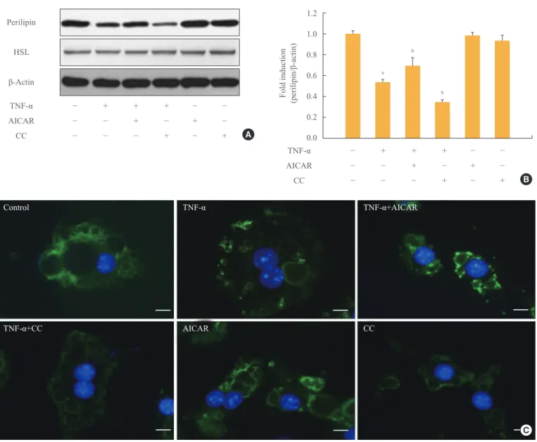
AMPK alleviates TNF-α induced diminution of perilipin Perilipin, which protects lipids on the surface of lipid droplets, is a key factor in the process of lipolysis by lipolytic enzymes.
The amount of perilipin protein was decreased in TNF-α- treated adipocytes, and was further diminished by treatment with CC, while perilipin protein was restored in adipocytes in- cubated with TNF-α and AICAR (Fig. 2A, B). In contrast, the amount of HSL was unchanged in TNF-α and/or AMPK-regu-
lated adipocytes. We also evaluated the amount of perilipin protein using an immunofluorescence assay (Fig. 2C). The in- tensity of green fluorescence indicating perilipin protein on the surface of lipid droplets decreased upon TNF-α and/or CC treatment. In adipocytes incubated with AICAR and TNF-α;
however, green fluorescence was remarkably increased in amount and intensity, although it was weaker than the level of intensity in control adipocytes.
Fig. 1. Chronic incubation with tumor necrosis factor (TNF)-α and AMP-activated protein kinase (AMPK) activation regulates lipolysis in cultured 3T3-L1 adipocytes. Adipocytes were incu- bated with or without TNF-α (10 ng/mL) for 24 hours in the pres- ence or absence of activator minoimidazole carboxamide ribonu- cleotide (AICAR; 1 mM) or compound C (CC; 20 µM). (A) Total and phosphorylated AMPK protein levels were examined by Western blot assay. (B) Lipolysis was quantified by determination of glycerol release into the media. Aliquots of the culture medium were collected at 24 hours, and the amount of released glycerol was measured. The results represent the mean±SE from at least three independent batches of 3T3-L1 adipocytes. The released glycerol level in control adipocytes was designated as 100%.
aP<0.001 compared with the control group; bP<0.01 compared with the TNF-α group.
AMPK-p
AMPK-t
β-Actin
TNF-α AICAR CC
− + + + − −
− − + − + −
− − − + − + A
300 250 200 150 100 50
% Released glycerol of control 0 TNF-α AICAR CC
− + + + − −
− − + − + −
− − − + − + B
a
b b
AMPK stimulates phosphorylation of PERK/eIF2α in TNF-α-treated adipocytes
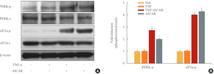
There is increasing evidence showing that UPR signaling, in- cluding PERK and eIF2α, is linked to adipocyte dysfunction [16,19,20]. Although PERK/eIF2α was not affected by TNF-α treatment in 3T3-L1 adipocytes, stimulation of AMPK with AICAR treatment significantly increased the activation of PERK/eIF2α in 3T3-L1 adipocytes, with or without TNF-α treatment (Fig. 3).
Antilipolytic effect of AMPK is dependent on UPR
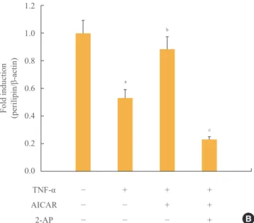
Next we investigated the involvement of the PERK/eIF2α pathway on the antilipolytic effect of AICAR, as described by perilipin protein and the amount of released glycerol. To this end, we used a concentration of 5 mM 2-AP, which inhibits eIF2α kinase, to repress AICAR-induced PERK/eIF2α activa- tion. Restoration of perilipin protein by AICAR was complete- ly abolished by 2-AP (Fig. 4A, B). Supplementation of adipo- cytes with 2-AP significantly overcame the antilipolytic effect of AICAR, describing a significant increase in lipolysis.
1.2 1.0 0.8 0.6 0.4 0.2 0.0 Fold induction (perilipin/β-actin)
Fig. 2. AMP-activated protein kinase (AMPK) attenuates the tumor necrosis factor (TNF)-α-induced decrease in perilipin. 3T3-L1 adi- pocytes were incubated with TNF-α (10 ng/mL) for 24 hours in the presence or absence of activator minoimidazole carboxamide ribonu- cleotide (AICAR; 1 mM) or compound C (CC; 20 µM). (A) Perilipin and hormone sensitive lipase (HSL) protein levels were examined by Western blot assay. (B) Quantification of blot shown in (A). (C) At the end of the incubation, cells were fixed, permeabilized, and then incubated with a specific antibody against perilipin (green fluorescence). Nuclei were visualized by DAPI staining (blue fluorescence).
Bars=10 µm. aP<0.001 compared with control group; bP<0.01 compared with TNF-α group.
Perilipin
HSL
β-Actin TNF-α AICAR CC
− + + + − −
− − + − + −
− − − + − +
TNF-α AICAR CC
− + + + − −
− − + − + −
− − − + − +
A
B
a b
b
Control
TNF-α+CC
TNF-α
AICAR
TNF-α+AICAR
CC
C
DISCUSSION
AMPK regulates cellular metabolism, as well as responses to a variety of signals not directly related to metabolism, such as ischemia [21], hypoxia [22,23], and oxidative stress [24]. In adipocytes it has been reported that AMPK is involved in tri- glyceride breakdown [25], with most reports indicating that AMPK antagonizes cAMP-mediated lipolysis [14,26]. Recent- ly, it has also been reported that a key factor in the antilipolytic effect of AMPK is HSL in cAMP-dependent lipolysis in ma- ture adipocytes [18]. However, because TNF-α-induced lipoly- sis is mediated by an intracellular pathway distinct from cAMP-dependent lipolysis, it is difficult to fully explain the mechanism of antilipolysis.
Here we demonstrated an increase in phosphorylation of AMPK by TNF-α in adipocytes. We also showed that addi- tional enhancement of AMPK activity by artificial manipula- tion repressed TNF-α-induced lipolysis (Fig. 1). These find- ings imply that an increase in AMPK activity in TNF-α-treated adipocytes is an antagonistic response to resist the lipolytic ef- fect of TNF-α. TNF-α can stimulate lipolysis by at least three separate mechanisms in adipocytes: inhibiting insulin receptor signaling, thereby counteracting the antilipolytic effect of the hormone; inhibiting signaling through the Gi-protein-coupled adenosine receptor to counteract the antilipolytic effect of ade- nosine; and stimulation of basal (nonhormonal) lipolysis through a decrease in the lipid-binding protein, perilipin [27].
In the present study, we found quantitative and morphologic
alteration of perilipin by TNF-α and AMPK. In addition, lipol- ysis was directly affected by fluctuation of perilipin expres- sion (Fig. 2). We concluded that perilipin is an essential factor in TNF-α-induced lipolysis, and that AMPK represses lipoly- sis via preservation of perilipin.
In response to a variety of environmental insults such as ac- cumulation of misfolded proteins, a family of protein kinases, including PERK and eIF2α, is phosphorylated to alleviate cel- lular injury or induce apoptosis. Phosphorylation of PERK/
eIF2α is a pivotal process of the UPR and reduces global trans- lation, allowing cells to conserve resources and initiate the re- configuration of gene expression to effectively manage stress conditions [17]. We therefore investigated variation in perilipin expression under conditions that inhibit eIF2α phosphorylation to determine whether PERK/eIF2α signaling induced lipolysis in adipocytes. We used 2-AP, a specific inhibitor of PERK/
eIF2α phosphorylation, to damage the UPR mechanism, which manages cell stress. In adipocytes treated with TNF-α, activa- tion of AMPK by AICAR preserved perilipin expression with stimulation of eIF2α phosphorylation. However, AICAR-in- duced preservation of perilipin was greatly diminished in 2-AP-treated adipocytes, which showed an increase in lipolysis (Fig. 4). Blockade of PERK/eIF2α signaling completely abro- gated protection of perilipin and repression of lipolysis by AICAR. These data suggested that PERK/eIF2α plays a central role in the antilipolytic effect of AMPK in TNF-α-induced li- polysis.
AMPK has been investigated as an essential target molecule
5
4
3
2
1
0
Fold induction (phosphorylated/total)
Fig. 3. AMP-activated protein kinase (AMPK) stimulates phosphorylation of protein kinase RNA-like endoplasmic reticulum kinase (PERK)/eukaryotic initiation factor 2 α (eIF2α). 3T3-L1 adipocytes were incubated with tumor necrosis factor (TNF)-α (10 ng/mL) for 24 hours in the presence or absence of activator minoimidazole carboxamide ribonucleotide (AICAR; 1 mM). (A) Total and phosphory- lated form of eIF2α, and PERK protein were detected by Western blot assay. (B) Quantification of blot shown in (A). aP<0.01; and
bP<0.001 compared with control group; cP<0.01; and dP<0.001 compared with TNF-α group.
PERK-p
PERK-t
elF2α-p
elF2α-t
β-Actin TNF-α AICAR
− + + −
− − + + A B
a
b
c
d
PERK-p elF2α-p
VehTNF TNF/AICAR AICAR
in a number of organs and cells, including pancreatic β-cells, muscle, liver, and brain, to cure diabetes and obesity [28]. TNF-α is one of the etiologic factors in a variety of diseases, and it is increased in obese individuals. In this study, we demonstrated an antilipolytic effect of AMPK in TNF-α-induced adipocytes through regulation of PERK/eIF2 signaling. The cytoprotec- tive effect of AMPK against TNF-α could be of practical use to treat diabetes and obesity, because the effect is expected to also exist in other organs and cells.
Based on these results, we suggest that AMPK represses TNF-α-induced lipolysis via protection of perilipin. It remains to be seen whether pharmacological manipulation of these pathway has the same effect in the therapeutic management of lipolysis in vivo.
CONFLICTS OF INTEREST
This work was supported by a grant from Chon Kun Dang Pharm (S-2013-0227-000). However the design and interpre- tation of the work are independent of the funding.
ACKNOWLEDGMENTS
This work was supported by a National Research Foundation of Korea (NRF) grant funded by the Korea government (NRF- 2013R1A1A2063069).
PERK-p
PERK-t
elF2α-p
elF2α-t
Perilipin
β-Actin TNF-α AICAR 2-AP
− + + +
− − + +
− − − + A
Fig. 4. Antilipolytic effect of AMP-activated protein kinase (AMPK) is abrogated by protein kinase RNA-like endoplasmic reticulum kinase (PERK)/eukaryotic initiation factor 2 α (eIF2α) inhibition. 3T3-L1 adipocytes were incubated with tumor necrosis factor (TNF)-α (10 ng/mL) for 24 hours in the presence or ab- sence of activator minoimidazole carboxamide ribonucleotide (AICAR; 1 mM) and/or 2-aminopurine (2-AP; 5 mM). (A) Total and phosphorylated form of PERK/eIF2α and perilipin protein were detected by Western blot assay. (B) Quantification of perili- pin protein from the blot shown in (A). (C) At the end of the incu- bation, the amount of released glycerol in the culture medium was measured. The released glycerol level in control adipocytes was set at 100%. aP<0.05 compared with control group; bP<0.05 compared with TNF-α group; cP<0.01 compared with TNF-α and AICAR group; dP<0.01 compared with the control group;
eP<0.001 compared with the TNF-α group; fP<0.001 compared with TNF-α and AICAR groups.
1.2
1.0
0.8
0.6
0.4
0.2
0.0 Fold induction (perilipin/β-actin)
TNF-α AICAR 2-AP
− + + +
− − + +
− − − + B
a
b
c
180 160 140 120 100 80 60 40 20 0
Released glycerol (% of control)
TNF-α AICAR 2-AP
− + + +
− − + +
− − − + C
d
e
f
REFERENCES
1. Qi C, Pekala PH. Tumor necrosis factor-alpha-induced in- sulin resistance in adipocytes. Proc Soc Exp Biol Med 2000;
223:128-35.
2. Hausman DB, DiGirolamo M, Bartness TJ, Hausman GJ, Martin RJ. The biology of white adipocyte proliferation. Obes Rev 2001;2:239-54.
3. Hotamisligil GS, Shargill NS, Spiegelman BM. Adipose expression of tumor necrosis factor-alpha: direct role in obesity-linked insulin resistance. Science 1993;259:87-91.
4. Hotamisligil GS, Arner P, Caro JF, Atkinson RL, Spiegel- man BM. Increased adipose tissue expression of tumor ne- crosis factor-alpha in human obesity and insulin resistance.
J Clin Invest 1995;95:2409-15.
5. Kern PA, Saghizadeh M, Ong JM, Bosch RJ, Deem R, Sim- solo RB. The expression of tumor necrosis factor in human adipose tissue: regulation by obesity, weight loss, and rela- tionship to lipoprotein lipase. J Clin Invest 1995;95: 2111-9.
6. Kawakami M, Murase T, Ogawa H, Ishibashi S, Mori N, Takaku F, Shibata S. Human recombinant TNF suppresses lipoprotein lipase activity and stimulates lipolysis in 3T3- L1 cells. J Biochem 1987;101:331-8.
7. Feingold KR, Doerrler W, Dinarello CA, Fiers W, Grun- feld C. Stimulation of lipolysis in cultured fat cells by tu- mor necrosis factor, interleukin-1, and the interferons is blocked by inhibition of prostaglandin synthesis. Endocri- nology 1992;130:10-6.
8. Hauner H, Petruschke T, Russ M, Rohrig K, Eckel J. Effects of tumour necrosis factor alpha (TNF alpha) on glucose transport and lipid metabolism of newly-differentiated hu- man fat cells in cell culture. Diabetologia 1995;38:764-71.
9. Orban Z, Remaley AT, Sampson M, Trajanoski Z, Chrou- sos GP. The differential effect of food intake and beta-ad- renergic stimulation on adipose-derived hormones and cy- tokines in man. J Clin Endocrinol Metab 1999;84:2126-33.
10. Ruderman N, Prentki M. AMP kinase and malonyl-CoA:
targets for therapy of the metabolic syndrome. Nat Rev Drug Discov 2004;3:340-51.
11. Zhou G, Myers R, Li Y, Chen Y, Shen X, Fenyk-Melody J, Wu M, Ventre J, Doebber T, Fujii N, Musi N, Hirshman MF, Goodyear LJ, Moller DE. Role of AMP-activated pro- tein kinase in mechanism of metformin action. J Clin Invest 2001;108:1167-74.
12. Fryer LG, Parbu-Patel A, Carling D. The Anti-diabetic drugs rosiglitazone and metformin stimulate AMP-activat-
ed protein kinase through distinct signaling pathways. J Biol Chem 2002;277:25226-32.
13. Yin W, Mu J, Birnbaum MJ. Role of AMP-activated protein kinase in cyclic AMP-dependent lipolysis In 3T3-L1 adipo- cytes. J Biol Chem 2003;278:43074-80.
14. Daval M, Diot-Dupuy F, Bazin R, Hainault I, Viollet B, Vaulont S, Hajduch E, Ferre P, Foufelle F. Anti-lipolytic ac- tion of AMP-activated protein kinase in rodent adipocytes.
J Biol Chem 2005;280:25250-7.
15. Xu L, Spinas GA, Niessen M. ER stress in adipocytes in- hibits insulin signaling, represses lipolysis, and alters the secretion of adipokines without inhibiting glucose trans- port. Horm Metab Res 2010;42:643-51.
16. Zhou QG, Zhou M, Lou AJ, Xie D, Hou FF. Advanced oxi- dation protein products induce inflammatory response and insulin resistance in cultured adipocytes via induction of endoplasmic reticulum stress. Cell Physiol Biochem 2010;
26:775-86.
17. Habinowski SA, Witters LA. The effects of AICAR on adi- pocyte differentiation of 3T3-L1 cells. Biochem Biophys Res Commun 2001;286:852-6.
18. Koh HJ, Hirshman MF, He H, Li Y, Manabe Y, Balschi JA, Goodyear LJ. Adrenaline is a critical mediator of acute ex- ercise-induced AMP-activated protein kinase activation in adipocytes. Biochem J 2007;403:473-81.
19. Lefterova MI, Mullican SE, Tomaru T, Qatanani M, Sch- upp M, Lazar MA. Endoplasmic reticulum stress regulates adipocyte resistin expression. Diabetes 2009;58:1879-86.
20. van der Kallen CJ, van Greevenbroek MM, Stehouwer CD, Schalkwijk CG. Endoplasmic reticulum stress-induced apoptosis in the development of diabetes: is there a role for adipose tissue and liver? Apoptosis 2009;14:1424-34.
21. Altarejos JY, Taniguchi M, Clanachan AS, Lopaschuk GD.
Myocardial ischemia differentially regulates LKB1 and an al- ternate 5’-AMP-activated protein kinase kinase. J Biol Chem 2005;280:183-90.
22. Esumi H, Izuishi K, Kato K, Hashimoto K, Kurashima Y, Kishimoto A, Ogura T, Ozawa T. Hypoxia and nitric oxide treatment confer tolerance to glucose starvation in a 5’- AMP- activated protein kinase-dependent manner. J Biol Chem 2002;277:32791-8.
23. Marsin AS, Bouzin C, Bertrand L, Hue L. The stimulation of glycolysis by hypoxia in activated monocytes is mediat- ed by AMP-activated protein kinase and inducible 6-phos- phofructo-2-kinase. J Biol Chem 2002;277:30778-83.
24. Horie T, Ono K, Nagao K, Nishi H, Kinoshita M, Kawamu-
ra T, Wada H, Shimatsu A, Kita T, Hasegawa K. Oxidative stress induces GLUT4 translocation by activation of PI3- K/Akt and dual AMPK kinase in cardiac myocytes. J Cell Physiol 2008;215:733-42.
25. Kim SJ, Nian C, McIntosh CH. Activation of lipoprotein li- pase by glucose-dependent insulinotropic polypeptide in adipocytes. A role for a protein kinase B, LKB1, and AMP- activated protein kinase cascade. J Biol Chem 2007;282:
8557-67.
26. Dagon Y, Avraham Y, Berry EM. AMPK activation regu-
lates apoptosis, adipogenesis, and lipolysis by eIF2alpha in adipocytes. Biochem Biophys Res Commun 2006;340:43-7.
27. Langin D, Arner P. Importance of TNFalpha and neutral li- pases in human adipose tissue lipolysis. Trends Endocrinol Metab 2006;17:314-20.
28. Viollet B, Mounier R, Leclerc J, Yazigi A, Foretz M, An- dreelli F. Targeting AMP-activated protein kinase as a nov- el therapeutic approach for the treatment of metabolic dis- orders. Diabetes Metab 2007;33:395-402.