INTRODUCTION
A wide range of pathologic insults to the brain induce depolarization of the neuronal cell membrane, and release neurotransmitters leading to the activation of aberrant cel- lular signaling pathways. Such insults include traumatic brain injury, ischemia, and seizures. Activation of voltage- sensitive ion channels and/or the N-methyl-D-aspartate (NMDA) subtype of glutamate receptor increase cellular calcium (Ca2+), which plays a major role in the development of neuronal injury (1-3). Increased intracellular calcium activates many calcium-dependent enzymes, including pro- tein kinase C (PKC), calmodulin kinase II (CaMKII, also often referred to as Ca2+/calmodulin kinase II or Ca2+/cal- modulin-dependent protein kinase II), phosphorylase A2, nitric oxide synthase (NOS), and various proteases and endonucleases. These may have direct effects on structural proteins, and may modify the function of enzymes, recep- tors or ion channels by altering phosphorylation. They may also produce toxic free radicals via various cascade mecha- nisms. Both PKC and CaMKII phosphorylate and activate the serum responsive factor (SRF) and Ca2+/cAMP response element binding protein (CREB), respectively (4). CREB can also be phosphorylated by cAMP-dependent kinase (protein kinase A, PKA). The increase in cellular levels of
phosphorylated SRF and/or CREB results in the induction of immediate early genes (IEGs) (4). Neuronal exposure to excitotoxic levels of glutamate cause either acute toxicity (osmotic lysis) or delayed Ca2+-dependent neuronal death (5-7).
CaMKII is a major calcium messenger component that regulates many calcium-dependent processes in neurons.
CaMKII phosphorylates and regulates receptor-gated ion channels (8, 9), neuroskeletal elements (10) and calcium- dependent ion channels (6). It is also involved in neuro- transmission (11). CaMKII constitutes 1% of total forebrain protein and up to 2% of total hippocampal protein (12). In addition, CaMKII is predominantly expressed in neurons rather than glial cells (13). The -subunit is homologous to the major postsynaptic density (PSD) protein which consti- tutes up to 50% of the total PSD protein (14). The high sy- naptic expression of CaMKII suggests that this enzyme may be important for normal synaptic function. Since CaMKII is a neuronally enriched enzyme that regulates many important cellular functions, it is easily speculated that the inhibition of this enzyme have important effects on neuronal function.
Significant inhibition of CaMKII activity has been obser- ved in many models of delayed neuronal cell death including animal models of ischemia (15) and glutamate excitotoxicity in neuronal cultures (16). Transient forebrain ischemia results
Min Cheol Lee, Sung Soo Ban, Young-Jong Woo*, Seung U Kim*
Departments of Pathology and Pediatrics*, Chonnam National University Medical School and Research Institute of Medical Sciences, Kwangju, Brain Disease Research Center*, Ajou University, Suwon, Korea
Received : 30 October 2000 Accepted : 12 April 2001
Address for correspondence Min Cheol Lee, M.D.
Department of Pathology, Chonnam National University Medical School and Hospital, 5 Hak- dong, Dong-ku, Kwangju 501-190, Korea Tel : +82.62-220-4300, Fax : +82.62-227-3429 E-mail : [email protected]
* The study was partly supported by a grant of the Brain Disease Research Center, KOSEF, and Chonnam National University Research Institute of Medical Sciences.
643
Calcium/Calmodulin Kinase II Activity of Hippocampus in Kainate-Induced Epilepsy
This study investigated calcium/calmodulin kinase II (CaMKII) activity related to long-standing neuronal injury of the hippocampus in kainate (KA)-induced experimental temporal lobe epilepsy. Epileptic seizure was induced by injection of KA (1 g/ L) dissolved in phosphate buffer (0.1 M, pH 7.4) into the left amyg- dala. Clinical seizures, histopathologic changes and CaMKII activity of the hip- pocampus were evaluated. Characteristic early limbic and late seizures were developed. Hippocampal CaMKII activity increased significantly 4 and 8 weeks after intra-amygdaloid injection of KA, when late seizures developed. The histopathologic changes of the hippocampus included swelling of neuronal cyto- plasm with nuclear pyknosis and loss of neurons in CA3 during this period. The increased activity of CaMKII may correlate with appearance of distant damage in the hippocampus. The above results indicate that intra-amygdaloid injection of KA produces excitatory signals for ipsilateral CA3 neurons in the hippocam- pus and that subsequently increased levels of CaMKII in postsynaptic neurons induce neuronal injury via phosphorylation of N-methyl-D-aspartate type gluta- mate receptor.
Key Words : Ca(2+)-Calmodulin Dependent Protein Kinase; Epilepsy; Hippocampus; Kainic Acid;
Neurons
in more than 50% inhibition of CaMKII activity in the hip- pocampus and cortex (17). The decrease in CaMKII activity observed after ischemia is an early (within 10 sec) and long- lasting phenomenon that precedes the development of de- layed neuronal cell death (17). This inhibition of CaMKII activity has been implicated in the delayed neuronal cell death (15). Understanding the cellular mechanisms regulat- ing the inhibition of CaMKII will provide an insight into the molecular mechanism of excitotoxicity-induced changes in neuronal transducing systems.
Kainate (KA) administration, an experimental model for human temporal lobe epilepsy (18), is known to engender abnormal excitation/inhibition in the limbic system (19), characteristic neuronal injury in the hippocampus, and spontaneous recurrent seizures (20). This study investigates CaMKII activity relative to long-standing neuronal injury in the hippocampus in KA-induced experimental temporal lobe epilepsy.
MATERIALS AND METHODS Experimental model for temporal lobe epilepsy
Adult male Wistar rats, 250-300 g, were divided into two groups; KA injection for biochemical and histopathologic studies (75 rats), and sham operated control (6 rats). The experimental rats were anesthetized with pentobarbital sodium (Nembutal, Abbott, Osaka, Japan, 50 mg/kg i.p.) fixed on a stereotactic frame (David-Kopf, USA), and stereotactic operations were performed. A stainless steel cannula with internal stylet for KA (Nacalai tesque, Kyoto, Japan) microinjection, 0.03 mm in internal diameter, was implanted in the left amygdala using sterile techniques.
Coordinates for the implantation target were: AP +5 mm, ML +5 mm and DV +2 mm with respect to the interaural zero point (20). The cannula was fixed with dental cement.
The experimental animals were left free for 7 days to recover from the operation.
KA was prepared immediately before each injection. KA crystals were dissolved in a 0.2 M phosphate-buffered solu- tion (pH 7.4) at a concentration of 1 mg/mL and sterilized through a 0.45 m microfilter. The injection was delivered while the animals were awake and resting under aseptic conditions. Removing the inner guide wire from the cannu- la, an injection needle was inserted. Following the injection of KA (1 g/ L) into the left amygdala, successful adminis- tration was determined by the induction of clinical seizures.
During the first hour after KA injection, animals exhibited
“staring spells”followed by repetitive head nodding and
“wet dog shakes”. During the next 2 hr, progressive motor seizures developed, including masticatory and facial move- ments, tremors of the forepaws, and rearing and loss of pos- tural control. Finally, animals suffered from limbic status
epilepticus with continuous convulsions, lasting 1 to 2 days.
The seizures disappeared spontaneously 3 days after KA injection, and motor seizures developed again at about 4 weeks after the injection. During the initial postictal peri- od, animals demonstrated reduced motor activity, but were otherwise normal. Each of ten rats, which developed clinical seizures successfully, were sacrificed by decapitation at 1, 2, 4, 8, and 16 weeks after the injection. Five whole brains were taken immediately and fixed in 10% neutral buffered formalin for histopathologic study, and the other five brains and six brains of sham operated control rats were kept frozen in liquid nitrogen for CaMKII assay.
Histopathologic examination
Routine paraffin blocks were made from both hippocampi and observed histopathologic features in the H&E and cre- syl violet stained slides. The right side of the brain was used as control.
Calcium/calmodulin kinase II (CaMKII) assay
CaMKII activity from the hippocampal tissue was mea- sured using a modification of Soderling’s method (21).
Frozen hippocampus, 100 mg, was washed and suspended with 1 mL ice-cold homogenization buffer comprised of 30 mM HEPES (pH 7.4), 1 mM ethylenediaminetetraacetic acid (EDTA), 27 TIU aprotinin, 0.1 mM [ethylene-bis- (oxyethylenenitrilo)]tetraacetic acid (EGTA), 1 mM phenyl- methylsulfonyl fluoride (PMSF) and 1 mM ATP. The sus- pension was transferred into a glass homogenizer (Kontes, Vineland, NJ) and was disrupted (16). Homogenates were normalized for protein and were studied for endogenous protein phosphorylation.
Standard phosphorylation reaction solutions for kinase assay contained 3 g protein, 30 mM HEPES (pH 7.4), 1 mM dithiothreitol, 10 mM magnesium acetate, 3.3 M syntide II and 1 mM [ -32P]ATP. Final reaction volume was 100 L. Standard reactions were performed in a shaking water bath at 30℃. To measure calcium-dependent activi- ty, reactions were initiated by the addition of 2 mM CaCl2 and 1.2 mM calmodulin; whereas, for calcium-independent activity, 2 mM EGTA was added instead of CaCl2and cal- modulin. The reactions continued for 1 min, and were ter- minated by the addition of 11 L of stop solution contain- ing 25 mM Tris, 10 mM Na+-pyrophosphate, and 10 mM -glycerophosphate. Aliquots (80 L) were spotted on pho- sphocellulose paper (Whatman p81). After washing the paper, the radioactivity of free, nonphosphorylated [ -32P]
ATP was quantified by liquid scintillation counter. CaMKII activity (cpm/ g protein/min, mean±SD) was measured three times from each tissue sample and the average value was calculated. The data were analyzed by Student t-test.
RESULTS Histopathologic features
The control hippocampus disclosed well defined, palisad- ing neuronal arrangements, consisting of cornu ammonis 1 (CA1), CA2, CA3, CA4, and dentate gyrus. A few neurons were pyknotic, but no neuronal swelling or partial loss was
noted. There were no identifiable histopathologic changes on the control side of the hippocampus until 16 weeks after the injection of KA (Fig. 1A and 2A).
The experimental hippocampus from the KA injection revealed no significant histopathologic change within the first week. Mild swelling of neuronal cytoplasm with a few pyknotic nuclei in CA3 was observed at 2 weeks after the injection. The changes progressed with time. A severe
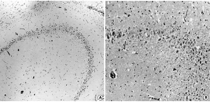
A B
Fig. 1.Coronal section of the brain examined by Nissl stain, 16 weeks after KA injection into the left amygdala, reveals relatively intact hippocampal neurons on the right side (A,×25) and selective, significant loss of CA3 neurons in the left hippocampus (B,×25).
A B
Fig. 2.Coronal section of the brain, at 8 weeks after KA injection, examined by H&E stain revealed relatively intact CA2 and CA3 neu- rons of the right hippocampus (A,×40), and moderate loss of CA3 neurons in the left hippocampus (B,×80).
swelling of neuronal cytoplasm was observed at 4 weeks (Fig. 2B) and a decrease in the number of neurons in CA3 at 8 weeks (Fig. 2B) after the injection. The neuronal loss in CA3 persisted up to 16 weeks after KA injection into the amygdala (Fig. 1B).
Calcium/calmodulin kinase II (CaMKII) activity
The CaMKII values (cpm/ g protein/min) on the control side of the hippocampus ranged from 39,725 to 48,173 regardless of the experimental period. Mean values were 42,107 at 1 week; 44,934 at 2 weeks; 45,505 at 4 weeks;
43,432 at 8 weeks; and 41,363 at 16 weeks after KA injec- tion. There were no statistically significant differences of CaMKII values in the hippocampus between the sham oper- ated and the control side of the KA-injected rats.
Mean CaMKII values on the experimental side of the hip- pocampus were 42,556 at 1 week; 46,037 at 2 weeks; 63,666 at 4 weeks; 65,448 at 8 weeks; and 50,050 at 16 weeks after KA injection. There was no significant difference in CaMKII activity between the control and lesional sides of the hippocampus at 1, 2 and 16 weeks after the KA injec- tion. However, significantly increased CaMKII activity was noted at 4 and 8 weeks (p<0.001) on the left (KA-injected) side of the hippocampus. CaMKII activity is summarized in Fig. 3.
DISCUSSION
Intra-amygdaloid injection of KA produces initial limbic
motor seizures lasting 2 or 3 days with staring, head nod- ding, wet-dog shakes, salivation, chewing, forepaw tremors, and rearing and loss of postural control (22). Continuous convulsions lasting over 30 sec are frequently seen. The seizures spontaneously disappear 3 days after KA injection.
These are more complex seizures, involving initial lip and facial movements, barrel rotation, and circling movements indicating the secondary involvement of extra-amygdaloid structures. A significant loss of CA3 neurons in the ipsilat- eral hippocampus was noted after intra-amygdaloid injec- tion of KA in this study, supporting results found in the lit- erature (20, 23).
A simple hypothesis to explain the distant lesions would be that KA diffuses from the amygdala either directly or through the cerebrospinal fluid (CSF), and that sufficient concentrations of KA directly damage vulnerable neurons, such as the CA3 neurons of the hippocampus. However, sufficient doses of diazepam administration blocked distant damage in the CA3 even in the presence of direct damage to the amygdala by KA injection (24). Therefore, the results suggest that KA-induced hippocampal damage following intra-amygdaloid injection stems from two sources-local damage due to the direct toxic action of KA and distant injury mediated by the paroxysmal discharge accompanying convulsions. Other electrophysiologic, autoradiographic and histopathologic studies support the hypothesis that distant damage in the hippocampus is mediated by paroxysmal epileptiform discharge (25-27). Collectively, these findings strongly suggest that paroxysmal discharge after intra- amygdaloid injection of KA is generated in the entorhinal cortex, where the amygdala heavily projects (28). This dis- charge may deliver a powerful excitatory action on CA3 neurons primarily via the granule cells with their mossy fibers, and the perforating pathways from the entorhinal cortex to granule cells (18). The early and late expression of c-FOS, c-JUN and heat shock protein (HSP) 72 in the entorhinal cortex and hippocampus also indicate indirect damage of the hippocampus (29).
It is well known that glutamate synapses are subject to various forms of prolonged enhancement (30), including long-term potentiation (LTP). The activation of subtypes of glutamate receptors plays a major role in the induction of LTP, and such changes contribute to the induction of epi- leptic seizures. A number of postsynaptic kinases, PKC, CaMKII, tyrosine kinase (PKT) and PKA, are believed to be important for the induction of LTP (31, 32). Long-term alterations in these protein kinases have also been reported in kindling models for epilepsy (33). Among these kinases, PKA appears to contribute significantly to the induction of the most persistent and long-lasting components of LTP in the hippocampus (32).
In the present study, CaMKII activity in the hippocampus increased significantly 4 and 8 weeks after an intra-amyg- daloid injection of KA, when late seizures developed clini-
CaMKII 80,000
60,000
40,000
20,000
0
1 2 4 8 16
Weeks
Fig. 3. Graphic presentation of calcium/calmodulin kinase II (CaMKII) activity (cpm/ g protein/min) in the hippocampus after microinjection of kainic acid (KA, 1 g/ L) into the left amygdala (n=5 rats in each week). Bar: means+SD. *, statistically significant (p<0.001).
Rt. Hippocampus Lt. Hippocampus
* *
cally. Among the histopathologic changes of the hippocam- pus, swelling of neuronal cytoplasm with nuclear pyknosis and loss of neurons developed in these periods. Increased CaMKII activity may be correlated with the appearance of distant damage in the hippocampus. But, it remains uncer- tain why CaMKII activity in the hippocampus became nor- malized 16 weeks after the KA injection. Possible explana- tions are that the decreased frequency of seizure attacks, from two to four times a day, and marked loss of hippocampal neurons might play a role. Therefore, the activity of CaMKII in the hippocampus after the intra-amygdaloid injection of KA may increase during the periods of active neuronal da- mage mediated by excitotoxic transmitters.
These findings were supported in part by other studies in which CaMKII appears to induce the phosphorylation of the NMDA-receptor channel domain in the PSD through excitatory transmitters in fetal rat cortical cultures (34).
This can then cause an enhancement of calcium influx through the channel (35). A specific cell-permeable inhibitor of CaMKII, KN-62 (1-[N,O-bis-(5-isoquinoline sulfonyl)- N-methyl-L-tyrosyl]-4-phenylpiperazine) protects the neu- rons from NMDA toxicity and hypoxia/hypoglycemia- induced neuronal injury (36). Therefore, the intra-amyg- daloid injection of KA sends excitatory signals to ipsilateral CA3 neurons in the hippocampus, and subsequently in- creases levels of CaMKII in the PSD. It reacts with subtypes of the glutamate receptor and enhances calcium influx, thus inducing neuronal injury.
REFERENCES
1. Choi DW. Excitotoxic cell death. J Neurobiol 1992; 23: 1261-76.
2. Meldrum BS. Amino acids as dietary excitotoxins: A contribution to understanding neurodegenerative disorders. Brain Res Rev 1993;
18: 292-314.
3. McIntosh TK. The neurochemical sequelae of traumatic brain injury: Therapeutic implications. Cerebrovasc Brain Metab Rev 1994; 6: 109-62.
4. Sheng M, Greenberg ME. The regulation and function of c-fos and other immediate early genes in the nervous system. Neuron 1990; 4:
477-85.
5. Olney JW, Price MT, Samson L, Labruyere J. The role of specific ions in glutamate toxicity. Neurosci Lett 1986; 65: 65-71.
6. Choi DW. Calcium-mediated neurotoxicity: Relationship to specific channel types and role in ischemic damage. Trends Neurosci 1988;
11: 465-9.
7. Ogura A, Miyamoto M, Kudo Y. Neuronal death in vitro: paral- lelism between survivability of hippocampal neurons and sustained elevation of cytosolic Ca2+after exposure to glutamate receptor agonist. Exp Brain Res 1988; 73: 447-58.
8. McGlade-McCulloh E, Yamamoto H, Tan SE, Brickey DA, Soder- ling TR. Phosphorylation and regulation of glutamate receptors by calcium/calmodulin-dependent protein kinase II. Nature 1993; 362:
640-2.
9. Machu TK, Firestone JA, Browning MD. Ca2+/calmodulin-depen- dent protein kinase II and protein kinase C phosphorylate a synthet- ic peptide corresponding to a sequence that is specific for the gamma 2L subunit of the GABAAreceptor. J Neurochem 1993; 61:
375-7.
10. DeLorenzo RJ, Gonzalez B, Goldenring J, Bowling A, Jacobson R.
Ca2+/calmodulin tubulin kinase system and its role in mediating the Ca2+signal in brain. Prog Brain Res 1982; 56: 257-86.
11. Llinas R, McGuinness TL, Leonard CS, Sugimori M, Greengard P.
Intra-terminal injection of synapsin I or calcium/calmodulin-depen- dent kinase II alters neurotransmitter release at the squid giant synapse. Proc Natl Acad Sci USA 1985; 82: 3035-9.
12. Goldenring JR, Gonzalez B, McGuire JS Jr, DeLorenzo RJ. Purifi- cation and characterization of a calmodulin-dependent kinase from rat brain cytosol able to phosphorylate tubulin and microtubule- associated proteins. J Biol Chem 1983; 258: 12632-40.
13. Erondu NE, Kennedy MB. Regional distribution of type II Ca2+/ calmodulin-dependent protein kinase in rat brain. J Neurosci 1985;
5: 3270-7.
14. Kennedy MB, Bennett MK, Erondu NE. Biochemical and immuno- chemical evidence that the “major postsynaptic density protein”is subunit of a calmodulin-dependent protein kinase. Proc Natl Acad Sci USA 1983; 80: 7357-61.
15. Aronowski J, Grotta JC, Waxham MN. Ischemia-induced translo- cation of Ca2+/calmodulin-dependent protein kinase II: possible role in neuronal damage. J Neurochem 1992; 58: 1743-53.
16. Churn SB, Sombati S, Taft WC, DeLorenzo RJ. Excitotoxicity affects membrane potential and calmodulin kinase II activity in cul- tured rat cortical neurons. Stroke 1993; 24: 277-8.
17. Churn SB, Taft WC, DeLorenzo RJ. Effects of ischemia on multi- functional calcium/calmodulin-dependent protein kinase type II in the gerbil. Stroke 1990; 21: III 112-6.
18. Ben-Ari Y. Limbic seizure and brain damage produced by kainic acid: Mechanisms and relevance to human temporal lobe epilepsy.
Neuroscience 1985; 14: 375-403.
19. Franck JE, Schwartzkroin PA. Do kainate-lesioned hippocampi become epileptogenic? Brain Res 1985; 329: 309-13.
20. Tanaka S, Kondo S, Tanaka T, Yonemasu Y. Long-term observa- tion of rats after unilateral intra-amygdaloid injection of kainic acid. Brain Res 1988; 463: 163-7.
21. Fukunaga K, Rich DP, Soderling TR. Generation of the Ca2+-inde- pendent form of Ca2+/calmodulin-dependent protein kinase II in cerebellar granule cells. J Biol Chem 1989; 264: 21830-6.
22. Ben-Ari Y, Tremblay E, Riche D, Ghilini G, Naquet R. Electro- graphic, clinical and pathological alterations following systemic administration of kainic acid, bicuculline and pentetrazole:
metabolic mapping using the deoxyglucose method with special ref- erence to the pathology of epilepsy. Neuroscience 1981; 6: 1361- 91.
23. Tanaka T, Tanaka S, Fujita T, Takano K, Fukuda H, Sako K, Yone- masu Y. Experimental complex partial seizures induced by a microinjection of kainic acid into limbic structures. Prog Neurobiol 1992; 38: 317-34.
24. Ben-Ari Y, Tremblay E, Ottersen OP, Naquet R. Evidence suggest- ing secondary epileptogenic lesions after kainic acid: pretreatment with diazepam reduces distant but not local brain damage. Brain Res 1979; 165: 362-5.
25. Ben-Ari Y, Tremblay E, Ottersen OP, Meldrum BS. The role of epileptic activity in hippocampal and “remote”cerebral lesions induced by kainic acid. Brain Res 1980; 191: 79-97.
26. Nadler JV, Shelton DL, Perry BW, Cotman CW. Regional distribu- tion of 3H kainic acid after intraventricular injection. Life Sci 1980;
26: 133-8.
27. Nadler JV, Cuthbertson GJ. Kainic acid neurotoxicity toward hip- pocampal formation: Dependence on specific excitatory pathways.
Brain Res 1980; 195: 47-56.
28. Price JL. The efferent connections of the amygdaloid complex in the rat, cat and monkey, In: Ben-Ari Y, editor, The Amygdaloid Com- plex, Amsterdam: Elsevier/North-Holland 1981; 125-32.
29. Lee JK, Kang SS, Lee MC. Stress protein expression in kainate- induced experimental temporal lobe epilepsy in rats. J Korean Neu- rosurg 1998; 27: 1641-52.
30. McNaughton BL. The mechanism of expression of long-term enhancement of hippocampal synapses: Current issues and theoret- ical implications. Ann Rev Physiol 1993; 55: 375-96.
31. Bliss TV, Collingridge GL. A synaptic model of memory: long-term potentiation in the hippocampus. Nature 1993; 361: 31-9.
32. Frey U, Huang YY, Kandel ER. Effects of cAMP stimulate a late stage of LTP in hippocampal CA1 neurons. Science 1993; 260:
1661-4.
33. Saitoh T, Masliah E, Jin LW, Cole GM, Wieloch T, Shapiro IP.
Protein kinases and phosphorylation in neurologic disorders and cell death. Lab Invest 1991; 64: 596-616.
34. Yakel JL, Vissavajjhala P, Derkach VA, Brickey DA, Soderling TR. Identification of a Ca2+/calmodulin-dependent protein kinase II regulatory phosphorylation site in non-N-methyl-D-aspartate gluta- mate receptors. Proc Natl Acad Sci USA 1995; 28: 1376-80.
35. Hajimohammadreza I, Probert AW, Coughenour LL, Borosky SA, Marcoux FW, Boxer PA, Wang KK. A specific inhibitor of calci- um/calmodulin-dependent protein kinase II provides neuroprotec- tion against NMDA- and hypoxia/hypoglycemia-induced cell death.
J Neurosci 1995; 15: 4093-101.
36. Kitamura Y, Miyazaki A, Yamanaka Y, Nomura Y. Stimulatory effects of protein kinase C and calmodulin kinase II on N-methyl-D- aspartate receptor/channels in the postsynaptic density of rat brain.
J Neurochem 1993; 61: 100-9.