2004;10:5123-5130.
Clin Cancer Res
Yoo Hong Min, June-Won Cheong, Mark Hong Lee, et al.
Tensin Homologue Protein and Poor Prognosis
with Constitutive Phosphorylation of Phosphatase and
Association
Expression in Acute Myelogenous Leukemia: Its
Elevated S-Phase Kinase-Associated Protein 2 Protein
Updated version
http://clincancerres.aacrjournals.org/content/10/15/5123
Access the most recent version of this article at:
Cited Articles
http://clincancerres.aacrjournals.org/content/10/15/5123.full.html#ref-list-1
This article cites by 60 articles, 35 of which you can access for free at:
Citing articles
http://clincancerres.aacrjournals.org/content/10/15/5123.full.html#related-urls
This article has been cited by 4 HighWire-hosted articles. Access the articles at:
E-mail alerts
Sign up to receive free email-alerts
related to this article or journal.
Subscriptions
Reprints and
.
[email protected]
Department at
To order reprints of this article or to subscribe to the journal, contact the AACR Publications
Permissions
.
[email protected]
Department at
Elevated S-Phase Kinase-Associated Protein 2 Protein Expression in
Acute Myelogenous Leukemia: Its Association with Constitutive
Phosphorylation of Phosphatase and Tensin Homologue
Protein and Poor Prognosis
Yoo Hong Min,
1,2June-Won Cheong,
1Mark Hong Lee,
4Ji Yeon Kim,
3Seung Tae Lee,
1Jee Sook Hahn,
1and Yun Woong Ko
11Department of Internal Medicine, Yonsei University College of
Medicine, Seoul;2Brain Korea 21 Project for Medical Science, and 3Clinical Research Center, Yonsei University College of Medicine,
Seoul; and4Department of Internal Medicine, Sungkyunkwan
University School of Medicine, Seoul, Korea
ABSTRACT
Purpose: The F-box protein S-phase kinase-associated protein 2 (Skp2) positively regulates the G1-S phase
transi-tion by controlling the stability of several G1 regulators,
such as p27Kip1. However, the clinical significance of Skp2 in patients with acute myelogenous leukemia (AML) re-mains unknown.
Experimental Design: We examined the clinical and biological significance of Skp2 expression in AML and eval-uated the relationship between Skp2 and p27Kip1 expres-sion and phosphatase and tensin homologue (PTEN) phos-phorylation.
Results: Western blot analysis showed that high Skp2 expression was observed in 57 (57.6%) cases and signifi-cantly correlated with unfavorable cytogenetics (Pⴝ 0.035) but not with age, white blood cell count, serum lactic dehy-drogenase level, and the French-American-British subtype. An inverse correlation was not observed between Skp2 and p27Kip1 expression. However, p27Kip1 protein was prefer-entially localized to cytoplasm in the high-Skp2-expression group. The cytoplasmic to nuclear ratio of p27Kip1 expres-sion was significantly correlated with the levels of Skp2 expression (P< 0.001). The frequency of PTEN
phospho-rylation was significantly higher in the high-Skp2-expres-sion group compared with the low- Skp2-expreshigh-Skp2-expres-sion group (Pⴝ 0.035). The Skp2 overexpression was significantly as-sociated with shorter disease-free survival and overall
sur-vival (Pⴝ 0.0386 and P ⴝ 0.0369, respectively). Multivariate analysis showed that Skp2 expression was an independent prognostic factor both in the disease-free survival and over-all survival.
Conclusion: These findings suggest that Skp2 expres-sion is an independent marker for a poor prognosis in AML. The presence of a positive correlation between Skp2 and phosphorylated PTEN suggests that an aberration in the PTEN/Skp2 signaling pathway might be operating in AML.
INTRODUCTION
The importance of the G1-S-phase progression in various
tumors has been highlighted by the frequent observations of aberrant regulation of molecules involved in this process. p27Kip1 is a potent inhibitor of the cyclin E– cyclin-dependent kinase-2 and cyclin A– cyclin-dependent kinase-2, which drive the cells from the G1to the S phase (1–2). Although p27Kip1 is
rarely mutated in human cancers, many studies have shown that the reduced expression of p27Kip1 is frequently observed in various cancers (3–7). Low p27Kip1 levels are associated with the high aggressiveness and reduced survival in many cancers including acute myelogenous leukemia (AML; refs. 3–10).
The expression of p27Kip1 is controlled both at the level of transcription and by multiple posttranslational mechanisms, in-cluding the ubiquitin-mediated proteasomal degradation (11– 14). S-phase kinase-associated protein 2 (Skp2) is a member of the F-box family of the specific substrate-recognition subunit of the Skp1/Cu11/F-box ubiquitin-protein ligase complexes (15). Skp2 is required for the ubiquitination and subsequent protea-somal degradation of p27Kip1 protein (16 –18). High levels of p27Kip1 and free cyclin E expression were observed in the Skp2 knockout cells (19). Recent studies have indicated a possible relationship between Skp2 and its oncogenic potential. Skp2 expression was increased in the transformed cell lines (15) and malignant tumors (9, 20 –25). Furthermore, a high Skp2 expres-sion level was significantly correlated with an advanced clinical stage (9, 24) and a poor prognosis in tumors (9, 23, 24, 26, 27). The p27Kip1 level was inversely related to the Skp2 level in various human cancers (9 –10, 21–23, 25, 26). Interestingly, this inverse correlation was not observed in a substantial proportion of aggressive cancers, although the high levels of Skp2 expres-sion correlated well with the cell proliferation and disease progression (27–29). Until now, an evaluation of Skp2 protein expression in relation to the p27Kip1 expression and the clinical outcome has not been undertaken in AML.
Skp2 has a complex relationship with the tumor suppressor protein phosphatase and tensin homologue (PTEN; refs. 30 –32). By dephosphorylating phosphatidylinositol 3,4,5-triphosphate (PIP3), PTEN antagonizes the phosphatidylinositol 3-kinase
Received 1/22/04; revised 4/19/04; accepted 4/26/04.
Grant support: Korea Research Foundation Grant
(KRF-2003-015-E00138).
The costs of publication of this article were defrayed in part by the payment of page charges. This article must therefore be hereby marked
advertisement in accordance with 18 U.S.C. Section 1734 solely to
indicate this fact.
Requests for reprints: Department of Internal Medicine, Yonsei
Uni-versity College of Medicine, Seodaemun-ku Shinchon-dong 134, Seoul 120-752, Korea. Phone: 82-2-361-5438; Fax: 82-2-393-6884; E-mail: [email protected].
(PI3-K)-mediated growth-promoting and antiapoptotic path-ways (33–34). A high frequency of PTEN mutations has been reported in several human cancers (35). The loss of PTEN also predisposes the malignant transformation in a variety of tissues (36). The downstream effectors that mediate the PTEN-induced growth arrest have been identified, including Akt (37–38) and p27Kip1 (39). The relationship between Skp2 expression and the loss of PTEN is particularly interesting in view of the finding that a deletion of PTEN in mouse fibroblasts leads to the increased Skp2 levels with concomitant reductions in the p27Kip1 levels (40). Skp2 expression was inversely correlated with PTEN expression in prostate cancer (9). These findings suggest that PTEN functions as a negative regulator of the Skp2 pathway. Therefore, it would be necessary to evaluate the func-tional implication of PTEN as a regulator of the Skp2 pathway in AML.
The mutations in the PTEN gene are very infrequent ge-netic aberrations in myeloid leukemia (41– 42). Recently, our group demonstrated that the COOH-terminal regulatory domain of the PTEN protein is constitutively phosphorylated in a sub-stantial proportion of AML cases (43). The phosphorylation of the PTEN protein was strongly associated with the activation of the downstream molecules, including Akt and the forkhead transcription factor in AML cells (43). However, the relation-ship between Skp2 expression and PTEN phosphorylation has not been evaluated in AML.
The aim of this study was to examine the expression of the Skp2 protein and evaluate its correlation with the patient char-acteristics and clinical outcome in AML. This study also exam-ined whether or not Skp2 expression is correlated with the expression and the localization of p27Kip1 protein, and the constitutive phosphorylation of PTEN protein in AML cells. We demonstrated for the first time that a high Skp2 expression is observed in a substantial proportion of AML cases and is associated with a poor prognosis. Skp2 expression was also strongly correlated with the mislocalization of the p27Kip1 protein and the constitutive phosphorylation of the PTEN pro-tein. These findings might provide additional insight into the molecular pathogenesis and potential therapeutic targets for adult AML.
MATERIALS AND METHODS
Patients and Treatment. A total of 99 consecutive adults patients with de novo AML who had not received anti-leukemia treatment were enrolled in the study. According to the French-American-British classification (63), four patients had the M0, 20 patients had the M1, 33 patients had the M2, 19 patients had the M4, 21 patients had the M5 and 2 patients had the M6 subtype (Table 1). Patients with the M3 subtype of AML were excluded from this study. All of the patients were treated according to the first-line induction and consolidation regimens of Severance Hospital. Eighty-four patients received induction chemotherapy comprising cytarabine (100 mg/m2
/d, continuous infusion for 7 days) and idarubicin (12 mg/m2
/d, i.v. bolus for 3 days). Complete remission was defined as the normalization of the blood counts and bone marrow morphology along with the disappearance of all signs of leukemia, lasting for 4 weeks or longer, in accordance with the recommendations of the National
Cancer Institute-sponsored Workshop (44). All patients achiev-ing complete remission received the same two courses of mi-toxantrone-etoposide-cytarabine (MEC) consolidation therapy consisting of cytarabine (1g/m2
/d, i.v. infusion for 2 hours, every 12 hours for a total of eight times), mitoxantrone (12 mg/m2
/d for 3 days), and (etoposide; 100 mg/m2
/d for 2 days), as described previously (45). The patients who relapsed also received a mitoxantrone-etoposide-cytarabine chemotherapy.
Isolation of Leukemic Cells. In conjunction with the institutional review board-approved treatment protocol, bone marrow aspirates were prospectively prepared from the patients before initiating chemotherapy. The bone marrow was sedi-mented on a Ficoll-Hypaque (Pharmacia Biotech, Uppsala, Sweden) density gradient. After washing the mononuclear cells collected from the upper interface, we performed the T-cell depletion using a high-gradient magnetic cell separation system/ anti-CD3 monoclonal antibody (Miltenyi Biotech, Auburn, CA) according to the manufacturer’s instructions. A morphological evaluation indicated that more than 95% of the isolated cells were leukemic blasts.
Antibodies and Reagents. The mouse monoclonal anti-bodies to Skp2 and p27Kip1 were purchased from ZYMED (San Francisco, CA). The rabbit polyclonal antibodies against PTEN and phosphorylated PTEN (pPTEN) were obtained from Cell Signaling Technology (Beverly, MA). The anti-pPTEN antibody detects PTEN protein that is phosphorylated at the Ser380/Thr382/Thr383 residues in the COOH-terminal regula-tory domain. The antihuman ␣-tubulin monoclonal antibody was acquired from Cedarlane (Ontario, Canada). The horserad-ish peroxidase-conjugated goat antimouse IgG and horseradhorserad-ish peroxidase-conjugated goat antirabbit IgG were purchased from PharMingen (San Diego, CA). Unless indicated otherwise, all other culture reagents were purchased from Life Technologies, Inc. (Grand Island, NY).
Cytogenetic Analysis. Chromosomal analysis was per-formed on the pretreated bone marrow cells. The samples were processed using short-term unstimulated cultures (24 –72 h). The clonality criteria and descriptions of the chromosomal ab-errations were in accordance with the International System for Human Cytogenetic nomenclature. The patients were divided into three prognostic groups based on the karyotype; the prog-nostic groups were favorable [t(8;21), inv(16)], intermediate (normal cytogenetics), and unfavorable (all other abnormali-ties).
Cell Cycle Analysis. After treatment, the cells were pel-leted and fixed in 70% ethanol on ice for 1 hour and resus-pended in 1 ml of a cell cycle buffer (0.38 mMsodium citrate, 0.5 mg/ml RNase A, and 0.01 mg/ml propidium iodide) at a concentration of 106
cells/ml. Cell cycle analysis was performed using a FACSCalibur flow cytometer (Becton Dickinson, San Jose, CA) equipped with CellQuest software (Becton Dickin-son).
Preparation of Nuclear and Cytoplasmic Fractions.
Cells were subfractionated as described previously (46) with minor modifications. Cells were pelleted by centrifugation (5 min; 12,000 rpm; 4°C) and incubated in a hypotonic buffer [10 mM HEPES (pH 7.2), 10 mM KCl, 1.5 mM MgCl2, 0.1 mM
EGTA, 20 mM NaF, 100 M Na3VO4, and 0.1% protease
inhibitor cocktail (Sigma Chemical, St. Louis, MO)] for 30
minutes at 4°C while rocking. Cells were broken using a Dounce homogenizer (30 strokes), after which nuclei were pelleted by centrifugation (10 min; 3,500 rpm; 4°C). The nuclei-free supernatant was subjected to a second centrifugation at 10,000⫻ g for 45 minutes at 4°C to separate membrane (pellet) from cytosolic (supernatant) fractions. Nuclear pellets (above) were resuspended in nuclear lysis buffer [10 nMTris-HCl (pH 7.5), 150 mMNaCl, 5 mMEDTA, and 1% Triton X-100] and were incubated for 1 minute in a sonicating water bath, followed by a 30-min incubation at 4°C while rocking. Twenty milli-grams of total cytosolic and nuclear protein were analyzed by Western blot.
Western Blotting. The cells were dissolved in 100l of a SDS-PAGE sample buffer containing-mercaptoethanol at a final concentration of 3⫻ 106
cells. The lysates were sonicated for 15 seconds with a Vibra Cell Sonicator, boiled for 10 minutes, and further analyzed by Western blotting. The protein yields were quantified using the Bio-Rad Dc protein assay kit (Hercules, CA) and equivalent amounts of the protein (40g) were applied to the 15% acrylamide gels. The proteins were separated by SDS-PAGE and were transferred to nitrocellulose membranes (Amersham Biosciences, Piscataway, NJ). The membranes were blocked at room temperature with 3% bovine serum albumin in TBST (1⫻ Tris-buffered saline, 0.1% Tween 20) for 16 h. After washing twice in TBST, the membranes were incubated with the primary antibodies for 2 hours at room temperature. The membranes were then washed four times in TBST and were incubated with the relevant horseradish perox-idase-conjugated secondary antibodies (1:3000 dilution with 3% bovine serum albumin in TBST) for 1 h. After washing four times in TBST, the reactive proteins were visualized by an enhanced chemiluminescence detection system (Amersham Biosciences). Densitometry was performed using the Molecular Dynamics Imaging system and ImageQuant 3.3 software (Am-ersham Biosciences) to quantify the relative amounts of the protein detected on Western blots.
Statistical Analysis. To perform the quantitative analy-sis, the Skp2 protein expression level in the AML cells (L) was evaluated by normalization as follows: corrected L (Lc)⫽ Skp2 (L)/␣-tubulin (L) as determined by Western blot analysis. The patients were divided into two groups (high and low) based on the median Lc value of Skp2 expression in bone marrow mono-nuclear cells of 10 normal candidates. The comparisons among characteristics of the subgroups were made using a2
test for the binary variables and a Mann-Whitney test for the continuous variables. The linear regression analysis and two-tailed Student t test were used to assess the association between Skp2 and p27Kip1 protein expression. The2
test was used to determine the relationship of Skp2 with pPTEN. The disease-free survival and overall survival probabilities were calculated using the Kaplan-Meier method. The log-rank statistic was used to test the difference in survival times between the groups. In addition to the Skp2 expression, the white blood cell count, age, and cyto-genetics were analyzed in the univariate and multivariate anal-ysis. Multivariate analysis was used to test for the independent prognostic significance of variables using the Cox proportional hazards regression model. The patients alive, and still in remis-sion at last follow-up examination, were censored in the analy-sis. All of the calculations were performed using the SPSS
software, version 11.0.1 (SPSS Inc, Chicago, IL). A P-value of
⬍0.05 was considered significant.
RESULTS
Skp2 Protein Expression in AML Cells. The expres-sion levels of the Skp2 protein in the AML cells varied among the patients (Fig. 1A). Western blot analysis revealed that Skp2 protein expression was present in 74 (74.7%) of the 99 cases. To perform the quantitative analysis, the Skp2 protein expression level was determined in the AML cells (L) by normalization as follows: Lc⫽ Skp2 (L)/␣-tubulin (L), as determined by Western blot analysis. In this study, the Lc ranged from 0 to 840.4. In the practical evaluations, a cutoff value (Lc, 0.70) was set for these protein expression levels in the AML cells, because the median Lc level of Skp2 protein expression in the 10 normal bone marrow mononuclear cells was 0.70 (range, 0 –1.02). Using this cutoff value, we classified the AML cases into two groups, a
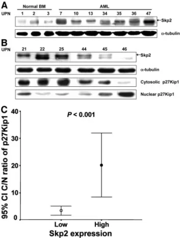
Fig. 1 Western blot analysis of Skp2 and p27Kip1 protein expression
in the representative AML and normal bone marrow (BM) samples. A, the levels of Skp2 protein expression were variable according to the samples.␣-tubulin was used for control of protein concentration on Western blot. UPN represents unique patient number. B, the subcellular localization and expression of the p27Kip1 protein in relation to the Skp2 protein expression were analyzed by SDS-PAGE of the nuclear and cytoplasmic fractions of the cell lysates as described in Materials and Methods. C, the mean C/N ratio of p27Kip1 protein was signifi-cantly higher in the AML cases showing a high Skp2 expression compared with the cases showing a low level of Skp2 expression (P⬍ 0.001). CI, confidence interval.
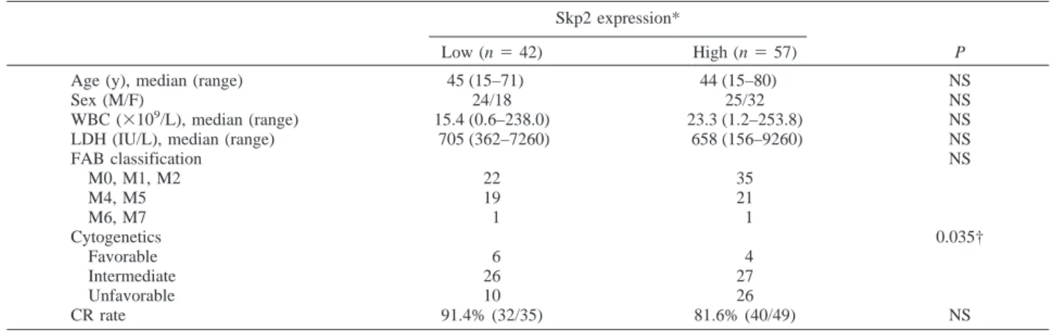
high-expression group [n⫽ 57 (57.6%)] and a low-expression group [n⫽ 42 (42.4%)].
The association between Skp2 expression and the various clinical parameters in AML were evaluated. As shown in Table 1, there was no significant difference regarding age, sex, white blood cell count, and the French-American-British subtype. A statistically significant correlation was observed between Skp2 expression and unfavorable cytogenetics (2
test, P⫽ 0.035). Cell cycle analysis demonstrated that the fraction of cells in the G0-G1phase was higher in the AML cells showing a high Skp2
expression (85.2⫾ 18.1%) compared with the cells showing a low Skp2 expression (75.9⫾ 24.0%) without statistical signif-icance (P ⫽ 0.435; Table 2). Likewise, the Skp2 expression levels were not correlated with the fractions of the cells in the G2-M or S phase, respectively (Table 2).
Association of Skp2 and p27Kip1 Protein Expression in AML. The relationship between Skp2 and p27Kip1 protein expression was examined. Linear regression analysis was used to assess the association between the Skp2 and p27Kip1 expres-sion levels. Overall, there was no inverse correlation between Skp2 and p27Kip1 protein expression (r⫽ 0.082, P ⫽ 0.613; data not shown). The subcellular localization of the p27Kip1 protein with respect to Skp2 expression was then examined. Western blotting of the fractionated cell lysates demonstrated that the subcellular localization of the p27Kip1 was predomi-nantly cytoplasmic in the group with high Skp2 expression level. In contrast, the p27Kip1 protein mainly located in the nucleus in the group with low Skp2 expression (Fig. 1B). To analyze the extent of subcellular localization of the p27Kip1
protein in relation to Skp2 protein expression, we estimated the cytoplasmic to nuclear (C/N) ratio of p27Kip1 protein expres-sion. The C/N ratio of p27Kip1 localization ranged from 0 to 127.21. The mean C/N ratio of p27Kip1 protein of the group showing high levels of Skp2 expression was significantly higher than that of the group showing low levels of Skp2 expression (3.35⫾ 1.99 versus 20.11 ⫾ 16.06, P ⬍ 0.001; Fig. 1C).
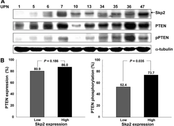
Correlation of Skp2 Expression with PTEN Phospho-rylation in AML. The status of PTEN phosphorylation in relation to Skp2 protein expression in the AML cells was next examined. The constitutive phosphorylation of PTEN protein was demonstrated in the majority of AML cases with high Skp2 expression (Fig. 2A). The frequency of PTEN phosphorylation was significantly higher in the group with high Skp2 protein expression (42 cases; 73.7%) compared with the cases with low Skp2 expression (22 cases; 52.4%; P⫽ 0.035; Fig. 2B). How-ever, Skp2 expression was not correlated with the levels of PTEN expression itself (Fig. 2B).
Prognostic Significance of Skp2 Expression in AML.
As shown in Table 1, the complete remission rate in the high Skp2 expression group was similar to that in the low Skp2 expression group (81.6% versus 91.4%, P⫽ 0.215). However, survival analysis using the Kaplan-Meier Method demonstrated that the high Skp2 expression group showed a significantly shorter disease-free survival compared with that of the low Skp2 expression group (Fig. 3A; P⫽ 0.0386 by log-rank test). The overall survival rate was also significantly lower in the high Skp2 expression group compared with the low Skp2 expression group (Fig. 3B; P⫽ 0.0369). The disease-free survival estimates at 5 years for the patients with or without Skp2 overexpression were 29.4% (SE⫽ 11.6%) and 57.9% (SE ⫽ 15.7%), respec-tively (P⫽ 0.0386; Fig. 3A). The overall survival estimates at 5 years for the patients with or without Skp2 overexpression were 15.8% (SE⫽ 6.8%) and 51.9% (SE ⫽ 10.8%), respec-tively (P⫽ 0.0369; Fig. 3B). Univariate analysis revealed that neither white blood cell count nor cytogenetics was a prognostic variable in our AML cases (Table 3). However, the Skp2 ex-pression level was a strong prognostic factor for disease-free survival and overall survival. Multivariate analysis also did not
Table 2 Cell cycle analysis according to Skp2 expression levels Skp2 expression P Low (n⫽ 42) High (n⫽ 57) G0-G1 75.9⫾ 24.0% 85.2⫾ 18.1% NS G2-M 11.9⫾ 17.6% 10.9⫾ 19.9% NS S phase 15.7⫾ 22.3% 8.5⫾ 17.1% NS Abbreviation: NS, not significant.
Table 1 Patient characteristics and complete remission (CR) rate according to Skp2 expression levels Skp2 expression*
P
Low (n⫽ 42) High (n⫽ 57)
Age (y), median (range) 45 (15–71) 44 (15–80) NS
Sex (M/F) 24/18 25/32 NS
WBC (⫻109/L), median (range) 15.4 (0.6–238.0) 23.3 (1.2–253.8) NS
LDH (IU/L), median (range) 705 (362–7260) 658 (156–9260) NS
FAB classification NS M0, M1, M2 22 35 M4, M5 19 21 M6, M7 1 1 Cytogenetics 0.035† Favorable 6 4 Intermediate 26 27 Unfavorable 10 26 CR rate 91.4% (32/35) 81.6% (40/49) NS Abbreviations: NS, not significant; WBC, white blood cell(s); LDH, lactic dehydrogenase; FAB, French-American-British (classification). * Skp2 expression关Lc ⫽ Skp2 (L)/␣-tubulin (L)兴, where L is expression level in the AML cells; low, Lc ⱕ 0.7; high, Lc ⬎ 0.7. † Unfavorable versus non-unfavorable (favorable and intermediate).
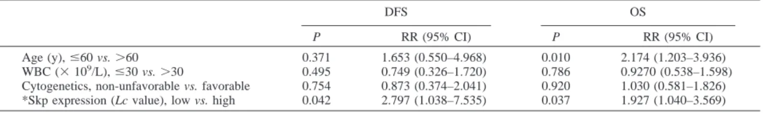
confirm the prognostic value of the white blood cell count nor of cytogenetics in the enrolled AML cases (Table 4). In contrast, Skp2 expression remained as an independent prognostic factor of disease-free survival [relative risk (95% confidence inter-val)⫽ 2.797 (1.038–7.535), P ⫽ 0.042], and the overall sur-vival [relative risk (95% confidence interval)⫽ 1.927 (1.040– 3.569), P⫽ 0.037] (Table 4). These findings suggest that Skp2 expression is an independent marker of a poor prognosis in adult AML.
DISCUSSION
This study demonstrated that the high levels of Skp2 pro-tein expression were observed in a substantial proportion of AML cases and were significantly correlated with a poor prog-nosis. Furthermore, it was shown for the first time that Skp2
expression was positively correlated with the constitutive phos-phorylation of the COOH-terminal regulatory domain of the PTEN protein, along with the cytoplasmic localization of p27Kip1 protein in AML cells.
This study supports a growing body of evidence demon-strating the role of Skp2 in an oncogenic process (21–23, 25, 26). Because Skp2 is required for the ubiquitination and pro-teasomal degradation of p27Kip1 protein (16 –18), the deregu-lation of Skp2 may contribute to the neoplastic transformation through accelerated p27Kip1 proteolysis. The Skp2-transfected cell lines expressed low levels of p27Kip1, and showed high levels of proliferation activity and of resistance to apoptosis and a high tumor-invasion potential (19, 26). Therefore, a low p27Kip1 expression in malignant cells may be caused by an increased expression of Skp2. Indeed, there was an inverse
Fig. 3 Kaplan-Meier survival
curve for disease-free survival and overall survival rate of pa-tients with AML according to the levels of Skp2 protein ex-pression. High levels of Skp2 protein expression were signif-icantly associated with lower disease-free survival (A) and overall survival rates (B; P⫽ 0.0386 and P⫽ 0.0369, respec-tively). The log-rank statistic was used to test the difference in survival times between the groups.
Fig. 2 A, Western blot
analy-sis of Skp2, PTEN, and pPTEN protein expression in the repre-sentative AML samples. Levels of protein loading were visual-ized by stripping the membrane and reprobing with an antibody for ␣-tubulin. B, the PTEN (left) and pPTEN (right) ex-pression according to levels of Skp2 expression. The fre-quency of PTEN phosphoryla-tion was significantly higher in the high Skp2 expression group compared with the low Skp2 expression group (P⫽ 0.035).
correlation between the p27Kip1 and Skp2 expression in the various human cancers (9 –10, 21–23, 25, 26).
However, an inverse correlation between the Skp2 and p27Kip1 protein expression was not observed in our AML cases. This finding was in agreement with the recent reports showing that an inverse correlation between these proteins was not observed in a substantial proportion of aggressive cancers, although high Skp2 level correlated well with the extent of cell proliferation and disease progression (27–29). The lack of an inverse correlation suggests that additional molecular events associated with the tumorigenic properties of Skp2 (22, 25) or the aberrant regulation of the p27Kip1 activity (47) may be operating in AML.
It was suggested that the oncogenic potential of Skp2 is not restricted to p27Kip1 proteolysis (17, 22, 25). The forced ex-pression of Skp2 in the quiescent fibroblasts induced DNA synthesis (17). Skp2 cooperated with N-Ras in the tumorigen-esis of a T-cell lymphoma in a transgenic mice model (25). Rodent fibroblasts primarily transformed by both Skp2 and H-Ras gene transfection could form tumors in nude mice (22). The Skp2-transfected gastric carcinoma cell lines showed sig-nificantly higher growth rates, invasion potential, and resistance to an apoptotic induction by chemotherapeutic agents (26). These findings suggest that Skp2 plays an important role in human oncogenesis and in the modulation of the phenotype of malignant tumors. However, the leukemogenic potential of Skp2 itself remains to be determined in AML.
In addition to degradation, p27Kip1 activity appears to be altered by the intracellular localization or sequestration. In tu-mors with abundant p27Kip1 expression, the protein is often mislocalized to the cytoplasm (48 –50). Because the
growth-restraining activity of p27Kip1 depends on its nuclear localiza-tion, the mislocalization effectively abrogates the p27Kip1 in-hibitory activity (48, 51–54). In fact, cytoplasmic p27Kip1 localization seems to directly correlate with a poor long-term survival and the tumor grade of esophagus and breast carcinoma (50, 55–57). Although an inverse correlation between Skp2 and the p27Kip1 protein was not observed in our AML cases, the high Skp2 level was found to be significantly associated with the extent of cytoplasmic localization of p27Kip1 protein. The C/N ratio of p27Kip1 protein significantly correlated with the level of Skp2 protein expression in AML. These findings sug-gest that the molecular mechanism, which is involved in the increased expression of Skp2, is also closely associated with the mislocalization of p27Kip1 protein in AML.
The mechanism by which the Skp2 protein expression is increased in AML cells needs to be understood. Skp2 expression is usually cell cycle dependent; it is low in the G1 phase,
increases in the S phase, and declines afterward (16, 19, 58). However, in this study, the cell cycle distribution was not different in relation to Skp2 expression. This finding is in line with the finding that p27Kip1 expression does not seem to reflect the cell cycle status of AML in this study, as many investigators have pointed this out in other types of cancer (59). It was also previously shown that the levels of Skp2 protein are not cell cycle dependent in malignant lymphoma (10). These findings suggest that the dysregulation of the cell cycle cannot explain the aberrant expression of Skp2 protein in AML cells.
PTEN regulates the ubiquitin-dependent Skp2-mediated p27Kip1 degradation (40). A deletion of PTEN in mouse fibro-blasts led to increased levels of Skp2 with concomitant reduc-tions in the p27Kip1 levels (40). Skp2 was down-regulated by the expression of the wild-type PTEN or by exposure to the PI3-K inhibitor in the human glioblastoma cell line U87MG (40). Skp2 expression is inversely correlated with PTEN expres-sion in prostate cancer cells (9). These findings suggest that PTEN functions as a negative regulator of the Skp2 pathway. However, it has been reported that mutations in the PTEN gene are very infrequent genetic aberrations in myeloid leukemia (41– 42). Loss of heterozygosity analysis and polymerase chain reaction single-strand conformation polymorphism (PCR-SSCP) of the entire coding region showed that none of the AML cases had a loss of heterozygosity or a mutation (41). We observed no correlation between the PTEN and Skp2 expression levels in AML cells.
It was shown that the phosphorylation of the serine and threonine residues in the COOH-terminal regulatory domain of PTEN protein regulates both PTEN stability and PTEN
enzy-Table 3 Univariate analysis of age, white blood cell count (WBC), cytogenetics, and Skp2 expression status for complete remission (CR)
rate, disease-free survival (DFS), and overall survival (OS) in AML patients P* CR DFS OS Age (y)ⱕ60 vs. ⬎60 1.0 0.6815 0.0126 WBC (⫻109/L),ⱕ30 vs. ⬎30 0.192 0.4519 0.8996 Cytogenetics, non-unfavorable vs. unfavorable 0.744 0.6187 0.7413 Skp2 expression (Lc value),† low vs. high 0.184 0.0386 0.0369
* Log-rank test.
† Skp2 expression 关Lc ⫽ Skp2 (L)/␣-tubulin (L)兴, where L is expression level in the AML cells.
Table 4 Multivariate analysis of disease free survival (DFS) and overall survival (OS) in AML patients
DFS OS
P RR (95% CI) P RR (95% CI) Age (y),ⱕ60 vs. ⬎60 0.371 1.653 (0.550–4.968) 0.010 2.174 (1.203–3.936) WBC (⫻ 109/L),ⱕ30 vs. ⬎30 0.495 0.749 (0.326–1.720) 0.786 0.9270 (0.538–1.598)
Cytogenetics, non-unfavorable vs. favorable 0.754 0.873 (0.374–2.041) 0.920 1.030 (0.581–1.826) *Skp expression (Lc value), low vs. high 0.042 2.797 (1.038–7.535) 0.037 1.927 (1.040–3.569) Abbreviations: RR, relative risk; CI, confidence interval.
* Skp2 expression关Lc ⫽ Skp2 (L)/␣-tubulin (L)兴, where L is expression level in the AML cells.
matic activity (60 – 61). The PTEN phosphorylation keeps the PTEN stable but makes it less active toward its substrate, PIP3
(62). Recently, we demonstrated that PTEN protein is constitu-tively phosphorylated in a substantial proportion of AML cases (43). PTEN phosphorylation was significantly associated with Akt phosphorylation and the activation of Akt downstream (43). The underlying mechanism for the constitutive PTEN phospho-rylation in AML remains unclear. Because it was demonstrated that a key enzyme regulating the phosphorylation of the COOH-terminal regulatory domain of PTEN seems to be the protein kinase casein kinase 2 (62), the casein kinase 2 activity of the AML cells in relation to PTEN phosphorylation needs to be evaluated. In this study, PTEN phosphorylation was strongly associated with the levels of Skp2 expression, but not with p27Kip1 protein. Because the phosphorylation of the PTEN protein is highly associated with Akt phosphorylation (43), PTEN phosphorylation is meant to be closely involved in the regulation of Skp2 expression via the PI3-K/Akt pathway. If the specific kinase that regulates PTEN phosphorylation is identi-fied, the direct contribution of PTEN phosphorylation to the Akt/protein kinase-B–mediated Skp2 expression will be under-stood.
This study demonstrated that the high Skp2 protein expres-sion is significantly associated with poor prognostic features in AML. The complete remission rate was not different according to the levels of Skp2 expression. However, the disease-free survival and overall survival were significantly shorter in AML cases showing high levels of Skp2 expression compared with the cases with low levels of Skp2 protein. Multivariate analysis using the Cox proportional hazard model revealed that Skp2 protein expression level is an independent prognostic factor for both disease-free survival and overall survival in the whole cohort.
In conclusion, we demonstrated for the first time that Skp2 overexpression is an independent prognostic factor in AML. In a majority of AML cases showing high levels of Skp2 expres-sion, the p27Kip1 protein was preferentially localized to the cytoplasm, suggesting that an aberrant regulatory pathway is operating in the Skp2-mediated p27Kip1 proteolysis in AML. In addition, the finding that Skp2 expression is significantly cor-related with the constitutive phosphorylation of the PTEN pro-tein suggests that the pPTEN-Skp2 axis may be a promising therapeutic target for adult AML.
REFERENCES
1. Philipp-Staheli J, Payne SR, Kemp CJ. p27(Kip1): regulation and function of a haploinsufficient tumor suppressor and its misregulation in cancer. Exp Cell Res 2001;264:148 – 68.
2. Slingerland J, Pagano M. Regulation of the Cdk inhibitor p27 and its deregulation in cancer. J Cell Physiol 2000;183:10 –7.
3. Tan P, Cady B, Wanner M, et al. The cell cycle inhibitor p27 is an independent prognostic marker in small (T1a,b) invasive breast carci-nomas. Cancer Res 1997;57:1259 – 63.
4. Catzavelos C, Bhattacharya N, Ung YC, et al. Decreased levels of the cell-cycle inhibitor p27Kip1 protein: prognostic implications in primary breast cancer. Nat Med 1997;3:227–30.
5. Fredersdorf S, Burns J, Milne AM, et al. High level expression of p27(Kip1) and cyclin D1 in some human breast cells: inverse correlation between the expression of p27(Kip1) and degree of malignancy in
human breast and colorectal cancers. Proc Natl Acad Sci USA 1997; 94:6380 –5.
6. Loda M, Cukor B, Tam SW, et al. Increased proteasome-dependent degradation of the cyclin-dependent kinase inhibitor p27 in aggressive colorectal cancers. Nat Med 1997;3:231– 4.
7. Yokozawa T, Towatari M, Lida H, et al. Prognostic significance of the cell cycle inhibitor p27Kip1in acute myeloid leukemia. Leukemia
(Baltimore) 2000;14:28 –33.
8. Esposito V, Baldi A, De Luca A, et al. Prognostic role of the cyclin-dependent kinase inhibitor p27 in non-small cell lung cancer. Cancer Res 1997;57:3381–5.
9. Yang G, Ayala G, De Marzo A, et al. Elevated Skp2 protein expres-sion in human prostate cancer: association with loss of the cyclin-dependent kinase inhibitor p27 and PTEN and with reduced recurrence-free survival. Clin Cancer Res 2002;8:3419 –26.
10. Chiarle R, Budel LM, Skolink J, et al. Increased proteasome deg-radation of cyclin-dependent kinase inhibitor p27 is associated with a decreased overall survival in mantle cell lymphoma. Blood 2000;95: 619 –26.
11. Pagano M, Tam SW, Theodoras AM, et al. Role of the ubiquitin-proteasome pathway in regulating abundance of the cyclin-dependent kinase inhibitor p27. Science (Wash DC) 1995;269:682–5.
12. Montagnoli A, Fiore F, Eytan E, et al. Ubiquitination of p27 is regulated by Cdk-dependent phosphorylation and trimeric complex for-mation. Genes Dev 1999;13:1181–9.
13. Nguyen H, Gitig DM, Koff A. Cell-free degradation of p27(kip1), a G1 cyclin-dependent kinase inhibitor, is dependent on CDK2 activity and the proteasome. Mol Cell Biol 1999;19:1190 –201.
14. Sheaff RJ, Groudine M, Gordon M, Roberts JM, Clurman BE. Cyclin E-CDK2 is a regulator of p27Kip1. Genes Dev 1997;11: 1464 –78.
15. Zhang H, Kobayashi R, Galaktionov K, Beach D. p19Skp1 and p45Skp2 are essential elements of the cyclin A-CDK2 S phase kinase. Cell 1995;82:915–25.
16. Carrano AC, Eytan E, Hershko A, Pagano M. SKP2 is required for ubiquitin-mediated degradation of the CDK inhibitor p27. Nat Cell Biol 1999;1:193–9.
17. Sutterluty H, Chatelain E, Marti A, et al. p45SKP2 promotes p27Kip1 degradation and induces S phase in quiescent cells. Nat Cell Biol 1999;1:207–14.
18. Tsvetkov LM, Yeh KH, Lee SJ, Sun H, Zhang H. p27(Kip1) ubiquitin and degradation is regulated by the SCF (Skp2) complex through phosphorylated Thr187 in p27. Curr Biol 1999;9:661– 4. 19. Nakayama K, Nagahama H, Minamishima YA, et al. Targeted disrup-tion of Skp2 results in accumuladisrup-tion of cyclin E and p27(Kip1), polyploidy and centrosome overduplication. EMBO J 2000;19:2069 – 81.
20. Chao Y, Shih YL, Chiu JH, et al. Overexpression of cyclin A but not Skp2 correlates with the tumor relapse of human hepatocellular carcinoma. Cancer Res 1998;58:985–90.
21. Hershko D, Bornstein G, Ben-Izhak O, et al. Inverse relation between levels of p27(Kip1) and of its ubiquitin ligase subunit Skp2 in colorectal carcinomas. Cancer (Phila) 2001;91:1745–51.
22. Gstaiger M, Jordan R, Lim M, et al. Skp2 is oncogenic and over-expressed in human cancers. Proc Natl Acad Sci USA, 2001;98:5043– 8. 23. Kudo Y, Kitajima S, Sato S, Miyauchi M, Ogawa I, Takata T. High expression of S-phase kinase-interacting protein 2, human F-box pro-tein, correlates with poor prognosis in oral squamous cell carcinomas. Cancer Res 2001;61:7044 –7.
24. Shigemasa K, Gu L, O’Brien TJ, Ohama K. Skp2 overexpression is a prognostic factor in patients with ovarian adenocarcinoma. Clin Can-cer Res 2003;9:1756 – 63.
25. Latres E, Chiarle R, Schulman BA, et al. Role of the F-box protein Skp2 in lymphomagenesis. Proc Natl Acad Sci USA, 2001;98:2515– 220.
26. Masuda T, Inoue H, Sonoda H, et al. Clinical and biological significance of S-phase kinase-associated protein (Skp2) gene expres-sion in gastric carcinoma: modulation of malignant phenotype by Skp2
overexpression, possibly via p27 proteolysis. Cancer Res, 2002;62: 3819 –25.
27. Oliveira AM, Okuno SH, Nascimento AG, Lloyd RV. Skp2 protein expression in soft tissue sarcomas. J Clin Oncol 2003;21:722–7. 28. Lim MS, Adamson A, Lin Z, et al. Expression of Skp2, a p27Kip1
ubiquitin ligase, in malignant lymphoma: correlation with p27Kip1 and proliferation index. Blood 2002;100:2950 – 6.
29. Penin RM, Fernandez-Figueras MT, Puig L, Rex J, Ferrandiz C, Ariza A. Over-expression of p45(SKP2) in Kaposi’s sarcoma correlates with higher tumor stage and extracutaneous involvement but is not directly related to p27(KIP1) down-regulation. Mod Pathol 2002;15:1227–35. 30. Li J, Yen C, Liaw D, et al. PTEN, a putative protein tyrosine phosphatase gene mutated in human brain, breast, and prostate cancer. Science (Wash DC) 1997;275:1943–7.
31. Steck PA, Pershouse MA, Jasser SA, et al. Identification of a candidate tumour suppressor gene, MMAC1, at chromosome 10q23.3 that is mutated in multiple advanced cancers. Nat Genet 1997;15:356 – 62.
32. Myers MP, Stolarov JP, Eng C, et al. P-TEN, the tumor suppressor from human chromosome 10q23, is a dual-specificity phosphatase. Proc Natl Acad Sci USA 1997;94:9052–7.
33. Maehama T, Dixon JE. The tumor suppressor, PTEN/MMAC1, dephosphorylates the lipid second messenger, phosphatidylinositol 3,4,5-trisphosphate. J Biol Chem 1998;273:13375– 8.
34. Furnari FB, Huang HJ, Cavenee WK. The phosphoinositol phos-phatase activity of PTEN mediates a serum-sensitive G1 growth arrest in glioma cells. Cancer Res 1998;58:5002– 8.
35. Ali IU, Schriml LM, Dean M. Mutational spectra of PTEN/ MMAC1 gene: a tumor suppressor with lipid phosphatase activity. J Natl Cancer Inst (Bethesda) 1999;91:1922–32.
36. Di Cristofano A, De Acetis M, Koff A, Cordon-Cardo C, Pandolfi PP. PTEN and p27KIP1 cooperate in prostate cancer tumor suppression in the mouse. Nat Genet 2001;27:222– 4.
37. Medema RH, Kops GJ, Bos JL, Burgering BM. AFX-like Forkhead transcription factors mediate cell-cycle regulation by Ras and PKB through p27Kip1. Nature (Lond) 2000;404:782–7.
38. Hyun T, Yam A, Pece S, et al. Loss of PTEN expression leading to high Akt activation in human multiple myelomas. Blood 2000;96: 3560 – 8.
39. Bruni P, Boccia A, Baldassarre G, et al. PTEN expression is reduced in a subset of sporadic thyroid carcinomas: evidence that PTEN-growth suppressing activity in thyroid cancer cells mediated by p27Kip1. Oncogene 2000;19:3146 –55.
40. Mamillapalli R, Gavrilova N, Mihaylova VT, et al. PTEN regulates the ubiquitin-dependent degradation of the CDK inhibitor p27(KIP1) through the ubiquitin E3 ligase SCF(SKP2). Curr Biol 2001;11:263–7. 41. Liu TC, Lin PM, Chang JG, Lee JP, Chen TP, Lin SF. Mutation analysis of PTEN/MMAC1 in acute myeloid leukemia. Am J Hematol 2000;63:170 –5.
42. Aggerholm A, Gronbaek K, Guldberg P, Hokland P. Mutational analysis of the tumor suppressor gene MMAC1/PTEN in malignant myeloid disorders. Eur J Haematol 2000;65:109 –13.
43. Cheong JW, Eom JI, Maeng HY, et al. Phosphatase and tensin homologue phosphorylation in the C-terminal regulatory domain is frequently observed in acute myeloid leukaemia and associated with poor clinical outcome. Br J Haematol 2003;122:454 – 6.
44. Cheson BD, Cassileth PA, Head DR, et al. Report of the National Cancer Institute-sponsored workshop on definitions of diagnosis and response in acute myeloid leukemia. J Clin Oncol 1990;8:813–9.
45. Lee ST, Jang JH, Min YH, Hahn JS, Ko YW. AC133 antigen as a prognostic factor in acute leukemia. Leuk Res 2001;25:757– 67. 46. Lenferink AE, Busse D, Flanagan WM, Yakes FM, Arteaga CL. ErbB2/neu kinase modulates cellular p27(Kip1) and cyclin D1 through multiple signaling pathways. Cancer Res 2001;61:6583–91.
47. Hara T, Kamura T, Nakayama K, Oshikawa K, Hatakeyama S, Nakayama KI. Degradation of p27Kip1 at the G0 –G1 transition medi-ated by a Skp2-independent ubiquitination pathway. J Biol Chem 2001; 276:48937– 43.
48. Baldassarre G, Belletti B, Bruni P, et al. Overexpressed cyclin D3 contributes to retaining the growth inhibitor p27 in the cytoplasm of thyroid tumor cells. J Clin Investig 1999;104:865–74.
49. Ciaparrone M, Yamamoto H, Yao Y, et al. Localization and ex-pression of p27KIP1 in multistage colorectal carcinogenesis. Cancer Res 1998;58:114 –22.
50. Singh SP, Lipman J, Goldman H, et al. Loss or altered subcellular localization of p27 in Barrett’s associated adenocarcinoma. Cancer Res 1998;58:1730 –5.
51. Jiang Y, Zhao RC, Verfaillie CM. Abnormal integrin-mediated regulation of chronic myelogenous leukemia CD34⫹ cell proliferation: BCR/ABL up-regulates the cyclin-dependent kinase inhibitor, p27Kip, which is relocated to the cell cytoplasm and incapable of regulating cdk2 activity. Proc Natl Acad Sci USA 2000;97:10538 – 43.
52. Soucek T, Yeung RS, Hengstschlager M. Inactivation of the cyclin-dependent kinase inhibitor p27 upon loss of the tuberous sclerosis complex gene-2. Proc Natl Acad Sci USA 1998;95:15653– 8. 53. Orend G, Hunter T, Ruoslahti E. Cytoplasmic displacement of cyclin E-cdk2 inhibitors p21Cip1 and p27Kip1 in anchorage-indepen-dent cells. Oncogene 1998;16:2575– 83.
54. Yaroslavskiy B, Watkins S, Donnenberg AD, Patton TJ, Steinman RA. Subcellular and cell-cycle expression profiles of CDK-inhibitors in normal differentiating myeloid cells. Blood 1999;93:2907–17. 55. Liang J, Zubovitz J, Petrocelli T, et al. PKB/Akt phosphorylates p27, impairs nuclear import of p27 and opposes p27-mediated G1 arrest. Nat Med 2002;8:1153– 60.
56. Shin I, Yakes FM, Rojo F, et al. PKB/Akt mediates cell-cycle progression by phosphorylation of p27Kip1at threonine 157 and
modu-lation of its cellular localization. Nat Med 2002;10:1145–52. 57. Viglietto G, Motti ML, Bruni P, et al. Cytoplasmic relocalization and inhibition of the cyclin-dependent kinase inhibitor p27Kip1 by
PKB/Akt-mediated phosphorylation in breast cancer. Nat Med 2002;8: 1136 – 43.
58. Bilodeau M, Talarmin H, Ilyin G, et al. Skp2 induction and phos-phorylation is associated with the late G1 phase of proliferating hepa-tocytes. FEBS Lett 1999;452:247–53.
59. Erlanson M, Portin C, Linderholm B, Lindh J, Roos G, Landberg G. Expression of cyclin E and the cyclin-dependent kinase inhibitor p27 in malignant lymphoma-prognostic implications. Blood 1998;92:770 –7. 60. Vazquez F, Ramaswamy S, Nakamura N, Sellers WR. Phosphoryl-ation of the PTEN tail regulates protein stability and function. Mol Cell Biol 2000;20:5010 – 8.
61. Vazquez F, Grossman SR, Takahashi Y, Rokas MV, Nakamura N, Sellers WR. Phosphorylation of the PTEN tail acts as an inhibitory switch by preventing its recruitment into a protein complex. J Biol Chem 2001;276:48627–30.
62. Miller SJ, Lou DY, Seldin DC, Lane WS, Neel BG. Direct identi-fication of PTEN phosphorylation sites. FEBS Lett 2002;528:145–53. 63. Bennett JM, Catovsky D, Daniel MT, et al. Proposals for the classification of the acute leukaemias (FAB cooperative group). Br J Maematol 1976;33:451– 8.