척추 경막외 출혈에 대한 수술적 치료성적 분석
울산대학교 의과대학 서울아산병원 신경외과 조영현∙박진훈∙김지훈∙노성우∙김창진∙전상룡
─ Abstract ─
Analysis of the Outcomes of Surgically-Treated Spinal Epidural Hematomas
Young Hyun Cho, M.D., Jin Hoon Park, M.D., Ji Hoon Kim, M.D., Sung Woo Roh, M.D., Chang Jin Kim, M.D., Sang Ryong Jeon, M.D., Ph.D.
Department of Neurological Surgery, Asan Medical Center, University of Ulsan College of Medicine, Seoul, Korea
Purpose: Spinal epidural hematoma (EDH) is a rare condition requiring an urgent diagnosis and manage- ment. We describe here the clinical features, magnetic resonance image (MRI) findings, and outcomes of surgery in six patients with spinal EDH.
Methods: We retrospectively analyzed six patients who underwent surgery for spinal EDH between April 2004 and May 2010. Preoperative MRI findings within 48 hours of symptom occurrence were analyzed for cord compression, extent of EDH, and presence of vascular abnormalities. Pre- and postoperative neurological status was also assessed comparatively.
Results: Our six patients consisted of three men and three women, with a mean age of 70 years (range: 54-88 years), who presented with the back pain or motor weakness. The mean follow-up period was 34 months (range:
2-72 months). Two patients had cardiovascular disease and were taking warfarin, but the others had no history of medical comorbidity. Those two patients taking warfarin had a history of trauma, another one experienced symp- toms during a strenuous effort, and the others developed spontaneously. Before surgery, motor power was grade III in three patients, grade 0 in two patients, and normal in one patient. Preoperative MRI showed no vascular abnormalities except for the EDH in any patient. At the last follow-up, all those five patients with motor weak- ness showed neurological improvement compared to their preoperative status. There were no complications related to surgery. All six patients were able to ambulate with or without an assistive device.
Conclusion: Spinal EDH can occur in patients without trauma, bleeding diathesis, or combined vascular pathology. The surgical outcomes of spinal EDH seem to be satisfactory, even in quadriplegic patients. (J Korean Soc Traumatol 2010;23:163-169).
Key Words: Spinal epidural hematoma, Surgical outcome, Magnetic Resonance Image
� Address for Correspondence : Sang Ryong Jeon, M.D., Ph.D.
Department of Neurological Surgery, Asan Medical Center, University of Ulsan College of Medicine 388-1 Poongnap-dong, Songpa-gu, Seoul 138-736, Korea
Tel : 82-2-3010-3562, Fax : 82-2-476-6738, E-mail : [email protected]
접수일: 2010년 10월 20일, 심사일: 2010년 11월 8일, 수정일: 2010년 12월 1일, 승인일: 2010년 12월 13일
I. Introduction
Spinal epidural hematoma (EDH) is a rare condition requiring an urgent diagnosis.(1-5) Although sometimes related to trauma, in 40-50% of patients, spinal EDH has no clear cause and is regarded as spontaneous.(6) Early diagnosis and management are necessary to prevent severe neurological deficits.(7) We analyzed the clinical features, magnetic resonance imaging (MRI) findings, and outcomes of surgery in six patients with spinal EDH.
II. Materials and Methods
We retrospectively analyzed six patients who underwent surgery for spinal EDH between April 2004 and May 2010.
Clinical characteristics of the patients were reviewed and preoperative MRI findings within 48 hours of symptom occurrence were analyzed for evidence of cord compression, extent of EDH, and presence of vascular abnormalities.
Pre- and postoperative neurological status was assessed comparatively. Presence of possible precipitating factors such as trauma or bleeding diathesis was also investigated.
III. Results
Our six patients consisted of three men and three women, with a mean age of 70 years (range: 54-88
years), who presented with the back pain or motor weak- ness. The mean follow-up period was 34 months (range:
2-72 months). Two patients had cardiovascular disease and were taking warfarin, but the others had no history of medical comorbidity. Those two patients taking warfarin had a history of trauma, another one patient experienced symptoms during a strenuous effort, and the others devel- oped spontaneously. Before surgery, motor power was grade III in three patients, grade 0 in two patients, and normal in one patient. Preoperative MRI showed no vascular abnor- malities except for the EDH in any patient (Fig. 1-6). At the last follow-up, all those five patients with motor weak- ness showed neurological improvement compared to their preoperative status. There were no complications related to surgery. All six patients were able to ambulate with or without an assistive device.
1. Case 1
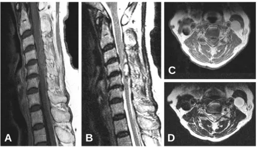
An 88-year-old woman visited the hospital with right hemiparesis of grade III and neck pain that had occurred 3 hours before. Her symptoms developed spontaneously with- out any trauma and she had no history of comorbidity or medication. MRI taken 36 hours after symptom onset showed spinal EDH without any vascular lesion on the C4- 5 levels (Fig. 1). Surgery for decompression was performed 48 hours after the onset of weakness. At the last follow up,
Fig. 1. MRI of Case 1
(A) Sagittal T1-weighted image showing the hematoma of iso signal intensity at the C4-5 levels. (B) Sagittal T2-weighted image showing a high signal intensity with a low signal rim at the C4-5 levels. (C) Axial T1-weighted image showing an iso signal intensity at the level of maximal cord compression. (D) Axial T2-weighted image showing a high signal intensity with a low signal rim at the same level.
A B D
C
her right hemiparesis was slightly improved into grade IV-.
2. Case 2
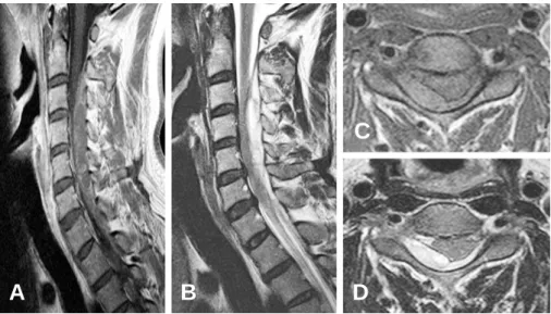
A 54-year-old man visited the hospital with acute severe back pain developed during a strenuous exertion. He had no neurological deficits and history of medication. MRI within 24 hours after symptom onset disclosed spontaneous
spinal EDH on the C2-T1 levels (Fig. 2). Decompressive surgery was undertaken 30 hours after the symptom onset.
Postoperatively, his back pain was alleviated immediately.
3. Case 3
A 70-year-old woman was admitted to the hospital due to the back pain and paraparesis of grade III developed
Fig. 2. MRI of Case 2
(A) Sagittal T1-weighted image showing a mixed iso and high signal intensity at the posterior aspect of the spinal canal at the C2-T1 levels. (B) Sagittal T2-weighted image showing a mixed high and low signal intensity at the C2-T1 levels. (C) Axial T1-weighted image showing an iso signal intensity at the level of maximal cord compression. (D) Axial T2-weighted image showing a high signal intensity at the same level.
A B D
C
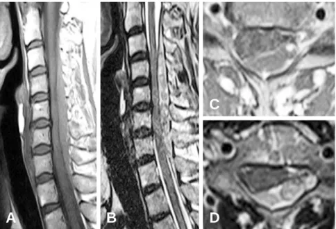
Fig. 3. MRI of Case 3
(A) Sagittal T1-weighted image showing an iso signal intensity at the posterior aspect of the C4-T1 levels. (B) Sagittal T2- weighted image showing a high signal intensity in the posterolateral epidural space of the C4-T1 levels. (C) Axial T1-weight- ed image showing an iso signal intensity in the posterior epidural space at the level of maximal cord compression. (D) Axial T2-weighted image showing a high signal intensity at the same level.
A B D
C
after a minor car accident 48 hours before. She had a his- tory of taking warfarin for mitral valve regurgitation. MRI showed an EDH in the posterior aspect of levels C4-T1 (Fig. 3). We delayed surgery for 10 days until her coagu- lopathy normalized, and there was no progression of weak- ness during this time. Postoperatively, her weakness
improved and she was able to ambulate with the assistance of a cane (motor power of grade IV).
4. Case 4
An 82-year-old man was admitted due to paraplegia
Fig. 4. MRI of Case 4
(A) Sagittal T1-weighted image showing an iso signal intensity at the C5-C7 levels. (B) Sagittal T2-weighted image showing a mixed high and low signal intensity at the C5-C7 levels. (C) Axial T1-weighted image showing an iso signal intensity in the posterolateral epidural space at the level of maximal cord compression. (D) Axial T2-weighted image showing a mixed high and low signal intensity at the same level.
A B D
C
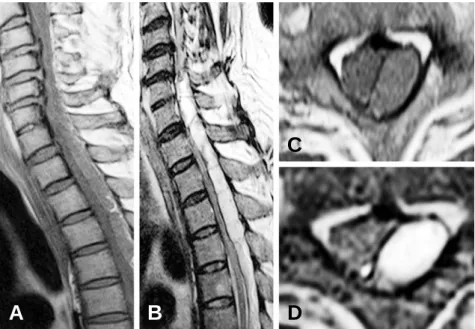
Fig. 5. MRI of Case 5
(A) Sagittal T1-weighted image showing an iso signal intensity at the C2-T1 levels. (B) Sagittal T1-weighted image a mixed high and low signal intensity at the C2-T1 levels. (C) Axial T1-weighted image showing an iso signal intensity in the pos- terolateral epidural space at the level of maximal cord compression. (D) Axial T2-weighted image showing a mixed high and low signal intensity at the same level.
A B D
C
occurred within 24 hours. He had not experienced any trauma and did not have a history of medication. MRI revealed an EDH located in the right posterolateral space on levels C5-C7 (Fig. 4). After a surgical decompression within 48 hours, he was able to ambulate with the assis- tance of a cane (motor power of grade IV).
5. Case 5
A 64-year-old woman presented with paraparesis of grade III developed 2 hours before. She had no history of trauma or medical illness. A spontaneous EDH was diagnosed on MRI at posterior aspect of the C2-T1 levels (Fig. 5). After an emergent evacuation of EDH, She recovered completely from the weakness.
6. Case 6
A 62-year-old man presented with paraplegia (motor weakness of grade 0) developed after a falling down 3 hours before. He had a history of taking warfarin. MRI revealed an EDH of the posterior aspect on levels C6-T10 (Fig. 6). With a decompressive surgery undertaken 48 hours after the trauma, his paraplegia recovered up to the motor power of grade IV
+.
The patient profiles and preoperative MRI findings of each case are summarized in Table 1.
The pre- and postoperative neurological status and time intervals between symptom onset and surgery are depicted in Table 2.
IV. Discussion
Spinal EDH occurring spontaneously or after minimal trauma has been attributed to various factors, including coagulopathy or anticoagulation, vascular anomaly, disc her- niation, Paget’ s disease of the bone, the Valsalva maneu- ver, and hypertension.(1,2,8-16) We retrospectively reviewed records for six patients who experienced spontaneous or trauma-related spinal EDH and were treated surgically. In three patients (50%), spinal EDH was not related to any previously described risk factors; in two patients (33%), spinal EDH was caused by trauma during anticoagulation treatment; and in one (17%), by the Valsalva maneuver.
Hematomas occur most commonly at the lower cervical and thoracolumbar spinal levels, with 86% of hematomas located on the dorsal aspect of the spinal cord, and the posterior epidural venous plexus is believed to be the source of dorsal bleeding.(17,18) In our patients, all of the hematomas were located on the dorsal aspect of the spinal
Fig. 6. MRI of Case 6
(A) Sagittal T1-weighted image showing an iso signal intensity at the C6-T10 levels. (B) Sagittal T1-weighted image show- ing a mixed high and low signal intensity at the C6-T10 levels. (C) Axial T1-weighted image showing an iso signal intensity in the posterolateral epidural space at the level of maximal cord compression. (D) Axial T2-weighted image showing a mixed high and low signal intensity at the same level.
A B D
C
cord, ranging from the mid- or lower cervical to the upper thoracic level.
Factors associated with good prognosis include a short interval from symptom onset to surgery, with patients who undergo evacuation of the hematoma within 24 hours hav- ing good neurological outcomes.(3) Surgical decompression within 36 hours in patients with complete sensorimotor loss, and within 48 hours in patients with incomplete deficits, has been associated with significantly better outcomes than surgical decompression performed after this time.(19) Although a rapid onset of motor deficits has been reported to result in unfavorable outcomes, others have found no association between functional outcome and the speed of neurological deterioration.(3,18)
Among five patients with preoperative weakness, all showed neurological improvement following surgery, and finally were able to ambulate with or without an assistive device. Even the patient who underwent delayed surgery (Case 3) performed 10 days later to correct a coagulopathy, showed functional recovery.
There have been no previous reports about the relation- ship between the extent of spinal cord compression and prognosis. Of our six patients, five showed compression of up to one-third of the spinal cord area on axial images
(Fig. 1-5), and another showed compression of half of the spinal cord (Fig. 6). All those patients, however, showed motor improvement after a decompressive surgery.
One report reviewed the outcome of surgery in 158 cases of spinal EDH and found that 95% of patients with incomplete deficit returned to normal neurology in contrast to 45.3% of patients with complete deficit.(3) Our two patients with paraplegia showed good clinical outcome.
V. Conclusion
Our findings indicate that spinal EDH can occur in patients without a trauma, bleeding diathesis, or combined vascular pathology. The surgical outcome of spinal EDH seems to be satisfactory even in those patients with com- plete motor deficit. Further study with a larger cohort and controlled design would facilitate our knowledge of this par- ticular kind of critical clinical entity.
REFERENCES
01) Holtås S, Heiling M, L?nntoft M. Spontaneous spinal epidural hematoma: findings at MR imaging and clini- cal correlation Radiology 1996;199:409-13.
Table 2. The pre- and postoperative motor power, severity of cord compression, and time intervals from symptom onset to surgery Preoperative Postoperative motor
F/U period Severity of cord Time from motor power power at last follow up
(months) compression at symptom onset axial image to surgery (hours)
Case 1 III IV- 72 ≤ one third 049
Case 2 V V 49 ≤ one third 030
Case 3 III IV 47 ≤ one third 240
Case 4 0 IV 40 ≤ one third 045
Case 5 III V 37 ≤ one third 008
Case 6 0 IV+ 02 One third to half 052
Table 1. The patient profiles and preoperative MRI findings Age (years)/
Extent of EDH Time from symptom MRI characteristics
Sex onset to imaging (hours) T1WI T2WI
Case 1 88/F C4-C5 36 Iso High with low signal rim
Case 2 54/M C2-T1 24 Mixed iso and high Mixed high and low
Case 3 70/F C4-T1 48 Iso High
Case 4 82/M C5-C7 24 Iso Mixed high and low
Case 5 64/F C2-T1 03 Iso Mixed high and low
Case 6 62/M C6-T10 08 Iso Mixed high and low
T1WI, T1-weighted images; T2WI, T2-weighted images; Iso, Iso signal intensity; High, high signal intensity; Low, low signal intensity
02) Fukui MB, Swarnkar AS, Williams RL. Acute sponta- neous spinal epidural hematomas AJNR Am J Neuroradiol 1999;20:1365-72.
03) Foo D, Rossier AB. Preoperative neurological status in predicting surgical outcome of spinal epidural hematomas Surg Neurol 1981;15:389-401.
04) McQuarrie IG. Recovery from paraplegia caused by spontaneous spinal epidural hematoma Neurology 1978;28:224-8.
05) Packer NP, Cummins BH. Spontaneous epidural haem- orrhage: A surgical emergency Lancet 1978;1:356-8.
06) Penar PL, Fischer DK, Goodrich I, Bloomgarden GM, Robinson F. Spontaneous spinal epidural hematoma Int Surg 1987;72:218-21.
07) Subbiah M, Avadhani A, Shetty AP, Rajasekaran S.
Acute spontaneous cervical epidural hematoma with neurological deficit after low-molecular-weight heparin therapy: role of conservative management Spine J 2010;10:e11-5.
08) Groen RJ, Ponssen H. The spontaneous spinal epidural hematoma. A study of the etiology J Neurol Sci 1990;98:121-38.
09) Groen RJ, Groenewegen HJ, van Alphen HA, Hoogland PV. Morphology of the human internal vertebral venous plexus: a cadaver study after intravenous Araldite CY 221 injection Anat Rec 1997;249:285-94.
10) Joseph AP, Vinen JD. Acute spinal epidural hematoma J Emerg Med 1993;11:437-41.
11) Lee KS, McWhorter JM, Angelo JN. Spinal epidural hematoma associated with Paget’s disease Surg Neurol
1988;30:131-4.
12) Brawn LA, Bergval UE, Davies-Jones GA.
Spontaneous spinal epidural haematoma with sponta- neous resolution Postgrad Med J 1986;62:885-7.
13) Gundry CR, Heithoff KB. Epidural hematoma of the lumbar spine: 18 surgically confirmed cases Radiology 1993;187:427-31.
14) Caldemeyer KS, Mocharla R, Moran CC, Smith RR.
Gadolinium enhancement in the center of a spinal epidural hematoma in a hemophiliac J Comput Assist Tomogr 1993;17:321-3.
15) Muhonen MG, Piper JG, Moore SA, Menezes AH.
Cervical epidural hematoma secondary to an extradural vascular malformation in an infant: case report Neurosurgery 1995;36:585-8.
16) David S, Salluzzo RF, Bartfield JM, Dickinson ET.
Spontaneous cervicothoracic epidural hematoma follow- ing prolonged valsalva secondary to trumpet playing Am J Emerg Med 1997;15:73-5.
17) Hussenbocus SM, Wilby MJ, Cain C, Hall D.
Spontaneous spinal epidural hematoma: a case report and literature review J Emerg Med 2009.
18) Shin JJ, Kuh SU, Cho YE. Surgical management of spontaneous spinal epidural hematoma Eur Spine J 2006;15:998-1004.
19) Groen RJ, van Alphen HA. Operative treatment of spontaneous spinal epidural hematomas: a study of the factors determining postoperative outcome Neurosurgery 1996;39:494-509.