Molecular Analysis of Carbapenem-Resistant Enterobacteriaceae at a South Korean Hospital
Miyoung Lee1,2 and Tae-Jin Choi1*
1Department of Microbiology, Pukyoung National University, Busan 48513, Republic of Korea
2Department of Laboratory Medicine, BHS Hanseo Hospital, Busan 48253, Republic of Korea
Received: February 17, 2020 / Revised: May 5, 2020 / Accepted: May 6, 2020
Introduction
Approximately 700,000 people die annually from anti- biotic-resistant infections, and this number will expect- edly surpass 10 million deaths per year by 2050 [1].
Antibiotic-resistant, gram-negative bacteria pose a seri- ous threat worldwide, and, owing to the lack of available therapeutic options, the Centers for Disease Control and Prevention (CDC) has listed carbapenem-resistant mem- bers of the family Enterobacteriaceae as an “immediate public health threat that requires urgent and aggressive
action” [2]. Two resistant forms of Enterobacteriaceae are of particular concern in the hospital setting: carbape- nem-resistant Enterobacteriaceae (CRE) and carbapene- mase-producing Enterobacteriaceae (CPE). Both CRE and CPE are associated with high mortality owing to their resistance to all available antibiotics.
In Korea, the first person infected with carbapenem- resistant Enterobacteriaceae was reported in December 2010. In 2014, a sudden increase in Klebsiella pneu- moniae carbapenemase (KPC) cases was observed nationwide, initially occurring as hospital-based out- breaks[3], which subsequently led to inter-hospital or inter-regional spread [4], thereby resulting in the endemic stage of KPC in 2015. Following this outbreak, numerous cases of CRE and CPE were reported in The prevalence of carbapenem-resistant Enterobacteriaceae (CRE) is increasing globally, resulting in high mortality rates. Although CRE is a relatively recent problem in Korea (the first case was not diagnosed until 2010), it is responsible for serious morbidities at an alarming rate. In this study, we carried out a molecular genetic analysis to determine the incidence of CRE and carbapenemase-producing Enterobacteriaceae (CPE) at a general hospital in Korea between August 2017 and August 2019. Forty strains of CPE were iso- lated from various clinical specimens and analyzed via antimicrobial susceptibility testing, polymerase chain reaction to detect β-lactamase genes, deoxyribonucleic acid sequencing, multilocus sequence typing, curing testing, and conjugal transfer of plasmids. The results demonstrated that all 40 isolates were multi- drug-resistant. The fluoroquinolone susceptibility test showed that 75% of the Enterobacteriaceae isolates were resistant to ciprofloxacin, whereas 72.5% were resistant to trimethoprim-sulfamethoxazole. Further, conjugation accounted for 57.5% of all resistant plasmid transfer events, which is 4.3-fold higher than that observed in 2010 by Frost et al. Finally, the high detection rate of transposon Tn4401 was associated with the rapid diffusion and evolution of CPE. Our results highlight the rapid emergence of extensively drug- resistant strains in Korea and emphasize the need for employing urgent control measures and protocols at the national level.
Keywords: Fluoroquinolone, blaKPC, Escherichia coli ST410, Klebsiella pneumoniae ST307, carbapenem-resistant Entero- bacteriaceae, carbapenemase-producing Enterobacteriaceae
*Corresponding author
Tel: +82-51-629-5617, Fax: +82-51-629-5619 E-mail: [email protected]
© 2020, The Korean Society for Microbiology and Biotechnology
Korea. As the mortality associated with invasive infec- tions caused by CPE is extremely high [5], the spread of CPE is of immense clinical concern.
β-Lactamases are grouped into four classes according to their amino acid sequence: class A includes KPC and Temoneira β-lactamase (TEM); class B consists of metallo- β-lactamases, such as Verona integron-encoded metallo-β- lactamases (VIM), Imipenem (IMP), and New Delhi metallo-β-lactamase-1(NDM-1); class C includes AmpC β- lactamases; and class D consists of oxacillinase (OXA)- type enzymes. Carbapenem resistance in K. pneumoniae has been reported in most countries and can be attributed to plasmid-mediated AmpC cephalospori- nases. AmpC cephalosporinases are associated with porin modifications [6, 7] and reduced outer membrane permeability owing to porin loss in combination with the production of either extended-spectrum β-lactamases (ESBLs) or β-lactamases, which are capable of hydrolyz- ing carbapenems (carbapenemases) [8, 9].
The dissemination of the blaKPC gene, encoding β-lac- tamase, occurs by the clonal spread of bacterial hosts harboring the gene or by horizontal transfer of trans- posons and plasmids carrying the gene [10, 11]. Plas- mids are transmissible by conjugation [12−14]. Smillie et al. reported that approximately 14% of full-sequenced plasmids were predicted to be conjugative [15]. Hence, conjugation is one of the most effective mechanisms used to spread genetic elements among bacteria [16]. The highly mobile 10-kb Tn3-type transposon Tn4401 was considered the primary cause of the successful dissemi- nation of blaKPC [17].
Carbapenems are β-lactam antibiotics employed as the last line of treatment during antibiotic therapy.
There are few treatment options for patients infected with carbapenem-resistant CRE and CPE. Therefore, it is of great clinical significance to effectively slow down the rate of resistance transfer until a new antibiotic is available. Taking this into consideration, it is possible to slow down the rate of infection through active monitor- ing of CPE and by strengthening clinical management programs. Management in the form of a coping protocol to identify the antibiotic resistance risk, guidelines for the effective use of antibiotics, and the identification of trends in carbapenem-resistant enterobacteria are required for effective prevention. Moreover, these mea- sures could play essential roles in understanding and
controlling the spread of carbapenem-resistant, gram- negative pathogens.
In this study, we evaluated the incidence of CRE and CPE in a single general hospital in South Korea and characterized the β-lactamases in CRE clinical isolates.
Materials and Methods
Patient description
The study was performed at a general hospital in Busan, South Korea, between August 2017 and August 2019. During the study period, all non-duplicated clini- cal isolates of Enterobacteriaceae obtained from inpa- tients that exhibited a reduced sensitivity to carbapenems were characterized. This prospective study was per- formed in accordance with the ethical standards given in the Declaration of Helsinki and was approved by the Institutional Review Board of the BHS Hanseo Hospital, Busan, Korea (approval number CTS-19-004).
Bacterial isolates and antimicrobial susceptibility testing Clinical isolates were identified by standard microbio- logical procedures and VITEK-2 (bioMérieux Vitek, USA). The identification of all isolates was confirmed by 16S ribosomal DNA (16S rDNA) sequencing [18]. Anti- microbial susceptibilities were determined using VITEK 2 AST N224 cards (bioMérieux Vitek) and disk diffusion tests on Mueller-Hinton agar (Becton Dickinson, USA) according to the Clinical and Laboratory Standards Institute (CLSI) guidelines [19]. Susceptibility testing was performed with the following antibiotics: ampicillin, aztreonam, cefotaxime, ceftazidime, cefoxitin, ciprofloxacin, amikacin, gentamicin, imipenem, meropenem, ertapenem, and colistin. The minimum inhibitory concentration (MICs) for colistin were assessed via the broth microdi- lution method using cation-adjusted Mueller-Hinton broth (Becton Dickinson) following the Clinical and Laboratory Standards Institute (CLSI) guidelines [19].
The susceptibility test results for tigecycline were con- firmed by the E-test (bioMérieux, France).
For the modified carbapenem inactivation method test, organisms were incubated with a meropenem disk in tryptic soy broth (Difco Laboratories, USA). For the ethylene diamine tetra acetic acid (EDTA)-modified car- bapenem inactivation method test, EDTA was added to the broth to chelate metal ions necessary for the metallo-
β-lactamase function. Following incubation, the disks were fished out and placed on a lawn of susceptible Escherichia coli (ATCC 25922) to determine whether the test organisms degraded meropenem. Zone diameters were measured and interpreted following the CLSI guidelines [19].
Genotyping of β-lactamases and the outer membrane protein
The β-lactamase-encoding gene was evaluated by poly- merase chain reaction (PCR). PCR was also performed to detect genes encoding carbapenemases (IMP-1-type, VIM-2-type, NDM, KPC, Guiana-extended-spectrum β- lactamase, and OXA-48-like) and ESBLs (CTX (cefotaxi- mase) -M-1-, CTX-M-9-, TEM-, and sulfhydryl variant (SHV)-type) [20]. CPE isolates were also examined for the presence of plasmid-mediated AmpCs (blaACT, blaACC, blaCMY, and blaDHA), aminoglycoside resis- tance determinants (ARD, including armA, rmtA, rmtB, and rmtD) [21, 22], and fluoroquinolone resistance determinants (including qepA, qnrA, qnrB, and qnrS) as previously described [23]. PCR was performed to detect the genes encoding the outer membrane protein (OmpK35, OmpK36) [24].
Isotyping the Tn4401 transposon
To determine the isotypes of Tn4401, isotype-specific forward primers (a−e) for the five most common isotypes [17] were newly designed (Table S1), and PCR was carried out with a universal reverse primer targeting blaKPC.
The amplicons were sequenced for verification [25].
Multilocus sequence typing (MLST)
PCR and sequencing (Table S2) were carried out for seven housekeeping genes: adk, fumC, gyrB, icd, mdh, purA, and recA for E. coli[26]; gapA, infB, mdh, pgi, phoE, rpoB, and tonB for K. pneumoniae[27]; and aspC, clpX, fadD, mdh, arcA, dnaG, and lysP for Citrobacter freundii. Nucleotide sequences were compared with those in the MLST database (http://bigsdb. web.pasteur.fr/
klebsiella for K. pneumoniae and http://mlst.war- wick.ac.uk/mlst/dbs/Ecoli for E. coli) to identify allelic types and STs.
Bacterial conjugation
For the standard agar mating method, 40 CPE strains
were used as the donor, and sodium azide-resistant E.
coli J53 was used as the recipient [28]. Following over- night mating at 37℃ on brain-heart infusion (BHI) agar (MB cell, USA), transconjugants were selected on BHI addition agar with 100μg/ml sodium azide and 0.5μg/ml IMP. The conjugated strains were identified via PCR, β-lactamase-encoding, ESBLs, plasmid-medi- ated AmpCs, ARD, and sensitivity to fluoroquinolones.
Curing test
KPC-carrying plasmid curing was performed via the temperature-mediated plasmid emission method. Briefly, CRE strains were subjected to elevated temperature- mediated plasmid elimination by sequential passages in BHI broth (MB cell) at 42℃ twice daily for 2 weeks.
After 2 weeks, cultures were diluted and plated onto tryptic soy infusion (TSI; Difco Laboratories Inc.) medium to obtain single colonies. Suspected cured colo- nies were identified by selecting colonies and replating them onto TSI medium containing 5μg/ml IMP (Sigma- Aldrich, USA) and a control TSI medium. Colonies that failed to grow in the presence of IMP were suspected to be cured and were further analyzed for blaKPC loss by PCR as well as antibiotic susceptibility testing [29].
Results
Patient characteristics
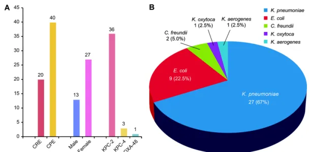
Carbapenemases were responsible for resistance in 40 isolates (66.7% of all 60 ertapenem-resistant Enterobac- teriaceae). The mean age of patients with CPE was 81.4 years (range, 57−92 years), and 27 patients (67.5%) were females (Fig. 1A). The 40 isolates comprised 27 K.
pneumoniae isolates (67.5%), 9 E. coli isolates (22.5%), 2 C. freundii isolates (5%), 1 Klebsiella oxytoca isolate (2.5%), and 1 Klebsiella aerogenes isolate (2.5%) (Fig. 1B).
Antimicrobial susceptibilities
All isolated strains showed a multidrug-resistant (MDR) phenotype. According to the antibiotic suscepti- bility profiles, all 40 isolates were resistant to ampicil- lin, amoxicillin/clavulanic acid, piperacillin/tazobactam, cefazolin, cefoxitin, cefotaxime, ceftazidime, cefepime, azt- reonam, ertapenem, and IMP. The results of fluoro- quinolone susceptibility testing showed that 32 isolates (80%) were resistant to ciprofloxacin, whereas the tri-
methoprim-sulfamethoxazole resistance rates were 72.5%. The aminoglycoside test results showed that 19 strains (47.5%) were resistant to gentamicin and two strains (5%) were intermediately resistant to amikacin.
Tigecycline and colistin resistance was confirmed in two strains (5%). The combination of β-lactamases relative to the susceptibility test results is shown in Table 1.
Distribution of molecular typing
Among the 40 CPE isolates, 36 (90%), 3 (7.5%), and 1 (2.5%) were KPC-2, KPC-4, and OXA-48 producers, respectively (Fig. 1A). We also observed a high preva-
lence of ESBLs. Other β-lactamases were observed as well, including CTX-M-1 group (57.5%), CTX-M-4 group (15%), SHV-like (50%), and TEM-like (32.5%) β-lact- amase isolates. Plasmid-mediated AmpC was rarely co- produced (4/40, 10%); in KPC-2 producers, co-production was observed with DHA (n = 2) and ACC (n = 1), whereas in KPC-4 producers, co-production was only observed with CIT (n = 1). Concerning the analysis of fluoroquinolone resistance determinants for the 40 car- bapenemase strains, 67.5% of the 27 Enterobacteriaceae isolates carried a fluoroquinolone resistance determi- nant. The 40 carbapenemase isolates comprised 22 qnrB Fig. 1. Distribution of species in carbapenemase-producing Enterobacteriaceae (CPE) isolates. (A) Distribution of carbapene- mase genotypes in carbapenem resistant Enterobacteriaceae isolates (n = 60), (B) Distribution of genotypes of CPE isolates (n = 40)
Table 1. Antimicrobial susceptibilities of CPE isolates and phenotypes.
CPE Isolates (%)
Antimicrobial susceptibility
Porin loss (%)
Conjugation No. (%) of isolates
FOX FEP ETP IMP AK GN CIP SXT TIG CST OmpK35 OmpK36
CPE strains (n = 40) 0 0 1 (2.5) 1 (2.5) 39 (97.5) 31 (77.5) 8 (20) 11 (27.5) 38 (95) 38 (95) 18 (45) 17 (42.5) 23 (57.5) KPC-2 0 0 0 0 31 (100) 17 (54.8) 6 (19.4) 8 (25.8) 26 (83.9) 25 (80.6) 15 (48.4) 16 (51.6) 20 (64.6) ΔTn4401a (n=26) 0 0 0 0 26 (100) 13 (50) 6 (23.1) 6 (23.1) 26 (100) 25 (3.85) 14 (53.8) 13 (50) 17 (65.4) ΔTn4401b (n = 5) 0 0 0 0 5 (100) 4 (80) 0 2 (40) 5 (100) 5 (100) 1 (20) 3 (60) 3 (60)
KPC-4 0 0 0 0 2 (66.7) 2 (66.7) 2 (66.7) 2 (66.7) 3 (100) 3 (100) 0 0 1 (33.3)
ΔTn4401b (n = 3) 0 0 0 0 2 (66.7) 2 (66.7) 2 (66.7) 2 (66.7) 3 (100) 3 (100) 0 0 1 (33.3) Total (n = 34) 0 0 0 0 33 (97.1) 19 (55.9) 8 (23.53) 10 (29.42) 29 (85.3) 28 (82.4) 15 (44.1) 16 (47.1) 21 (61.8) Transconjugant
strains (n = 23) 0 0 0 0 23 (100) 14 (60.9) 6 (26.1) 7 (30.4) 22 (95.7) 23 (100) - - - Cured strains (n = 2)1 (50) 1 (50) 2 (100) 1 (50) 2 (100) 2 (100) 1 (50) 1 (50) 2 (100) 2 (100) - - - Abbreviations: CPE, carbapenemase-producing Enterobacteriaceae; FOX, cefoxitin; FEP, cefepime; ETP, ertapenem; IMP, imipenem; AK, amik- acin; GN, gentamicin; CIP, ciprofloxacin; SXT, trimethoprim/sulfamethoxazole; TIG, tigecycline; CST, colistin.
(55%), 8 qnrA (20%), 8 qnrS (20%), and 6 qepA (15%) iso- lates. The analysis of aminoglycoside resistance in these strains showed that 18 strains (45%) displayed resis- tance: 11 rmtB (27.5%), 4 rmtD (10%), 2 rmtC (5%), 2 armA (5%), and 1 rmtA (2.5%) isolates (Table 2). In addi- tion to the production of ESBLs or AmpC enzymes, the deletion or mutation of porins, such as OmpK35 and OmpK36, was reportedly correlated with increased car- bapenem MICs [30]. A multi-agency survey conducted in 2013 confirmed that porins, including OmpK35 and OmpK36, were observed in 27 isolates (67.5%) [31]. In our study, the porins OmpK35 and OmpK36 were
observed in 18 (45%) and 17 (42.5%) isolates, respec- tively. OmpK35 and OmpK36 were both deleted or mutated at the same rate (20%) (Table 3).
MLST
K. pneumoniae was analyzed by MLST based on the analysis of the seven housekeeping genes from the 27 clinical isolates of KPC-producing K. pneumoniae. Using this approach, we observed five sequence types (STs).
Using the K. pneumoniae MLST database, we detected the clones ST307 (18/27, 66.7%) and ST11 (6/27, 22.2%).
Furthermore, three types were identified: ST429 (1/27, 3.7%), ST2521 (1/27, 3.7%), and ST3660 (1/27, 3.7%) (Table 4). E. coli MLST demonstrated that the STs ST410 and ST3520 were the most common among the carbapenemase-producing strains (2/9, 22.2%), followed by ST117 (1/9, 11.1%), ST617 (1/9, 11.1%), ST720 (1/9, 11.1%), ST457 (1/9, 11.1%), and ST52 (1/9, 11.1%).
Isotyping the Tn4401 transposon
The Tn4401 isotype was detected in 34 of the 40 ana- lyzed strains (34/40, 85%). The major isotypes associated with Tn4401 bracketing the blaKPC gene were Tn4401a (26/40, 65%) and Tn4401b (8/40, 20%). Tn4401b was identified in all three strains of blaKPC-4, which is con- sistent with the results of Jeong et al. [25]. For the six non-isotypable transposons (15%) by the Tn4401-isotyp- ing PCR, amplification between the 3′-end of the upstream ISKpn7 and the blaKPC gene was performed, and no amplicons were produced. Regarding strains car- rying Tn4401, 20 strains were identified (20/34, 59%), which increases the risk of carbapenem resistance trans- mission. The transposon isolates are listed in Table 4.
Conjugation
Carbapenem resistance was successfully transferred from CPE strains to E. coli J53 by conjugation (23/40, 57.5%). All transconjugants exhibited significantly reduced carbapenem susceptibility, with ertapenem and imipenem MICs of 4−8 mg/l and 8−16 mg/l, respectively.
In addition, the transconjugants exhibited MDR pheno- types similar to those of the clinical CPE isolate donors.
The transconjugants were also resistant to cephalospo- rins but were susceptible to aminoglycosides (Table 1).
Importantly, the transconjugant assays enabled the simultaneous transfer of blaKPC-2, blaKPC-4, blaOXA- Table 2. Identification of resistance determinants among
the 40 CPE isolates by PCR.
Antibiotic class/
Resistance gene
CPE strains N = 40 (%)
Transconjugants strains N = 23 (%)
Cured strains N = 2 (%) Carbapenems
KPC-2 36 (90) 16 (69.6) 0
KPC-4 3 (7.5) 3 (13.0) 0
OXA-48 1 (2.5) 1 (4.3) 0
Cephalosporins
SHV 20 (50) 11 (47.8) 0
TEM 13 (32.5) 2 (8.7) 0
CTX-M-1 23 (57.5) 7 (30.4) 1 (50)
CTX-M-9 6 (15) 1 (4.3) 0
AmpC
DHA 2 (5) 0 0
CIT 1 (2.5) 0 0
ACC 1 (2.5) 0 0
Aminoglycoside
rmtA 1 (2.5) 0 0
rmtB 11 (27.5) 3 (13.0) 0
rmtC 2 (5) 0 0
rmtD 4 (10) 0 0
armA 2 (5) 2 (8.7) 0
Fluoroquinolone
qnrA 8 (20) 0 0
qnrB 17 (42.5) 12 (52.2) 2 (100)
qnrS 8 (20) 7 (30.4) 0
qepA 6 (15) 1 (4.3) 0
Abbreviations: CPE, carbapenemase-producing Enterobacteriaceae;
KPC, Klebsiella pneumonia carbapenemase; OXA, oxacillinase; SHV, sulfhydryl variant; TEM, Temoneira β-lactamase; CTX-M, cefotaxi- mase.
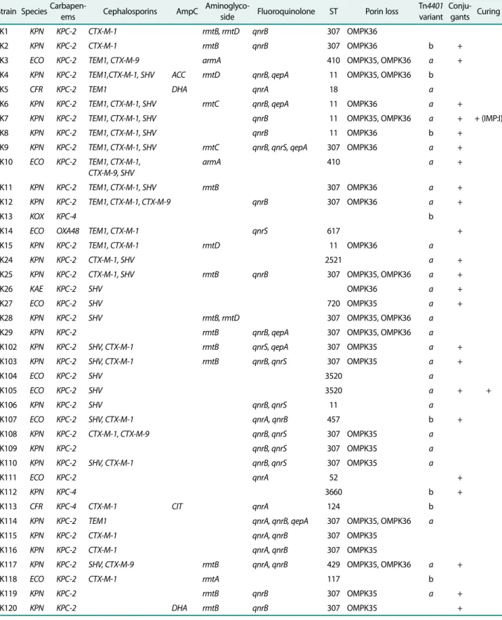
Table 3. Possible genes and outbreaks identified in the CPE collection.
Strain SpeciesCarbapen-
ems Cephalosporins AmpC Aminoglyco-
side Fluoroquinolone ST Porin loss Tn4401 variant
Conju- gants Curing
K1 KPN KPC-2 CTX-M-1 rmtB, rmtD qnrB 307 OMPK36
K2 KPN KPC-2 CTX-M-1 rmtB qnrB 307 OMPK36 b +
K3 ECO KPC-2 TEM1, CTX-M-9 armA 410 OMPK35, OMPK36 a +
K4 KPN KPC-2 TEM1,CTX-M-1, SHV ACC rmtD qnrB, qepA 11 OMPK35, OMPK36 b
K5 CFR KPC-2 TEM1 DHA qnrA 18 a
K6 KPN KPC-2 TEM1, CTX-M-1, SHV rmtC qnrB, qepA 11 OMPK36 a +
K7 KPN KPC-2 TEM1, CTX-M-1, SHV qnrB 11 OMPK35, OMPK36 a + + (IMP:I)
K8 KPN KPC-2 TEM1, CTX-M-1, SHV qnrB 11 OMPK36 b +
K9 KPN KPC-2 TEM1, CTX-M-1, SHV rmtC qnrB, qnrS, qepA 307 OMPK36 a +
K10 ECO KPC-2 TEM1, CTX-M-1, CTX-M-9, SHV
armA 410 a +
K11 KPN KPC-2 TEM1, CTX-M-1, SHV rmtB 307 OMPK36 a +
K12 KPN KPC-2 TEM1, CTX-M-1, CTX-M-9 qnrB 307 OMPK36 a +
K13 KOX KPC-4 b
K14 ECO OXA48 TEM1, CTX-M-1 qnrS 617 +
K15 KPN KPC-2 TEM1, CTX-M-1 rmtD 11 OMPK36 a
K24 KPN KPC-2 CTX-M-1, SHV 2521 a +
K25 KPN KPC-2 CTX-M-1, SHV rmtB qnrB 307 OMPK35, OMPK36 a +
K26 KAE KPC-2 SHV OMPK36 a +
K27 ECO KPC-2 SHV 720 OMPK35 a +
K28 KPN KPC-2 SHV rmtB, rmtD 307 OMPK35, OMPK36 a
K29 KPN KPC-2 rmtB qnrB, qepA 307 OMPK35, OMPK36 a
K102 KPN KPC-2 SHV, CTX-M-1 rmtB qnrS, qepA 307 OMPK35 a +
K103 KPN KPC-2 SHV, CTX-M-1 rmtB qnrB, qnrS 307 OMPK35 a +
K104 ECO KPC-2 SHV 3520 a
K105 ECO KPC-2 SHV 3520 a + +
K106 KPN KPC-2 SHV qnrB, qnrS 11 a
K107 ECO KPC-2 SHV, CTX-M-1 qnrA, qnrB 457 b +
K108 KPN KPC-2 CTX-M-1, CTX-M-9 qnrB, qnrS 307 OMPK35 a
K109 KPN KPC-2 qnrB, qnrS 307 OMPK35 a
K110 KPN KPC-2 SHV, CTX-M-1 qnrB, qnrS 307 OMPK35 a
K111 ECO KPC-2 qnrA 52 +
K112 KPN KPC-4 3660 b +
K113 CFR KPC-4 CTX-M-1 CIT qnrA 124 b
K114 KPN KPC-2 TEM1 qnrA, qnrB, qepA 307 OMPK35, OMPK36 a
K115 KPN KPC-2 CTX-M-1 qnrA, qnrB 307 OMPK35
K116 KPN KPC-2 CTX-M-1 qnrA, qnrB 307 OMPK35
K117 KPN KPC-2 SHV, CTX-M-9 rmtB qnrA, qnrB 429 OMPK35, OMPK36 a +
K118 ECO KPC-2 CTX-M-1 rmtA 117 b
K119 KPN KPC-2 rmtB qnrB 307 OMPK35 a +
K120 KPN KPC-2 DHA rmtB qnrB 307 OMPK35 +
Abbreviations: CPE, carbapenemase-producing Enterobacteriaceae; KPN, Klebsiella pneumonia; ECO, Escherichia coli; CFR, Citrobacter freundii; KOX, Klebsiella oxytoca; KAE, Klebsiella aerogenes; ST, sequence type; IMP, imipenem.
48, ESBL, and AmpC, as well as aminoglycoside and flu- oroquinolone resistance-determining genes (Table 2).
Curing test
A curing test was performed to artificially remove the plasmid containing the KPC-2 gene. However, curing was successfully performed in only two strains, K7 and K105 (2/40, 5%). In each of these strains, curing signifi- cantly reduced carbapenem resistance, with imipenem and ertapenem MICs of ≤ 0.5 mg/l and 0.5−2 mg/l, respectively. Other antibiotics that showed similar effects on the clinical CPE isolate donors are also shown in Table 1.
Discussion
In the 1940s, the first antibiotics were derived from mold; however, since then, advances in antibiotic devel- opment has relied on chemical synthesis. However, with the emergence of multidrug resistance, the end of the antibiotic era may be near. The World Health Organiza- tion warns about a post-antibiotic era, in which no anti- biotics will be able to kill bacteria because of their widespread resistance. Currently, hospitals face emerg- ing resistance to third-generation cephalosporins and fluoroquinolones by Staphylococcus aureus and Entero- coccus. Recently, there has been growing speculation about the impossibility of treating CRE in a post-antibi- otic era. To the best of our knowledge, this is the first study to conduct molecular genetic analysis of CPE at a single hospital in Korea since the conduction of the CRE survey by the Korea CDC in June 2017. Here, we described the distribution of major β-lactamases in a group of extensively drug-resistant (XDR) Enterobacteri- aceae isolates from a single hospital, presenting several important results.
Antibiotic resistance is increasing, and, therefore, a new classification termed “XDR” has been created. Fluo- roquinolones are important therapeutics used to treat both human and animal infections [32]. The introduction of fluoroquinolones has offered clinicians the ability to treat human cases of complicated urinary tract, gastro- intestinal, and respiratory tract infections as well as sex- ually transmitted diseases [33]. In veterinary medicine, fluoroquinolones have served as effective therapeutics for treating enteric infections and respiratory diseases in food-producing and companion animals [34]. Owing to their antimicrobial activity against a broad spectrum of pathogenic bacteria, advantageous pharmacokinetic characteristics, and low toxicity, fluoroquinolones have become attractive for use in farm animals [35, 36]. Since the late 1980s, fluoroquinolones used in human medi- cine have differed from those used in veterinary medi- cine [37, 38]. Nonetheless, a public health concern remains, i.e. the use of fluoroquinolones in livestock may result in bacterial resistance that can be transmitted through the food chain. This can be explained by cross- resistance between fluoroquinolones and enrofloxacin, a commonly used agent in farm animals, which is par- tially metabolized to ciprofloxacin within animals [39, 40]. Thus, resistance to antibiotics has continuously remained in the food chain and may be permanent; there- fore, close management and restrictions are required in the future. Additionally, there is also an emerging resis- tance to colistin and tigecycline, last-resort antibiotics for CRE treatment.
Interestingly, our study showed that CRE is more sus- ceptible to amikacin, an antibiotic of the aminoglycoside family. The EUCAST(European Committee on Antimi- crobial Susceptibility Testing) guidelines state that the amikacin susceptibility status must be revised to “inter- mediate” should a member of the Enterobacteriaceae test Table 4. blaKPCmobile elements carried by KPC producers.
Strain Sequence type
Klebsiella pneumoniae Escherichia coli Citrobacter
freundii
Klebsiella oxytoca
Klebsiella aerogenes
307 11 429 2521 3660 410 720 3520 457 117 124 18 - -
blaKPC-2 Tn4401a 13 (32.5) 4 (10) 1 (2.5) 1 (2.5) - 2 (5) 1 (2.5) 2 (5) - - - 1 (2.5) - 1 (2.5)
blaKPC-2 Tn4401b 1 (2.5) 2 (5) - - - 1 (2.5) 1 (2.5) - - - -
blaKPC-4 Tn4401b - - - - 1 (2.5) - - - 1 (2.5) - 1 (2.5) -
Abbreviations: KPC, Klebsiella pneumoniae carbapenemase.
as tobramycin intermediate or resistant, and gentamicin and amikacin susceptible [41].
Therefore, we recommend the sparingly use of amika- cin to treat intestinal bacterial infections resistant to other aminoglycosides.
The most predominant KPC-producing clone reported globally is K. pneumoniae ST258 [42, 43]; however, this was not the case in South Korea. Rather, the predomi- nant South Korean KPC producer was the K. pneumo- niae ST307 clone. ST11 was also observed in Korea at a significantly lower prevalence than ST307. According to a study by Jeong et al., the four K. pneumoniae sub- clones ST307/Tn4401a[blaKPC-2], ST307/Tn4401b[bla KPC-4], ST392/Tn4401b[blaKPC-4], and ST11/ND[bla KPC-2] triggered inter-regional dissemination of KPC producers in Korea [25]. In this study, various K. pneu- moniae subclones and E. coli subclones were isolated;
the primary K. pneumoniae subclones observed were ST307/Tn4401a[blaKPC-2], ST307/Tn4401b[blaKPC-4], ST11/Tn4401a[blaKPC-2], and Tn4401b[blaKPC-2], while the observed E. coli clones were ST410/Tn4401a [blaKPC-2] and ST3520/Tn4401a[blaKPC-2]. Impor- tantly, the Tn4401 isotypes differed in the promoter of the blaKPC gene, which was closely associated with the level of gene expression and caused variation in carbape- nem susceptibility [12]; however, there was no signifi- cant difference in susceptibility in this study.
K. pneumoniae ST307 and E. coli ST410 have gar- nered attention as potentially high-risk CPE producers and are associated with a notorious clone, CTX-M-15 [44]. Furthermore, clones co-producing CPE and CTX- M-15 were recently identified in Italy [45] and Denmark [46]. The high distribution of CTX-M-1 reported in this study highlights the risk of emergence and spread of high-risk clones (Table 1). High-risk clones are globally distributed and associated with various antimicrobial resistance determinants, such as ease of transmission, persistence in hosts, and effective transmission between hosts. These high-risk clones possess enhanced pathoge- nicity and are more prone to cause severe and recurrent infections. Thus, considering the clonal expansion over the past decades and increased antimicrobial resistance, ST307 and ST410 should be prospectively monitored in South Korea.
The CPE incidence and molecular genetic traits observed at this hospital revealed trends similar to those
reported in the Disease Control Headquarters survey conducted in 2017 [47]. However, the CPE incidence rate increased by 25% and that of CRE increased by 5% in 2018 relative to those in 2017. While the previous CRE occurrence in Korea was characterized by ESBL, AmpC, or porin loss, the current study reveals that carbapene- mase-producing bacteria currently represent the great- est risk to public health [31]. Transfer by conjugation accounted for 57.5% of resistant isolates, which is 4.3- fold higher than that found in the 2010 study [14]. Fur- thermore, the high detection rate found for transposon Tn4401 revealed the rapid spread and evolution of CPE.
In summary, our study highlights that carbapenem resistance can be transferred among strains through transconjugation processes, which results in a higher number of strains with reduced carbapenem susceptibil- ity. We found that the causes of carbapenem resistance spread were plasmids and transposons; therefore, more efforts are needed to control the spread of CPE in hospi- tals. The treatment of CRE infections often involves a combination therapy, including carbapenem, aminogly- cosides, and fosfomycin; however, this therapeutic option is not yet available in South Korea. Thus, medical insti- tutions should monitor whether carbapenem-resistant enterobacteria-infected patients are isolated, and if so, these institutions should prevent the spread through infection control measures, such as patient contact trac- ing, thorough use of personal protective equipment, and contact inspection. Systematic monitoring and regula- tion alone will enable better control and prevent the spread of KPC producers.
Acknowledgment
This research was supported by the BHS Hanseo Hospital in 2019.
Conflict of Interest
The authors have no financial conflicts of interest to declare.
References
1. O’Neill J. 2014. Antimicrobial resistance: Tackling a crisis for the health and wealth of nations. Review of antimicrobial resistance.
Available from https://amr-review.org/sites/default/files/AMR%20 Review%20Paper%20-%20Tackling%20a%20crisis%20for%20the
%20health%20and%20wealth%20of%20nations_1.pdf. Accessed
Nov. 20, 2019.
2. Centers for Disease Control and Prevention. 2013. Antibiotic resis- tance threats in the United States, 2013. Available from https://
www.cdc.gov/drugresistance/pdf/ar-threats-2013-508.pdf. Accessed Nov. 17, 2019.
3. Kim JO, Song SA, Yoon EJ, Shin JH, Lee H, Jeong SH, et al. 2017.
Outbreak of KPC-2-producing Enterobacteriaceae caused by clonal dissemination of Klebsiella pneumoniae ST307 carrying an IncX3-type plasmid harboring a truncated Tn4401a. Diagn.
Microbiol. Infect. Dis. 87: 343-348.
4. Jeong SH, Kim HS, Kim JS, Shin DH, Kim HS, Park MJ, et al. 2016.
Prevalence and molecular characteristics of carbapenemase- producing Enterobacteriaceae from five hospitals in Korea. Ann.
Lab. Med. 36: 529-535.
5. Mouloudi E, Protonotariou E, Zagorianou A, Iosifidis E, Karapa- nagiotou A, Giasnetsova T, et al. 2010. Bloodstream infections caused by metallo-β-lactamase/Klebsiella pneumoniae carbap- enemase-producing K. pneumoniae among intensive care unit patients in Greece: Risk factors for infection and impact of type of resistance on outcomes. Infect. Control Hosp. Epidemiol. 31: 1250- 1256.
6. Bradford PA, Urban C, Mariano N, Projan SJ, Rahal JJ, Bush K.
1997. Imipenem resistance in Klebsiella pneumoniae is associated with the combination of ACT-1, a plasmid-mediated AmpC β-lac- tamase, and the foss of an outer membrane protein. Antimicrob.
Agents Chemother. 41: 563-569.
7. Kaczmarek FM, Dib-Hajj F, Shang W, Gootz TD. 2006. High-level carbapenem resistance in a Klebsiella pneumoniae clinical isolate is due to the combination of blaACT-1 β-lactamase production, porin OmpK35/36 insertional inactivation, and down-regulation of the phosphate transport porin PhoE. Antimicrob. Agents Chemother. 50: 3396-3406.
8. Nordmann P, Dortet L, Poirel L. 2012. Carbapenem resistance in Enterobacteriaceae: here is the storm! Trends Mol. Med. 18: 263- 272.
9. Giani T, Pini B, Arena F, Conte V, Bracco S, Migliavacca R, et al.
2013. Epidemic diffusion of KPC carbapenemase-producing Klebsiella pneumoniae in Italy: Results of the first countrywide survey, 15 May to 30 June 2011. Euro. Surveill. 18: 20489.
10. Lee CR, Lee JH, Park KS, Kim YB, Jeong BC, Lee SH. 2016. Global dissemination of carbapenemase-producing Klebsiella pneumo- niae: Epidemiology, genetic context, treatment options, and detection methods. Front. Microbiol. 7: 895.
11. Chen YT, Lin JC, Fung CP, Lu PL, Chuang YC, Wu TL, et al. 2014.
KPC-2-encoding plasmids from Escherichia coli and Klebsiella pneumoniae in Taiwan. J. Antimicrob. Chemother. 69: 628-631.
12. Frost L, Leplae R, Summers AO, Toussaint A. 2005. Mobile genetic elements: The agents of open source evolution. Nat. Rev. Micro- biol. 3: 722-732.
13. Sota M, Top E. 2008. Horizontal gene transfer mediated by plas- mids, pp. 111-181. In Lipps G (ed.), Plasmids: Current Research and Future Trends. Caister Academic Press, Horizon Scientific Press, Norfolk, VA.
14. Frost LS, Koraimann G. 2010. Regulation of bacterial conjugation:
Balancing opportunity with adversity. Future Microbiol. 5: 1057- 1071.
15. Smillie C, Garcillán-Barcia MP, Francia MV, Rocha EP, de la Cruz F.
2010. Mobility of plasmids. Microbiol. Mol. Biol. Rev. 74: 434-452.
16. Guglielmini J, Quintais L, Garcillán-Barcia MP, de la Cruz F, Rocha EP. 2011. The repertoire of ICE in prokaryotes underscores the unity, diversity, and ubiquity of conjugation. PLoS Genet. 7:
e1002222.
17. Naas T, Cuzon G, Truong HV, Nordmann P. 2012. Role of ISKpn7 and deletions in blaKPC gene expression. Antimicrob. Agents Chemother. 56: 4753-4759.
18. Cicek AC, Duzgun AO, Saral A, Sandalli C. 2014. Determination of a novel integron-located variant (blaOXA-320) of Class D β-lact- amase in Proteus mirabilis. J. Basic Microbiol. 54: 1030-1035.
19. Clinical and Laboratory Standards Institute. 2018. Methods for dilution antimicrobial susceptibility tests for bacteria that grow aerobically. Wayne, Pennsylvania.
20. Jeong S, Kim JO, Jeong SH, Bae IK, Song W. 2015. Evaluation of peptide nucleic acid-mediated multiplex real-time PCR kits for rapid detection of carbapenemase genes in gram-negative clini- cal isolates. J. Microbiol. Methods. 113: 4-9.
21. Pérez-Pérez FJ, Hanson ND. 2002. Detection of plasmid-mediated AmpC β-lactamase genes in clinical isolates by using multiplex PCR. J. Clin. Microbiol. 40: 2153-2162.
22. Ryoo NH, Kim EC, Hong SG, Park YJ, Lee K, Bae IK, et al. 2005. Dis- semination of SHV-12 and CTX-M-type extended-spectrum β- lactamases among clinical isolates of Escherichia coli and Klebsi- ella pneumoniae and emergence of GES-3 in Korea. J. Antimicrob.
Chemother. 56: 698-702.
23. Yamane K, Wachino J, Suzuki S, Arakawa Y. 2008. Plasmid-medi- ated qepA gene among Escherichia coli clinical isolates from Japan. Antimicrob. Agents Chemother. 52: 1564-1566.
24. Landman D, Bratu S, Quale J. 2009. Contribution of OmpK36 to carbapenem susceptibility in KPC-producing Klebsiella pneumo- niae. J. Med. Microbiol. 58: 1303-1308.
25. Yoon E-J, Kim JO, Kim D, Lee H, Yang JW, Lee KJ, et al. 2018. Klebsi- ella pneumonia carbapenemase producers in South Korea between 2013 and 2015. Front. Microbiol. 9: 56.
26. Wirth T, Falush D, Lan R, Colles F, Mensa P, Wieler LH, et al. 2006.
Sex and virulence in Escherichia coli: an evolutionary perspective.
Mol. Microbiol. 60: 1136-1151.
27. Diancourt L, Passet V, Verhoef J, Grimont PA, Brisse S. 2005. Multi- locus sequence typing of Klebsiella pneumoniae nosocomial iso- lates. J. Clin. Microbiol. 43: 4178-4182.
28. Jeong SH, Lee KM, Lee J, Bae IK, Kim JS, Kim HS, et al. 2015. Clonal and horizontal spread of the blaOXA-232 gene among Enterobacte- riaceae in a Korean hospital. Diagn. Microbiol. Infect. Dis. 82: 70-72.
29. Leavitt A, Chmelnitsky I, Ofek I, Carmeli Y, Navon-Venezia S. 2010.
Plasmid pKpQIL encoding KPC-3 and TEM-1 confers carbapenem resistance in an extremely drug-resistant epidemic Klebsiella pneumoniae strain. J. Antimicrob. Chemother. 65: 243-248.
30. Tsai YK, Fung CP, Lin JC, Chen JH, Chang FY, Chen TL, et al. 2011.
Klebsiella pneumoniae outer membrane porins OmpK35 and OmpK36 play roles in both antimicrobial resistance and viru- lence. Antimicrob. Agents Chemother. 55: 1485-1493.
31. Kim SY, Shin J, Shin SY, Ko KS. 2013. Characteristics of carbape- nem-resistant Enterobacteriaceae isolates from Korea. Diagn.
Microbiol. Infect. Dis. 76: 486-490.
32. Schulz J, Kemper N, Hartung J, Janusch F, Mohring SAI, Hamscher G. 2019. Analysis of fluoroquinolones in dusts from intensive live- stock farming and the co-occurrence of fluoroquinolone-resis- tant Escherichia coli. Sci. Rep. 9: 5117.
33. Dalhoff A. 2012. Global fluoroquinolone resistance epidemiol- ogy and implications for clinical use. Interdiscip. Perspect. Infect.
Dis. 2012: 976273.
34. Sárközy G. 2001. Quinolones: a class of antimicrobial agents. Vet.
Med. 46: 257-274.
35. Grobbel M, Lübke-Becker A, Wieler LH, Froyman R, Friederichs S, Filios S. 2007. Comparative quantification of the in vitro activity of veterinary fluoroquinolones. Vet. Microbiol. 124: 73-81.
36. Frye JG, Jackson CR. 2013. Genetic mechanisms of antimicrobial resistance identified in Salmonella enterica, Escherichia coli, and Enteroccocus spp. isolated from U.S. food animals. Front. Micro- biol. 4: 135.
37. Hopkins KL, Davies RH, Threlfall EJ. 2005. Mechanisms of quino- lone resistance in Escherichia coli and Salmonella: Recent devel- opments. Int. J. Antimicrob. Agents 25: 358-373.
38. Endtz HP, Ruijs GJ, van Klingeren B, Jansen WH, van der Reyden T, Mouton RP. 1991. Quinolone resistance in campylobacter iso- lated from man and poultry following the introduction of fluoro- quinolones in veterinary medicine. J. Antimicrob. Chemother. 27:
199-208.
39. García Ovando H, Gorla N, Luders C, Poloni G, Errecalde C, Prieto G, et al. 1999. Comparative pharmacokinetics of enrofloxacin and
ciprofloxacin in chickens. J. Vet. Pharmacol. Ther. 22: 209-212.
40. van den Bogaard AE, London N, Driessen C, Stobberingh EE.
2001. Antibiotic resistance of faecal Escherichia coli in poultry, poultry farmers and poultry slaughterers. J. Antimicrob. Chemo- ther. 47: 763-771.
41. Leclercq R, Cantón R, Brown DF, Giske CG, Heisig P, MacGowan AP, et al. 2013. EUCAST expert rules in antimicrobial susceptibility testing. Clin. Microbiol. Infect. 19: 141-160.
42. Sheppard AE, Stoesser N, Wilson DJ, Sebra R, Kasarskis A, Anson LW, et al. 2016. Nested Russian doll-like genetic mobility drives rapid dissemination of the carbapenem resistance gene blaKPC. Antimicrob. Agents Chemother. 60: 3767-3778.
43. Pitout JD, Nordmann P, Poirel L. 2015. Carbapenemase-produc- ing Klebsiella pneumoniae, a key pathogen set for global nosoco- mial dominance. Antimicrob. Agents Chemother. 59: 5873-5884.
44. Villa L, Feudi C, Fortini D, Brisse S, Passet V, Bonura C, et al. 2017.
Diversity, virulence, and antimicrobial resistance of the KPC-pro- ducing Klebsiella pneumoniae ST307 clone. Microb. Genom. 3:
e000110.
45. Geraci DM, Bonura C, Giuffrè M, Saporito L, Graziano G, Aleo A, et al. 2015. Is the monoclonal spread of the ST258, KPC-3-produc- ing clone being replaced in southern Italy by the dissemination of multiple clones of carbapenem-nonsusceptible, KPC-3-pro- ducing Klebsiella pneumoniae? Clin. Microbiol. Infect. 21: e15-e17.
46. Roer L, Overballe-Petersen S, Hansen F, Schønning K, Wang M, Røder BL, et al. 2018. Escherichia coli sequence type 410 is caus- ing new international high-risk clones. mSphere 3: e00337-18.
47. Korea Centers for Disease Control and Prevention. Distribution of carbapenem-resistant Enterobacteriaceae (CRE) in Korea, 2017.
Available from https://is.cdc.go.kr/upload_comm/syview/doc.
html?fn=156811210482900.pdf&rs=/upload_comm/docu/0034/.
Accessed Dec. 11, 2019.